
Double-targeting fluorescent probe capable of simultaneously visualizing plasma membrane and lysosome and application of double-targeting fluorescent probe

A fluorescent probe and dual-targeting technology, applied in the field of probes for displaying plasma membranes and lysosomes, can solve problems such as inability to meet imaging requirements, and achieve universal, low-cost, and easy-to-scale production Effect

Active Publication Date: 2022-02-11
QILU UNIV OF TECH
View PDF5 Cites 3 Cited by
  • Summary
  • Abstract
  • Description
  • Claims
  • Application Information

AI Technical Summary

Problems solved by technology

The inventor previously designed a single-molecule fluorescent probe with controllable spatial distribution, and the staining position is the plasma membrane and mitochondria, but further research found that this single-molecule fluorescent probe cannot meet the requirements of imaging plasma membrane and lysosome at the same time

Method used


Image

  • Double-targeting fluorescent probe capable of simultaneously visualizing plasma membrane and lysosome and application of double-targeting fluorescent probe
  • Double-targeting fluorescent probe capable of simultaneously visualizing plasma membrane and lysosome and application of double-targeting fluorescent probe
  • Double-targeting fluorescent probe capable of simultaneously visualizing plasma membrane and lysosome and application of double-targeting fluorescent probe

Examples


Embodiment 1

[0041] MEM-LYSO synthesis and characterization

[0042] Compound 2 is 4-methyl-1-octadecylpyrid-1-iodide (0.357 g, 0.754 mmol) and anhydrous methanol (25 mL), add three flasks, 55 ° C is heated to all dissolved, and then Compound 4, 6- (dimethylamino) -2-nolds (0.177 g, 0.754 mmol) and piperidine (6 droplets). After the addition is completed, the temperature is increased and refluxed to reddish brown solid precipitation, resulting in (E) -4- (2- (6- (dimethylamino) naphthalene-2-yl) vinyl) -1-octadecylpyridine - 1-ammonium iodide, MEM-LYSO, 0.22 g, and a yield of 45%.

[0043] 1 H NMR (DMSO-D 6 ., 300MHz): δ (PPM): 8.80 (D, J = 8 Hz, 2H), 8.11 (D, J = 8 Hz, 2H), 8.02 (D, J = 16 Hz, 1H), 7.91 (S, 1H), 7.67 (m, 3H), 7.39 (D, J = 16Hz, 1H), 7.17 (m, 1H), 6.88 (S, 1H), 4.37 (D, J = 16 Hz, 2H), 2.98 (S, 6H), 1.16 (m, 32H), 0.76 (D, J = 12 Hz, 3H) .hrms (m / z): Calcd for c 37 Hide 55 In 2 : 654.34; Found: 527.44 (m-i) + .

Embodiment 2

[0045] MEM-LYSO Optical Performance Test

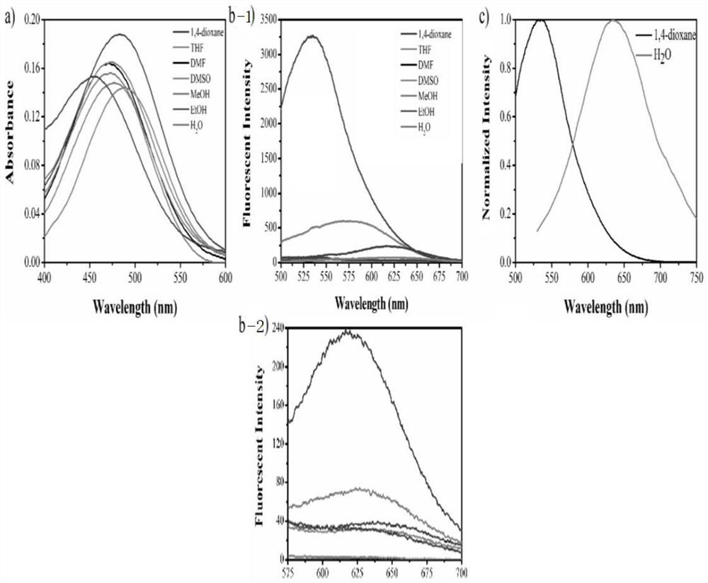
[0046] First, select the chromatographic pure solution of polarity as a solvent, and seven 6 solvents such as 1,4-dioxane, THF and DMF were selected. 5 μl of probe molecules were added thereto, and a probe molecular solution was formulated into a final concentration of 10 μm. It is then collected, and its absorption and fluorescence spectroscopy are collected, and the maximum emission peak is analyzed to obtain data of its absorption and emission spectrum, and the state of the Stokes displacement, the molar absorbance coefficient, the fluorescence quantum efficiency and other light physical parameters are calculated.

[0047] See figure 1 .

[0048] figure 1 : MEM-LYSO absorption spectroscopy (A), fluorescence spectrum (B) and normalized fluorescence spectroscopy (C) in different solvents. Concentration: 10 μm.

Embodiment 3

[0050] Stained living cell

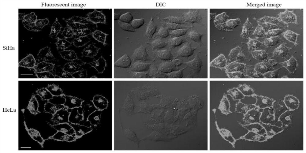
[0051] According to the initial design, the probe MEM-LYSO can target the plasma film and lysosomes in the cell. To verify this feature, first use MEM-Lyso to dye the living cells to see if it can simultaneously imaging the plasma membrane and lysosomes. figure 2 Indicated. from figure 2 In the contour of the cells, the contour of the cells have green fluorescently drawn, which demonstrates that MEM-Lyso can be accurately imaging film.

[0052] See figure 2 .

[0053] figure 2 : Fluorescent images of SiHA and HeLa cells stained with MEM-LYSO, DIC and their superimposed images (2 μm, 10 min). λ ex = 488 nm, λ em = 500-600 nm, BAR = 20 μm.


PUM


Abstract

The invention belongs to the field of fluorescence detection, and relates to a double-targeting fluorescent probe capable of simultaneously visualizing plasma membranes and lysosomes and application of the double-targeting fluorescent probe, the probe is superior to the application of the conventional probe which can only display one observation substance, and the probe can simultaneously mark the plasma membranes and the lysosomes and enable the plasma membranes and the lysosomes to display different colors. Mem-Lyso can be used for marking plasma membranes and lysosomes with red and yellow respectively, so that the relative positions of two organelles of the zebra fish are displayed through real color imaging. More importantly, the Mem-Lyso has the capability of spatially distinguishing the content of cytomembrane cholesterol (CL) due to the double-targeting characteristic, and a potential detection tool is provided for biological research and diagnosis of related diseases, so that research on the relationship between the plasma membrane and the lysosome has important significance for related biological research.

Description

Technical field [0001] The present invention relates to a probe for displaying a plasma film and a lysosomer, and more particularly to a double-targeting probe that simultaneously displays a plasma film and a lysosoma and its probe imaging that can only be observed to show a substance. Applications. Background technique [0002] The information disclosed in the background is disclosed only to increase the understanding of the overall background of the present invention, and is not necessarily considered to admit or imply that the information is intended to be a prior art known to those skilled in the art. [0003] It is well known that cells are basic units of organisms, many biologists and medical scares to analyze the structure of cells. The cells are composed of a plasma film and a variety of orpatipulary to form an organelle interaction network as a functional area. In the interaction network, the plastic film is a barrier that prevents extracellular substances from entering ...

Claims


Application Information

Patent Timeline
Patent Type & Authority Applications(China)
IPC IPC(8): C07D213/38C09K11/06G01N1/31G01N21/64A61K49/00
CPCC07D213/38C09K11/06G01N21/6486G01N21/6428G01N1/31A61K49/0021G01N2021/6439C09K2211/1029C09K2211/1011
Inventor 李学晨吴士宁闫梦琪于诗摩崔月芝
Owner QILU UNIV OF TECH
Learn More