Magnetofluid composite developing agent and application thereof in field of intraosseous angiography

A technology of contrast agent and magnetic fluid, which is applied in the field of magnetic fluid composite contrast agent and intraosseous blood vessel imaging and intraosseous blood vessel imaging, which can solve the problems of local image vacancies and inability to meet the needs of clinical diagnosis and treatment.

Pending Publication Date: 2022-03-22
青岛山大齐鲁医院(山东大学齐鲁医院(青岛))
View PDF2 Cites 0 Cited by
  • Summary
  • Abstract
  • Description
  • Claims
  • Application Information

AI Technical Summary

Problems solved by technology

For the above method ③, steady-state (SS) magnetic resonance angiography with MR susceptibility weighting and submillimeter resolution and three-dimensional reconstruction, we use Gd-DTPA vascular enhancer. For bone arteries, we repeated this experiment, and the results were basically the same as experiment ②. Only the segment with a relatively large diameter (diameter 1mm) just entering the bone can be imaged. We tried subtraction to remove the bone, and the result was that the blood vessels in the bone were simultaneously reduced. drop, there is a partial image vacancy ( Figure 5 Middle c-d), this result does not meet the needs of clinical diagnosis and treatment

Method used


Image

  • Magnetofluid composite developing agent and application thereof in field of intraosseous angiography
  • Magnetofluid composite developing agent and application thereof in field of intraosseous angiography
  • Magnetofluid composite developing agent and application thereof in field of intraosseous angiography

Examples


Embodiment 1

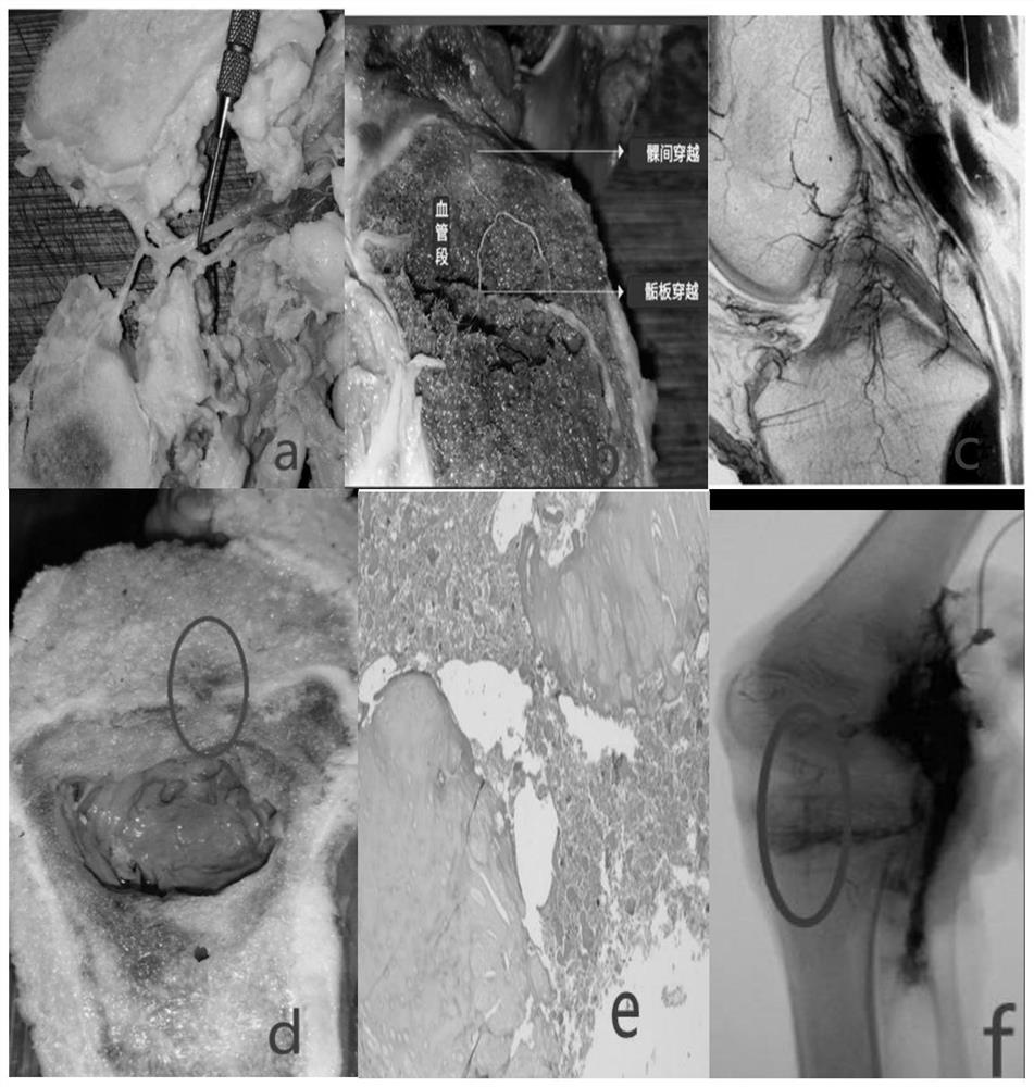
[0029] Previous studies used methods such as dissection and tissue sectioning to clarify the existence and distribution of blood vessels in the knee joint. However, it is difficult to realize intraosseous blood vessel imaging in vivo with existing hardware, software and methods. In this embodiment, by enhancing the concentration of the local contrast agent, it is intended to realize the development of the original development restricted area, such as intraosseous blood vessel imaging.

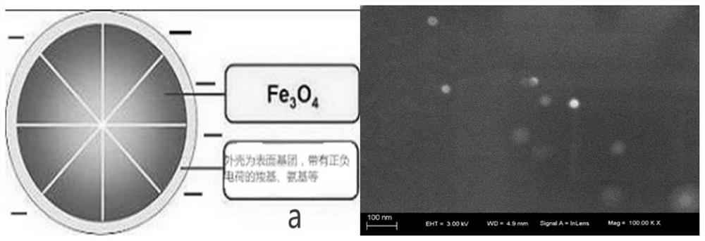
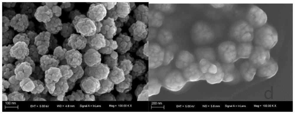
[0030] The raw material of this embodiment is selected diatrizoate meglumine compound injection (Guoyao Zhunzi H37023984, 20nm, 20ml, each containing diatrizoate sodium 32mg, diatrizoate meglumine 268mg, Lunan Beite Pharmaceutical Co., Ltd.) and amino trioxide Ferromagnetic nano-microsphere dispersion (100nm, 10ml: 50mg, Tianjin Basele Chromatography Technology Development Center).

[0031] Prepare 10ml of diatrizoate meglumine injection at 20°C, containing 16mg of sodium diatrizoate and 134mg ...

experiment example

[0039] research object

[0040] Inclusion criteria: ①SPF grade New Zealand rabbits, weight 2.6±0.4kg, age 9±0.5 months; ②healthy appearance, normal skeletal development and normal renal function; ③clear ear veins, convenient for intravenous general anesthesia; ④basic experimental research.

[0041] Exclusion criteria: ①Death during anesthesia; ②Artifacts produced by neodymium magnets in CT scans affecting the observation area; ③During CT scanning, the experimental rabbit excreted urine and caused diffusion of the contrast agent; ④Poor quality of tissue sections.

[0042] research object

[0043] According to the above inclusion and exclusion criteria, 20 experimental rabbits were purchased from Qingdao Kangda Biotechnology Co., Ltd. In the end, 1 case died during the anesthesia process, 2 cases were affected by artifacts caused by neodymium magnets in CT scanning, 1 case caused the contrast agent to disperse due to excretion of rabbit urine during CT scanning, and 1 case had ...


PUM

PropertyMeasurementUnit
particle diameteraaaaaaaaaa

Abstract

The invention belongs to the technical field of intraosseous angiography, and particularly relates to a magnetofluid composite developing agent and application thereof in the field of intraosseous angiography of living bodies. According to the magnetic fluid composite developing agent, a method of modifying magnetic microspheres is utilized, ferroferric oxide particles with shells being negative charges are used for adsorbing the surface of an ionic contrast agent to form a magnetic fluid composite structure in a coating mode, and the magnetic fluid composite structure is formed based on the characteristics that magnetic fluid has strong paramagnetism under the action of a magnetic field and can achieve directional movement and local aggregation in a human body; under the action of an external magnetic field, the developing agent can be concentrated in vivo, so that blood vessel imaging in a living body bone is realized, and the developing quality and effect of the blood vessel in the living body bone can be improved.

Description

technical field [0001] The invention belongs to the technical field of intraosseous blood vessel imaging, and in particular relates to a magnetic fluid composite imaging agent and its application in the field of intraosseous blood vessel imaging. Background technique [0002] Every bone is an organ, and exploring the distribution of blood vessels in the bone is the basis for exploring bone growth, disease, and injury mechanisms. Supply and transmission pathways, surgical approaches and methods, blood loss prediction and preoperative design for complex orthopedic surgery cover almost all branches of orthopedics, all of which have a fundamental role and are of positive significance. At present, the anatomical method of intraosseous blood vessels and the imaging technology of intraosseous blood vessels in vivo are still difficult problems and technical gaps in orthopedics, and are one of the bottlenecks in the basic and clinical development of orthopedics. [0003] For the stu...

Claims


Application Information

Patent Timeline
Patent Type & Authority Applications(China)
IPC IPC(8): A61K49/18B82Y15/00B82Y5/00
CPCA61K49/1824B82Y15/00B82Y5/00
Inventor 程坤李秋尧高晓斐杨志成冯琨邵显昊侯海青李建民
Owner 青岛山大齐鲁医院(山东大学齐鲁医院(青岛))
Why Eureka
Try Eureka