Multi-modality imaging microbubble structure, preparation method and applications

A multi-modal imaging and micro-bubble technology, applied in the field of materials and nano-medicine, biomedicine, can solve the problem that the resolution and accuracy of ultrasonic imaging cannot meet the requirements of advanced clinical diagnosis, and achieve the convenience of repeated inspection and enhanced ultrasonic imaging. Simple effect of image and synthesis process

Inactive Publication Date: 2012-11-14
CHONGQING MEDICAL UNIVERSITY
View PDF4 Cites 5 Cited by
  • Summary
  • Abstract
  • Description
  • Claims
  • Application Information

AI Technical Summary

Problems solved by technology

However, due to the limitations of the ultrasonic diagnostic method itself, the resolution and accuracy of ultrasonic imaging are far from meeting the requirements of advanced clinical diagnosis.

Method used


Image

  • Multi-modality imaging microbubble structure, preparation method and applications
  • Multi-modality imaging microbubble structure, preparation method and applications
  • Multi-modality imaging microbubble structure, preparation method and applications

Examples


Embodiment 1

[0033] Example 1 Preparation of multimodal microbubbles of the present invention

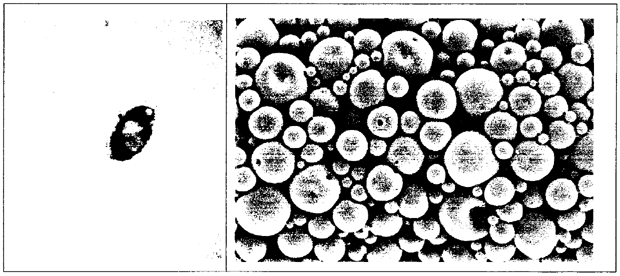
[0034]Dissolve 50mg PLGA (50:50, MW 12,000) and 0.15mg PE in 2ml dichloromethane solvent with an electrolytic balance, add 100μl water-soluble quantum dots after complete dissolution, and vibrate for 35-45s to obtain the primary emulsion (W / O ); the primary emulsion is mixed with 6ml 4% polyvinyl alcohol solution, and acts on 9600r / min dispersion homogeneous equipment 5min, obtains microsphere (W / O / W), adds 5ml 2% isopropanol solution, room temperature Stir with a magnetic stirrer at a constant speed for 2-5 hours to solidify the surface of the microspheres and volatilize the dichloromethane naturally as much as possible. The above liquid was divided into 5ml centrifuge tubes, and the supernatant was discarded by high-speed centrifugation (3500rpm, 5min). Add an appropriate amount of double distilled water again, mix thoroughly with a vortex mixer, wash, centrifuge, and discard the supernatant....

Embodiment 2A10

[0036] Example 2A10-PLGA QDs Coupling experiments targeting microbubbles

[0037] Get the white powdery PLGA that makes in embodiment 1 QDs Dissolve the microbubbles in 10 μg / μL RNase-free aqueous solution (DNase RNase-free water), add 400 μL EDC / NHS (4:1 molar ratio), and incubate for 45 minutes on a constant temperature incubation shaker . The microbubble suspension activated by NHS was washed three times by centrifugation with buffer (centrifugal speed: 3000r / min), then dissolved in DNase RNase-free water with a concentration of 1 μg / μL, and 50 μL 3’-NH2 modified A10PSMA was added aptamer, incubated at low temperature for 2-4 hours, and centrifuged and washed three times with ribonuclease-free deoxyribonuclease aqueous solution to obtain the covalent coupling product of A10 aptamer and microbubbles, which was stored in the form of suspension. Detect 0.5mg / mL 3'-NH2 modified A10PSMA and PLGA by flow cytometry and fluorescence microscope QDs -COOH microbubble coupling pro...

Embodiment 3

[0039] Example 3 In vivo MRI imaging experiment of multimodal microbubbles according to the present invention

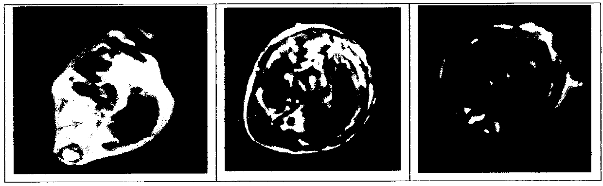
[0040] After the SD rats were anesthetized by intraperitoneal injection of 3% pentobarbital sodium (1ml / kg), they were scanned with a clinical GE 3.0T superconducting magnetic resonance instrument, using a head orthogonal circle with a relatively uniform internal magnetic field, SE sequence, T1W1, parameters: TR / TE=824ms / 10ms, field of view FOV=80mm*80mm. Using the control method of two rats with a body weight of 220g, inject the multimodal microbubble PLGA prepared in Example 1 through the tail vein of the rats at a dose of 5ml / kg during imaging QDs and blank microbubble PLGA, observe the imaging time and imaging effect after liver and kidney contrast (such as image 3 As shown), it can be clearly seen that the multimodal non-targeting microbubbles protected by the present invention have the purposes of preparing MRI contrast agents, and predictably the multimodal ...


PUM


Abstract

The invention relates to the cross field of chemical engineering, materials and nanomedicines, and specifically discloses a multi-modality imaging probe structure and a preparation method thereof. A multi-modality imaging probe is a microbubble with a putamen structure, a shell membrane material is composed of a high polymer material and a phospholipid material which are biodegradable and good in biocompatibility, water-solubility quantum dot solutions with different fluorescent characteristics are embedded at the center, a perfluor carbon alkyl gas is fed, and the multi-modality probe which is controllable in grain diameter, good and stable in dispersion and easy to store are prepared by a double-emulsion-freeze-drying gas feeding method. According to the multi-modality imaging probe structure the preparation method thereof, the microbubble is a reticuloendothelial system specificity contrast agent, the fluorescent imaging is achieved, the enhancement for ultrasonoscopy and magnatic resonance imaging (MRI) display and the microbubble multi-mode imaging can be achieved, and the multi-modality imaging probe structure the preparation method thereof have a wide application prospect.

Description

technical field [0001] The invention relates to the interdisciplinary fields of biomedicine, materials and nanomedicine, and in particular to a construction, preparation method and application of multimodal targeted imaging microbubbles. Background technique [0002] With the development of medical imaging, various contrast agents are more and more widely used in clinical practice. Contrast agents can increase tissue contrast, improve the ability of qualitative positioning of images, and improve the accuracy of diagnosis. At present, various imaging techniques have their own contrast agents, such as microbubble contrast agents for ultrasound imaging, iodine contrast agents for CT imaging, Gd-DTPA and superparamagnetic iron oxide for magnetic resonance imaging, etc. In order to confirm the diagnosis, the same patient often needs to receive a large amount of contrast agents in a short period of time, which not only increases the metabolic burden of the body and the risk of si...

Claims


Application Information

Patent Timeline
Patent Type & Authority Applications(China)
IPC IPC(8): A61K49/18A61K49/22
Inventor 郝兰王志刚冉海涛
Owner CHONGQING MEDICAL UNIVERSITY
Why Eureka
Try Eureka