Corynebacterium and application thereof in ocular surface fungal infection resistance

A technology for coryneform bacteria and fungal infection, applied in antifungal agents, bacteria, sensory diseases, etc., can solve problems such as immune disorders, tissue damage, antibiotic abuse, etc., to enhance ocular surface immunity and protect against fungal infections.

Pending Publication Date: 2022-07-22
EYE INST OF SHANDONG FIRST MEDICAL UNIV
View PDF0 Cites 0 Cited by
  • Summary
  • Abstract
  • Description
  • Claims
  • Application Information

AI Technical Summary

Problems solved by technology

[0005] At present, antifungal drugs are widely used in the treatment of fungal keratitis, but long-term or inappropriate use of antifungal drugs can lead to immune disorders and

Method used


Image

  • Corynebacterium and application thereof in ocular surface fungal infection resistance
  • Corynebacterium and application thereof in ocular surface fungal infection resistance
  • Corynebacterium and application thereof in ocular surface fungal infection resistance

Examples


Embodiment 1

[0028] Example 1: Bacterial isolation and detection

[0029] In 2021, at the Institute of Ophthalmology Affiliated to Shandong First Medical University, according to the ethical regulations of biological sampling, the target strains were isolated from the conjunctival sac of healthy people who visited the Department of Corneal and Refractive Surgery.

[0030] The specific screening and separation steps are as follows:

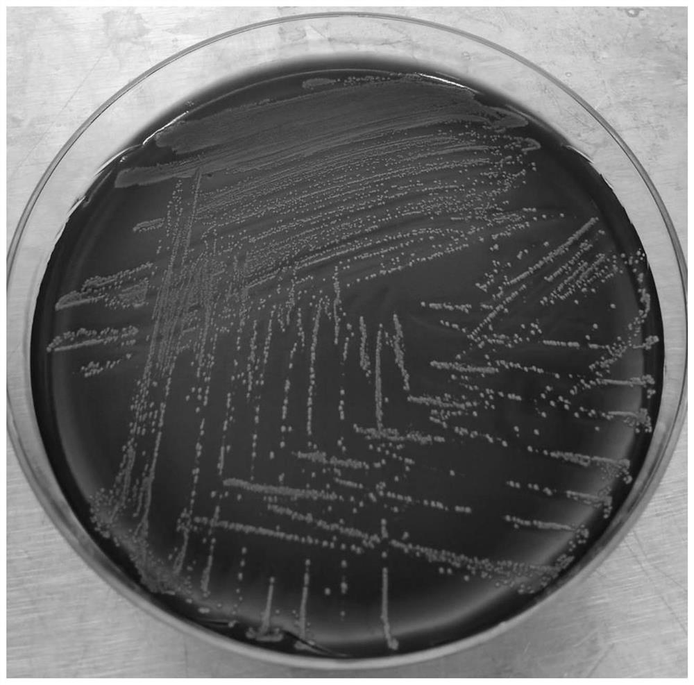
[0031] Use a disposable sterile cotton swab to gently wipe the conjunctival sac of a healthy person, immediately seal the cotton swab aseptically, put the cotton swab in the LB broth, soak and stir in the UV-sterilized microbiological operation table, and then put it in a constant temperature of 37°C. Culture in the box, observe once a day, when the broth is turbid, under sterile conditions, dip the broth bacterial solution with a disposable inoculating ring, spread it evenly on the blood agar plate, and put it into a 37°C constant temperature incubator cultiv...

Embodiment 2

[0042] Example 2: The process and effect of C.mac2 strain colonization on the ocular surface

[0043] The live bacteria preparation of the Corynebacterium C.mac2 strain, the inactivated preparation of the Corynebacterium, the relevant single or mixed bacterial components, and the bacterial derivative products were tested for the control effect of the ocular surface pathogenic strains.

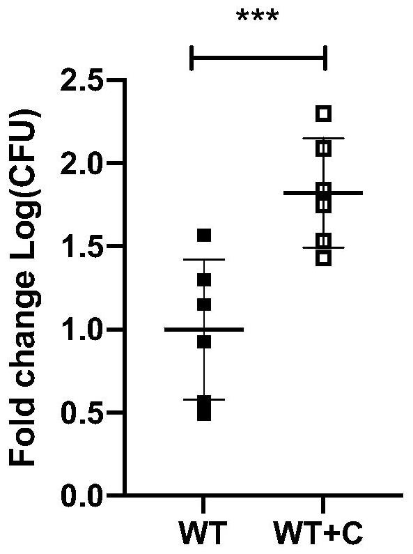
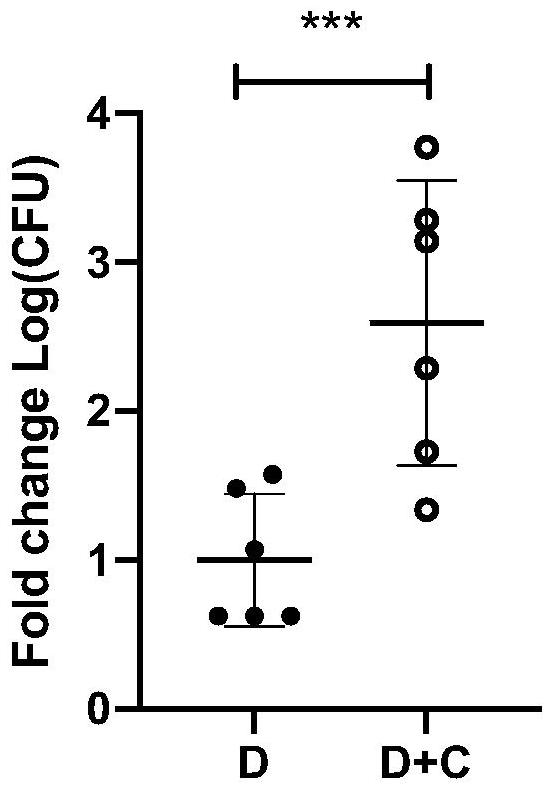
[0044] By colonizing the ocular surface, the concentration was 10 8 cfu / mL of Corynebacterium C.mac2 strain viable bacterial preparation was instilled into the ocular surface of wild-type C57BL / C mice for a total of 9 days of colonization. After the colonization was completed, the colonization success rate was evaluated. The colonization effect of wild-type C57BL / C mice was detected by PCR after 2 weeks of colonization with C.mac2. Figure 2A As shown, the results of PCR detection of C.mac2 colonization in diabetic C57BL / C mice for 2 weeks were as follows: Figure 2B shown.

Embodiment 3

[0045] Example 3: Promoting effect of C.mac2 strain on repair of ocular surface damage in mice

[0046] In wild-type C57BL / C mice (WT group), colonized C.mac2 wild-type mice (WT+C group), diabetic C57BL / C mice (D group) and colonized C.mac2 diabetic mice (D+C group) to conduct epithelial healing experiments, mice were cured under the state of chloral hydrate anesthesia with a 2.5 mm diameter epithelial layer in the center of the corneal epithelium, and then the ocular surface of the mice was treated at 0 hours, 12 hours, 24 hours, 36 hours and other time points. Fluorescein sodium staining was used to take a general slit lamp photo. The results are shown in Figure 3. It was found that after colonization, C.mac2 wild-type mice (WT+C group) and diabetic mice (D+C group) were compared to their non-colonized counterparts. group had faster epithelial healing.


PUM


Abstract

The invention provides a corynebacterium and an application of the corynebacterium in ocular surface fungal infection resistance, the corynebacterium is a corynebacterium macginleyi C.mac2 strain, the corynebacterium is preserved in the China General Microbiological Culture Collection Center on November 16, 2021, and the preservation number is CGMCC No.23924. The invention further provides a preparation method of the corynebacterium and an application of the corynebacterium in ocular surface fungal infection resistance. The invention further provides a new application of the corynebacterium, namely application of the corynebacterium in preparation of products for treating ocular surface diseases of different individuals. The invention further provides a product capable of being colonized on the ocular surface. The product is used for preventing and treating ocular surface diseases caused by fungal infection. Wherein the culture medium contains the corynebacterium and the product thereof. The corynebacterium provided by the invention does not induce eye infection under normal conditions, and when the corynebacterium is colonized on the ocular surface, the immunity of the ocular surface can be enhanced, and the cornea can be protected from fungal infection.

Description

technical field [0001] The invention belongs to the technical field of ophthalmic treatment products, in particular to a coryneform bacterium and its application in anti-ocular surface fungal infection. [0002] technical background [0003] The ocular surface is the barrier between the inner eye tissue and the external environment. Anatomically, the ocular surface refers to the entire mucosal epithelium originating between the upper and lower lid margins, including the corneal epithelium and conjunctival epithelium, as well as the tear film covering the surface of the eyeball. The ocular surface tissue is susceptible to various stimuli in the external environment and abnormality occurs. The stability of the ocular surface tissue is an important condition for maintaining the health of the eye and clear vision. The cornea is an important part of the ocular surface and is essential for maintaining a normal ocular surface microenvironment. [0004] The prevalence of corneal bl...

Claims


Application Information

Patent Timeline
IPC IPC(8): C12N1/20A61K35/74A61P27/02A61P31/10C12R1/15
CPCC12N1/20A61K35/74A61P27/02A61P31/10
Inventor 张碧凝邵征黄钰森
Owner EYE INST OF SHANDONG FIRST MEDICAL UNIV
Why Eureka
Try Eureka