

Low antigen hetero stroma of cornea treated by frozen, and its prepn. method
A corneal stroma, xenogeneic technology, applied in medical science, eye implants, prostheses, etc., can solve problems such as hindering the application of xenogeneic corneas, and achieve the effect of good biocompatibility and low immunogenicity
- Summary
- Abstract
- Description
- Claims
- Application Information
AI Technical Summary
Problems solved by technology
Method used
Image


Examples
Embodiment 1
[0018] Example 1 Preparation of frozen porcine corneal stroma
[0019] Select healthy donor pigs in qualified breeding farms, anesthetize with intramuscular injection of sodium thiopental, remove the pig’s eyeballs, trim the accessory tissues on the surface of the eyeballs, soak the eyeballs in 5% povidone-iodine for 3 minutes, and then use Wash with 500 ml of balanced salt solution of phosphoric acid, and store it in a freezer at -70°C. The xenogeneic eyeballs in the cryopreserved state were irradiated with gamma rays at a dose of 25 kGy to kill possible pathogenic microorganisms. It was then stored at -70°C until use.
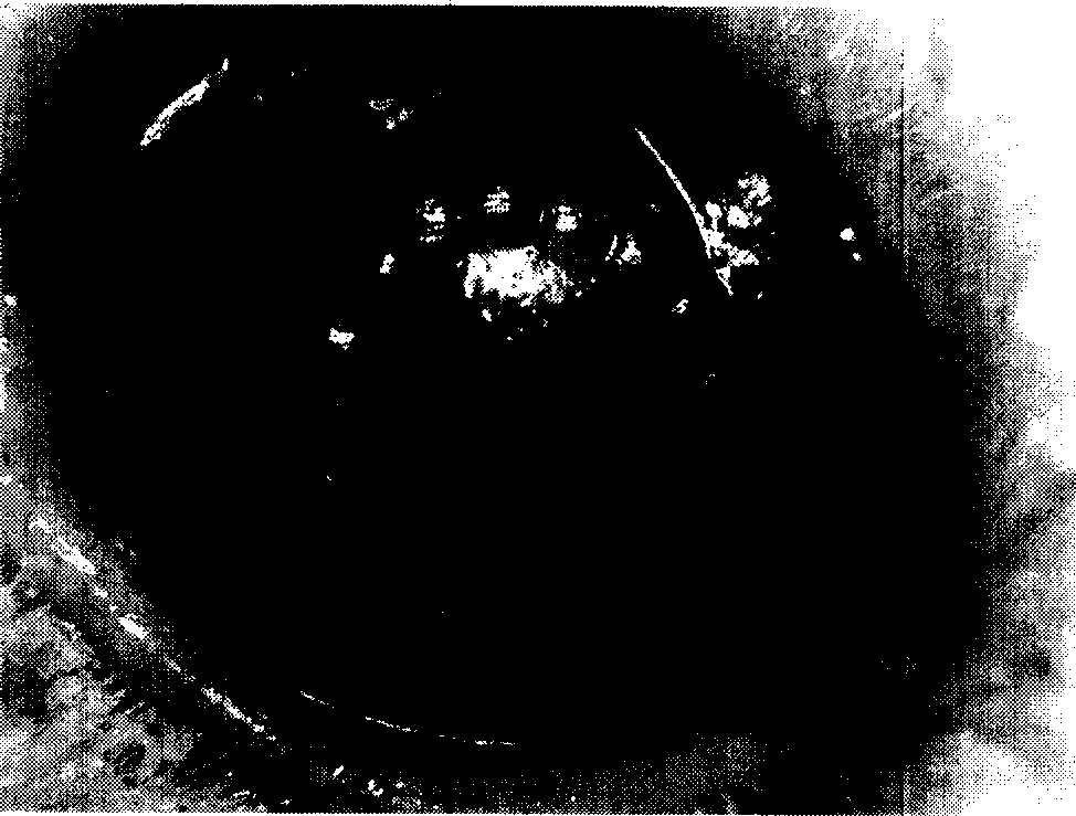
[0020] After the eyeball was thawed at normal temperature, the whole cornea was cut along the corneal limbus under the operating microscope, and the corneal epithelial layer and Descemet's membrane were torn off to obtain porcine corneal stroma with low antigen content. The cells in the corneal stroma are all dead, and the fluorescent staining agent 5-CMFDA...
Embodiment 2
[0021] Example 2 Preparation of frozen bovine corneal stroma
[0022] Choose healthy donor cattle in qualified farms, anesthetize with thiopental sodium needle intramuscularly, remove the eyeballs, trim the accessory tissues on the surface of the eyeballs, soak the eyeballs in 5% povidone-iodine for 3 minutes, and then use Rinse with 500 ml of normal saline, transport to the laboratory at 4°C, and store in -70°C normal saline.
[0023] The xenogeneic eyeballs in the cryopreserved state were irradiated with gamma rays at a dose of 25 kGy to kill possible pathogenic microorganisms. It was then stored at -20°C until use. When in use, after the eyeball is thawed at normal temperature, the whole cornea is cut along the corneal limbus under an operating microscope, and the corneal epithelial layer and Descemet's membrane are torn off to obtain a low-antigen bovine corneal stroma.
Embodiment 3
[0024] Example 3 New Zealand Rabbit Therapeutic Corneal Transplantation Experiment
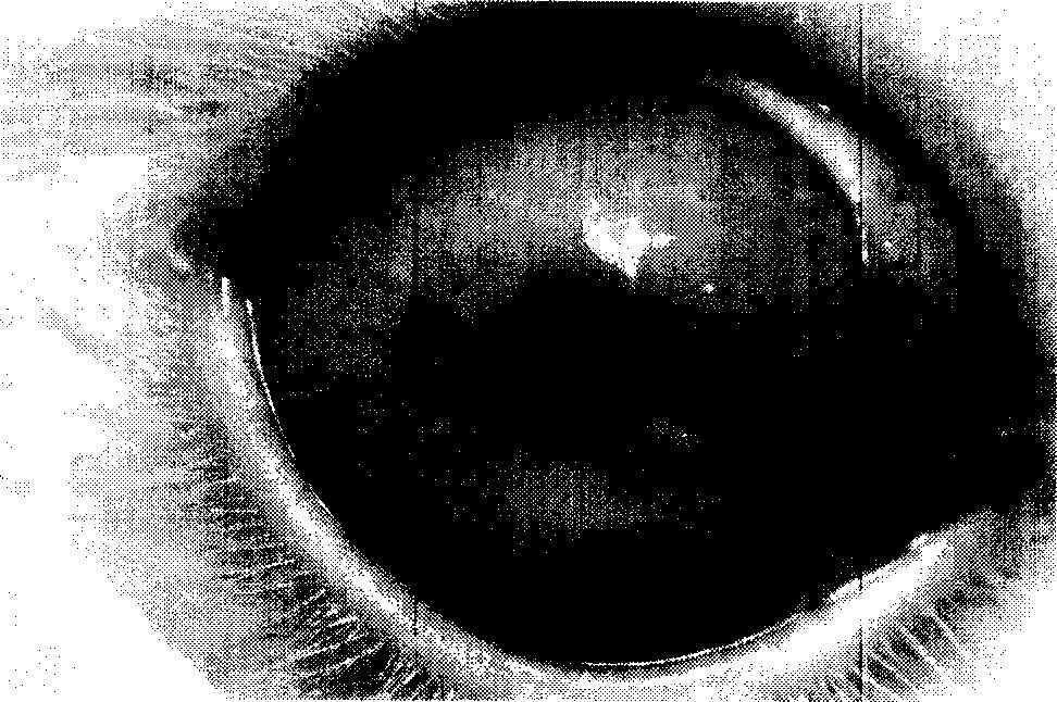
[0025] The low-antigen bovine corneal stroma prepared by the method of Example 2 is the medical donor material (hereinafter referred to as the donor), and the healthy New Zealand rabbit is selected as the recipient for therapeutic penetrating keratoplasty, and the rabbit is intramuscularly injected with thiopental sodium After successful general anesthesia, routine PVP iodine disinfection of the operated eye, laying a drape, opening the eyelids with a lid speculum, and fixing the upper and lower rectus muscle sutures, the donor cornea was rinsed with normal saline and placed on the corneal pad with the inner stroma facing upward, and a 7.5 mm The trephine was drilled in the center of the recipient cornea to the anterior chamber, and the viscoelastic agent was injected into the front. Corneal scissors were used to cut off the recipient cornea along the edge of the drill to prepare the implant be...
PUM
 Abstract
Description
Claims
Application Information