Implantable human liver tissue constructs and uses thereof

a human liver and construct technology, applied in the field of human liver tissue constructs, can solve the problems of liver model mice generated by tissue engineers, additional surgical steps, and time-consuming, and achieve the effect of sufficiently stabilizing the desired morphology and reducing the amount of time, cost and labor

Active Publication Date: 2018-06-26
MASSACHUSETTS INST OF TECH
View PDF15 Cites 4 Cited by
  • Summary
  • Abstract
  • Description
  • Claims
  • Application Information

AI Technical Summary

Benefits of technology

[0004]The present invention features tissue engineering approaches to establish humanized animal models of human tissue (e.g., liver, pancreas, neuronal tissue, muscle tissue, adipose tissue, and the like) with minimal time, cost and labor, compared to existing art. The invention features implantable scaffolds that are stabilized ex vivo using an engineered biomaterial (e.g., a biocompatible, polymeric hydrogel, tuned with key soluble and / or adhesion-promoting factors or other important biochemical cues and standard tissue culture techniques. Cells can be cultured for short periods (e.g., −1 week) to sufficiently stabilize the desired morphology, phenotype and / or function, (or can be cultured in order to obtain the desired morphology, phenotype and / or function, for example, using progenitor or stem cells as a parenchymal cell source) and then inserted into an ectopic site in an animal (e.g., a mouse), for example, in the intraperitoneal space, subcutaneous space, in a tissue, in a body cavity, etc. The constructs of the invention are amenable to facile manipulation, implantation and integration, removal and assessment in a normal animal setting.
[0006]In exemplary aspects, the invention features a highly tunable implantable scaffold to stabilize the phenotype of human hepatocytes, which mitigates the need for complicated mouse genetics, or a lengthy engraftment and repopulation time period (−6 weeks) in vivo associated with art-recognized humanized mouse approaches for studying liver biology and disease. The human hepatocytes are not required to repopulate the mouse liver in order to rescue the mouse from liver failure; therefore, systemic, in vivo studies can be performed readily, and species-specificity confirmed on a per construct basis (i.e., upon explanting liver tissues).
[0008]Unlike existing tissue engineered platforms, this invention uses an encapsulating hydrogel which is optimized to support hepatic functions of a homogenously distributed cell-matrix suspension, and which can be implanted directly in vivo for efficient integration and rapid probing of human liver responses. Using non-invasive imaging technologies, it has been shown that engineered tissues survive in vivo for up to several weeks, a sufficient period of time to dose animals and assess human cytochrome P450-mediated drug-drug interactions and drug-induced human liver toxicity (See e.g., Brandon et al. (2003) Toxicol Appl Pharmacol 189(3): p. 233-46; and Wienkers and Heath (2005) Nat. Rev. Drug. Discov. 4(10): p. 825-33.). It has further been shown that engineered tissues can be infected in vitro or in vivo with virus (e.g., HCV) and that viral infection can persist for up to several weeks. This model therefore allows non-expert researchers to effectively evaluate human liver responses to pharmacologic drugs (e.g., in toxicology testing and / or therapeutic testing) in vivo. In certain embodiments, the engineered liver tissue constructs include hepatocytes encapsulated an appropriate density or concentration such that cells have sufficient oxygen and / or nutrients to survive for a period of time preceding vascularization of the tissue. The skilled artisan will appreciate the highly metabolic nature of hepatocytes and recognize the benefits of the systems of the invention in promoting survival and / or preventing necrosis of the parenchymal cells of the constructs.
[0009]The human liver model of the invention results in long term survival of human liver tissues for the study of viral life cycles, liver pathologies, and the development of new treatments (e.g., anti-viral treatments, etc.) Furthermore, the engineered tissue model has the potential to provide several unique advantages not feasible using cell transplantation techniques alone. Engineered human tissues may be used to generate human liver models in immune-competent hosts, as well as immune-compromised hosts. The humanized mice generated by the methods of the invention are also conducive to multiplexing, whereby hepatocytes from different liver donors may be simultaneously implanted and compared in the same host animal. Only in an engineered human liver mouse model can both the liver tissue and host background be so readily exchanged, tested and compared. Without requiring researchers to perform complex mouse genetics or to wait months for human hepatocytes to repopulate a damaged liver, this invention allows academic and commercial scientists to more efficiently study human-specific liver pathologies, infectious diseases, treatment therapies, dosing regimes, drug-drug interactions, and toxicity.

Problems solved by technology

Tissue engineering techniques combining scaffolds and cells have been previously used to deliver hepatocytes in vivo; however, prior work has focused on the long-term treatment of liver disease and has been challenged by the high metabolic needs of primary hepatocytes.
In particular, previous work using biodegradable polymer scaffolds for hepatocyte delivery has focused on acellular scaffolds, which have proven to be limited in assuring homogenous seeding, achieving high engraftment efficiency and maintaining the hepatocyte phenotype (See e.g., Kaufmann et al.
Indeed, hepatocytes transplanted into rats on such biodegradable polymer matrices were found to be inferior to liver grafts of equivalent liver mass in compensating for metabolic deficiencies (See e.g., Uyama et al.
Tissue engineers have also struggled with challenges in vascularizing and oxygenating transplanted hepatic tissues.
Despite moderate success using these strategies for therapeutic liver tissue engineering applications, these protocols add additional surgical steps and add technical complexity, as well as time (weeks) to the generation of liver model mice.

Method used


Image

  • Implantable human liver tissue constructs and uses thereof
  • Implantable human liver tissue constructs and uses thereof
  • Implantable human liver tissue constructs and uses thereof

Examples


example 1

Materials and Methods

Fabrication and Culture of Implantable Liver Mimetics.

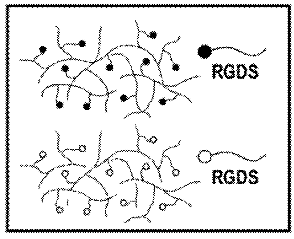
[0193]Liver mimetics were fabricated using a hydrogel polymerization apparatus. In brief, cellular pre-polymer solution was loaded into a 20-mm diameter, 250 mm-thick silicone spacer, and the solution exposed to UV light from a spot curing system with collimating lens (320-390 nm, 10 mW / cm2, 20-30 s; EXFO Lite). Pre-polymer solution comprised of polyethylene glycol diacrylate (PEGDA 20 kDa at 10% w / v; Laysan Bio, Inc.), 0.1% w / v Irgacure 2959 photoinitiator, and 15 mmol / ml acrylate-PEG-peptide monomers. Acrylate-PEG-peptide monomers were synthesized by conjugating RGDS or RGES to acrylate-PEG-N-hydroxysuccinimide (3.4 kDa) at a 1:1 molar ratio in 50 mM sodium bicarbonate buffer (pH 8.5). Reactions were dialyzed overnight against 1000 kDa MWCO cellulose ester membrane and lyophilized for long-term storage at −80° C. Hepatocyte / fibroblast co-cultures were encapsulated at a final concentration of 8×10⁶ hepatocyt...

example 2

Implantable Human Liver Mimetics

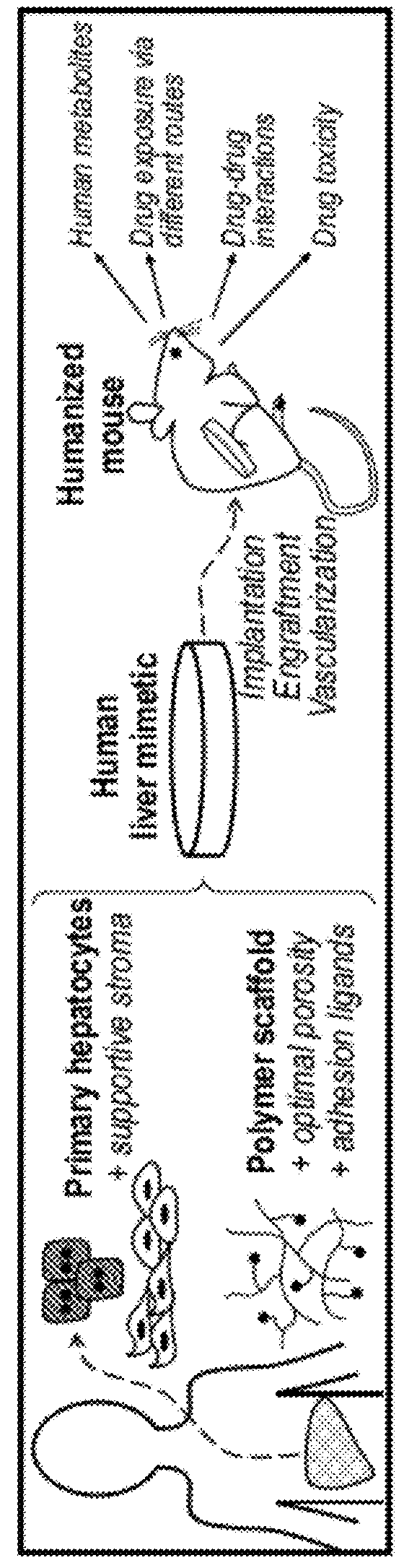
[0211]A tissue engineering approach to establish a novel humanized liver mouse model which can be generated rapidly and reproducibly among mice with diverse backgrounds, and which is broadly enabling for research and drug development was used in this study. (FIG. 1a) This approach leverages a micro-engineered hydrogel scaffold capable of functionally stabilizing primary hepatocytes ex vivo, delivering hepatocytes to accessible ectopic sites in vivo, and integrating with host mouse circulation (FIGS. 1b and 1c).

[0212]Primary hepatocytes representing the full complement of liver functions and drug metabolism pathways are ideal cells for building implantable human liver mimetics but are challenging to maintain upon isolation. To engineer an implantable microenvironment for stabilizing primary human hepatocytes ex vivo, the effects of hepatocyte-nonparenchymal cell interactions with stromal fibroblasts in 2D and 3D culture models were analyzed. Co-cultiva...

example 3

Characterization of Human Liver Mimetics

[0215]In order to assess the utility of tissue-engineered hepatocyte cultures for drug metabolism studies, we characterized human liver mimetics, or HEALs, for the expression and function of human drug-metabolizing enzymes, comparing 3D-encapsulated HEP / FIB HEALs to same-donor 2D HEP / FIB cultures on day 10 of culture. The 2D condition acts as reference for a stable hepatocyte coculture model, previously shown to express a number of genes pertinent to ADME / Tox in vitro. However, to date, assessment of hepatocyte models has hinged on the measurement of only a small handful of drug-metabolizing enzymes. Noting that drug-metabolizing enzymes are regulated primarily at the level of transcription, we hypothesized that we could comprehensively assess drug-metabolizing enzyme expression levels in a low-cost, high-throughput assay based on the multiplexed ligation mediated amplification (LMA) of transcripts coupled to detection on Luminex beads. Accord...


PUM

PropertyMeasurementUnit
thicknessaaaaaaaaaa
thickaaaaaaaaaa
thicknessaaaaaaaaaa

Abstract

Engineered human tissue constructs are provided that are suitable for use in making humanized animals for use in pharmaceutical development. Humanized animals having the constructs implanted in vivo are provided. Methods of making and using the tissue-engineered constructs and humanized animals are also provided.

Description

STATEMENT OF FEDERALLY SPONSORED RESEARCH[0001]This invention was made with government support under grant no. ROI-DK056966-06 6914791 awarded by the National Institutes of Health (NIDDK). The government has certain rights in the invention.BACKGROUND[0002]In recent years, there has been enormous interest in developing “humanized” mouse models as tools for the study, treatment and prevention of human disease. Humanized mice are typically either immune-deficient mice into which human cells are injected and then coaxed to engraft in vivo via regenerative stimuli, or they may be chimeric mice genetically-engineered to express human genes. The explosion of conference meetings sponsored by both academic and pharmaceutical efforts has highlighting the rapid growth of the humanized mouse field. Within basic research settings, humanized mice have been critical to our understanding of many human biological processes that are not easily recapitulated in cell culture. For example, hematopoetic ...

Claims


Application Information

Patent Timeline
Patent Type & Authority Patents(United States)
IPC IPC(8): A61L33/18A61L27/36A61L27/18A61L27/38C12N5/071A01K67/027C12N5/00G01N33/50
CPCA61L27/3604A01K67/0271A61L27/18A61L27/362A61L27/3804A61L27/3886C12N5/0062C12N5/0671G01N33/5088C08L71/02G01N2500/10A01K2207/15A01K2267/035C12N2502/13C12N2533/30C12N2533/50C12N2533/74
Inventor BHATIA, SANGEETA N.CHEN, ALICE A.NG, SHENGYONG
Owner MASSACHUSETTS INST OF TECH
Why Eureka
Try Eureka