Functional pigmented skin equivalent

a pigmented skin and equivalent technology, applied in the field of reconstructive skin, can solve the problems of not being able to evaluate a modulation, not being able to provide an understanding, and not being able to keep the melanocytes in a normal phenotyp

Active Publication Date: 2020-04-07
LOREAL SA
View PDF10 Cites 0 Cited by
  • Summary
  • Abstract
  • Description
  • Claims
  • Application Information

AI Technical Summary

Problems solved by technology

However, these epidermis equivalents are obtained on an inert support which does not reproduce the physiological interactions with the dermis, and which does not contain fibroblasts or a matrix environment.
Furthermore, the culture conditions do not make it possible to keep the melanocytes in a normal phenotype owing to the use of a tumor promoter (TPA or PMA).
However, none of these models makes it possible to provide an understanding of the modulation of pigmentation by the participants of the dermis (fibroblasts, extracellular matrix, dermo-epidermal junction components) since they do not comprise a live dermis.
Even when human organotypic models comprising live fibroblasts are developed, they do not make it possible to evaluate a modulation of pigmentation in a physiological context because they have functionality deficiencies (absence of constitutive pigmentation or pigmentation that can be induced in particular by UV exposure).
However, the localization of the melanocytes therein is often imperfect and these models exhibit functionality deficiencies.
However, these models have drawbacks which do not enable them to be used to evaluate the modulation of pigmentation.
Among the drawbacks of such a technique, exemplary is the lack of control of the epidermal cells and the fact that only one skin reconstruction is possible with a biopsy: no experiment is therefore comparable to the other (no reproducibility).

Method used


Image

  • Functional pigmented skin equivalent
  • Functional pigmented skin equivalent
  • Functional pigmented skin equivalent

Examples


example 1

Preparation of the Skin Equivalents

[0164]Preparation of the Lattices (D-4):

[0165]The dermis equivalents are prepared with bovine collagen I and human fibroblasts derived from dermis.

[0166]The dermis equivalents are prepared with bovine collagen I and human fibroblasts derived from dermis according to the techniques described by Asselineau et al., 1985 (Exp Cell Res., vol. 159, 536-539) and Bernerd and Asselineau 1997 (Dev Biol., vol. 183, 123-138). The Petri dish was precoated with MEM containing 10% of serum.

[0167]Before pouring the collagen preparation containing the cells, a solution of laminin (3% / final volume), collagen IV (1.5% / final volume) and entactin (0.35% / final volume) is added to the mixture, which is stirred vigorously. This suspension is placed in an incubator (37° C.-5% CO2) so as to allow the lattice to contract.

[0168]Seeding of the Keratinocytes and the Melanocytes (D0):

[0169]The keratinocytes and melanocytes are seeded on the dermis equivalent after contraction of...

example 2

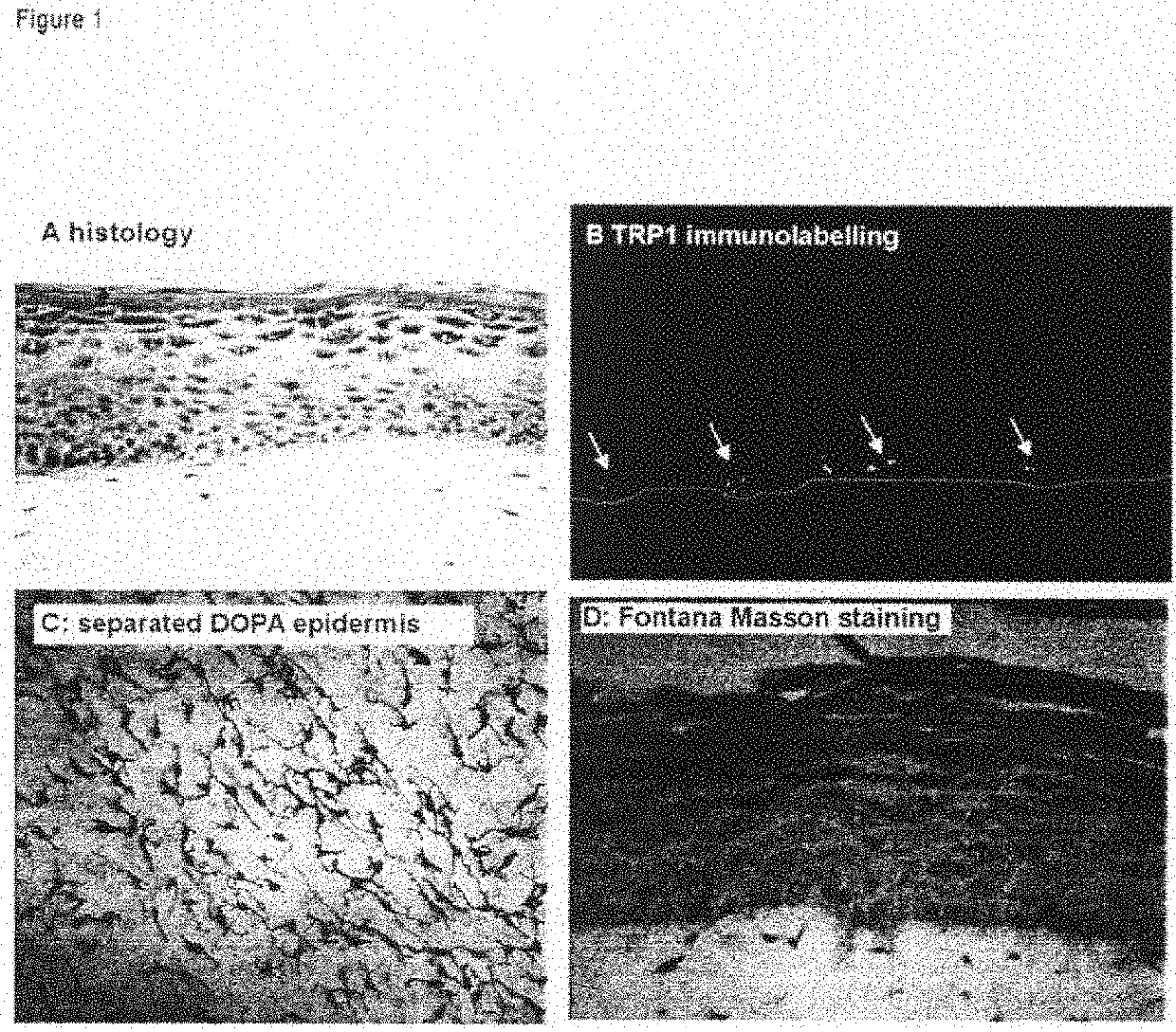
Human Pigmented Reconstructed Skin Containing the 3 Cell Types: Melanocytes, Keratinocytes and Fibroblasts (FIG. 1)

[0181]After 7 days of immersion, the skin exhibits a histology and a three-dimensional architecture close to that of human skin.

[0182]It has a stratified and differentiated epidermis made up of the various cell layers (basal, prickle-cell, granular) and a horny layer.

[0183]The dermis comprises live fibroblasts embedded in the collagen matrix (FIG. 1A).

[0184]The melanocytes are integrated into the epidermis and show a morphology and a density similar to those of normal skin (FIG. 1C, DOPA reaction on separated epidermis). They are clearly localized in the basal layer (FIG. 1B, TRP1 immunolabeling, the dashed white line indicating the dermo-epidermal junction), and produce melanosomes and melanin which are transferred into the neighboring keratinocytes (FIG. 1D, staining of melanin with Fontana-Masson).

example 3

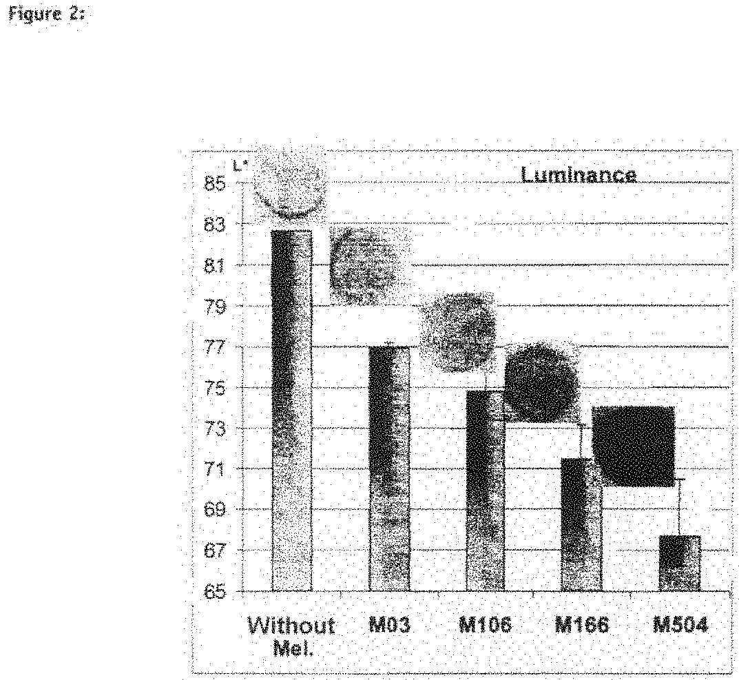
Human Pigmented Reconstructed Skin with Constitutive Pigmentation Linked to the Melanocyte Type (FIG. 2)

[0185]The model is capable of integrating various melanocyte strains, from the least pigmented to the most pigmented (sound model) and the phenotype of the reconstructed skin, which is more or less pigmented, corresponds to the melanocyte type used (physiology maintained): in fact, the more pigmented the melanocyte strain (from M03 to M504), the stronger the macroscopic color of the skin and the greater the amount of melanin visible on the section. This macroscopic pigmentation is reflected by a decrease in luminance (FIG. 2, Luminance).


PUM

PropertyMeasurementUnit
thicknessaaaaaaaaaa
thicknessaaaaaaaaaa
thicknessaaaaaaaaaa

Abstract

An in vitro skin equivalent includes at least one epidermis equivalent and at least one dermis equivalent, and further includes melanocytes constitutively producing melanin and fibroblasts.

Description

CROSS-REFERENCE TO PRIORITY / PCT / PROVISIONAL APPLICATIONS[0001]This application claims priority under 35 U.S.C. § 119 of FR 0851705, filed Mar. 17, 2008, FR 0852886, filed Apr. 29, 2008, and of Provisional Application No. 61 / 046,119, filed Apr. 18, 2008 and Provisional Application No. 61 / 053,890, filed May 16, 2008, each hereby expressly incorporated by reference in its entirety and each assigned to the assignee hereof.BACKGROUND OF THE INVENTION[0002]Technical Field of the Invention[0003]The present invention relates to reconstructed skin having a constitutive pigmentation, and to the use thereof in methods for evaluating skin pigmentation phenomena; it thus relates to methods for evaluating agents capable of modulating this pigmentation, whether it is constitutive or induced. It also relates to a method for preparing such skin models.[0004]Description of Background and / or Related and / or Prior Art[0005]Skin color is principally due to the presence of a pigment, melanin, in the epide...

Claims


Application Information

Patent Timeline
Patent Type & Authority Patents(United States)
IPC IPC(8): C12N5/071G01N33/50
CPCC12N5/0698G01N33/5088C12N2533/50C12N2501/11C12N2502/094C12N2502/091C12N2501/117C12N2501/39C12N2533/52C12N2533/54C12N2502/1323A61L27/3813A61L27/3891A61L27/60A61L27/38
Inventor DUVAL, CHRISTINEBERNERD, FRANÇOISE
Owner LOREAL SA
Why Eureka
Try Eureka