
Paper based diagnostic test

a diagnostic test and paper-based technology, applied in the field of paper-based diagnostic tests, can solve the problems of large and expensive laboratory instruments, difficult implementation of most current diagnostic assays, and large volume of biological samples

Active Publication Date: 2021-02-02
THE ADMINISTRATORS OF THE TULANE EDUCATIONAL FUND
View PDF5 Cites 0 Cited by
  • Summary
  • Abstract
  • Description
  • Claims
  • Application Information

AI Technical Summary

Benefits of technology

Enables rapid, low-cost, and portable diagnostics capable of analyzing whole blood samples directly, improving accessibility and accuracy for diseases like sickle cell disease, and allowing for automated quantification and differentiation between healthy and affected individuals.

Problems solved by technology

Most current diagnostic assays typically require large and expensive laboratory instruments that must be operated by trained personnel, and further require considerable volumes of biological samples.
Thus, most current diagnostic assays can be difficult to implement in remote regions, and arc therefore inaccessible for developing countries.
Additionally, most current diagnostic assays are not useful for emergency situations or home health care situations.
However, traditional colorimetric assays are limited to optically transparent samples (e.g., water, urine, pre-separated blood plasma).
If a non-transparent sample is used, then the color of the sample can interfere with the detection of the developed color.
Blood plasma separation is a particularly important step for a colorimetric assay because the intense color of the red blood cells (“RBCs”) in the whole blood may interfere with quantification of the results of the diagnostic colorimetric assays.
Plasma purification methods based on the fluid dynamics and rheological behavior of whole blood at the microscale require specifically designed microfluidic devices with fine features to achieve separation; and, thus, are not suitable for use in most clinical situations (and are particularly unsuited for use in the field or home health care situations).
Performing these diagnostic tests, however, requires a clinical laboratory equipped with specialized instruments, consumable materials and highly-trained technicians, which is expensive and largely unavailable in resource-limited settings of low-income countries where SCD is most prevalent.
Thus, in most countries in Africa, universal newborn screening remains prohibitively expensive, and most of the affected individuals are not diagnosed at birth or during their lifetime.
In addition, the diagnostic tests currently used for SCD in the high-income countries (e.g. HE, HPLC and IEF) require the transfer of blood samples from the point of care to a centralized hospital laboratory, which makes definitive diagnosis of SCD using these tests nearly impossible in the emergency room setting.
Although the standard Hb solubility test is a low-cost and rapid assay, it cannot distinguish between SCT and SCD because both types of blood samples contain Hb S. Previous modifications of the Hb solubility assay addressing this limitation require extra sample preparation steps, use additional laboratory equipment (e.g. centrifuge, membrane filters) and rely on analytical instruments (e.g. spectrophotometer) to differentiate between SCT and SCD, which makes the test significantly more expensive, complex, time consuming and largely impractical for either the resource-limited or emergency care settings.

Method used


Image

  • Paper based diagnostic test
  • Paper based diagnostic test
  • Paper based diagnostic test

Examples


example 1

Determination of Plasma Glucose Concentration in Whole Blood Samples

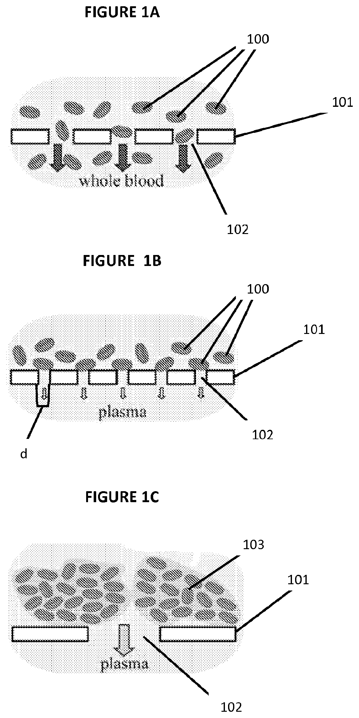
[0057]A μPAD with RBC agglutination-based plasma separation was tested using an assay for plasma glucose as an example. In this assay, glucose oxidase catalyzes oxidation of glucose present in the sample of plasma to yield hydrogen peroxide (H2O2). Horseradish peroxidase then catalyzes the reaction of H2O2 with potassium iodide, which results in brown color. The intensity of the color change is proportional to the amount of H2O2 produced, and thus to the amount of glucose.

[0058]To calibrate the sensitivity of the colorimetric assay to plasma glucose, 3.5 μL of plasma with different known concentrations of glucose was spotted onto square-patterned regions of chromatography paper (the same paper used to fabricate the μPADs and with observed pores of approximately 2-200 μm in diameter) that were pre-treated with the reagents of the assay. The plasma was prepared by centrifugation (800×g, 15 minutes) of whole blood samp...

example 2

Classification of Blood Samples as Healthy, SCT or SCD

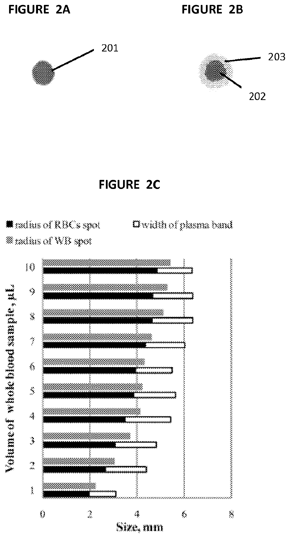
[0075]The μPAD of the present invention was used on one normal (Hb AA), one SCT (Hb AS) and one SCD (Hb SS) blood sample as representative examples. We gently mixed a small volume, approximately 10-50 μL, of each sample of whole blood with the SickleDex solution at a 1:20 ratio by volume, waited 5 minutes and deposited a 20 μL droplet of each of the mixtures onto the center of a μPAD. Normal human venous blood (Hb AA)) was collected from healthy consenting volunteers; SCD (Hb SS) and SCT (Hb AS) blood samples were obtained at the Sickle Cell Center of Southern Louisiana (New Orleans, La.). Blood samples from SCD patients who received blood transfusion in the previous three months were excluded. The Hb A, F, C and S content of SCD samples was determined via hemoglobin electrophoresis as a part of standard patient care. SCT blood samples were collected from biological parents (usually mothers) of SCD patients. SCT samples with hema...


PUM


Abstract

The present invention relates to simple, low-cost, rapid paper-based diagnostic devices and their methods of use.

Description

COPENDING APPLICATIONS[0001]This application claims priority benefit of U.S. Provisional Application No. 61 / 692,994 filed Aug. 24, 2012; U.S. Provisional Application No. 61 / 558,009 filed Nov. 10, 2011; PCT Application No. PCT / US2012 / 064856 filed Nov. 11, 2012; and U.S. Non-Provisional application Ser. No. 14 / 357,074 filed May 8, 2014 and all of said applications are incorporated herein by reference as if set forth in full below.BACKGROUND OF THE INVENTION[0002]I. Field of the Invention[0003]The present invention relates to simple, low-cost, rapid paper-based diagnostic devices and their methods of use.[0004]II. General Background[0005]The analysis of biological fluids is useful for diagnosing a disease or condition and for monitoring the health of individuals and populations. Most current diagnostic assays typically require large and expensive laboratory instruments that must be operated by trained personnel, and further require considerable volumes of biological samples. Thus, most...

Claims


Application Information

Patent Timeline
Patent Type & Authority Patents(United States)
IPC IPC(8): G01N30/90G01N33/52G01N33/80G01N30/92G01N33/66G01N33/86
CPCG01N33/523G01N30/90G01N30/92G01N33/526G01N33/66G01N33/80G01N33/86G01N2333/805G01N30/02G01N33/00G01N33/49G01N33/52
Inventor SHEVKOPLYAS, SERGEY S.YANG, XIAOXIWASHKO, JULIE KANTERPIETY, NATHANIEL ZANE
Owner THE ADMINISTRATORS OF THE TULANE EDUCATIONAL FUND
Learn More