Device and method for measuring blood oxygen level and/or detecting brain hematoma

a blood oxygen level and brain hematoma technology, applied in the field of brain oxygen level determination and brain hematoma detection, can solve the problems of increasing mortality, worsening the outcome of survivors, and each of these techniques has its own limitation, so as to improve the accuracy and efficiency of measurement

Active Publication Date: 2021-03-02
MACKAY MEMORIAL HOSPITAL
View PDF12 Cites 0 Cited by
  • Summary
  • Abstract
  • Description
  • Claims
  • Application Information

AI Technical Summary

Benefits of technology

[0020]Compared with traditional detecting and imaging technology, the present device is advantageous in that the probe and detecting means can be disposed at different positions (such as head, nose, ear, and mouth of the subject) in accordance with the requirements of application, thereby improving the accuracy and efficiency of measurement.

Problems solved by technology

During the resuscitation, time is crucial as the expanding mass lesion can cause death from brainstem compression or cause ischemic injury of brain.
It has been generally accepted that a delay of more than 4 hours between brain injury and the evacuation of hematoma would increase mortality and worsen outcome in survivors.
However, each of these techniques has its own limitation when it comes to determine brain hematoma.
However, the efficacy of a normal CT is usually limited by several drawbacks: (1) long preparation and examination time; (2) failure to accurately distinguish hematoma from hydrocephalus; (3) discontinuous monitoring; (4) failure to exclude underlying injury, such as, ischemia and hypoxia; (5) poor visualization in brainstem and posterior fossa; (6) radiation exposure; and (7) the possibility of contrast-induced allergy or nephropathy.
All of which impedes the clinical application of CT.
Nevertheless, the clinical application of MRI might be restricted due to several reasons as indicated below.
First, the presence of an implant or other metallic object within the subject would render it difficult to obtain clear images.
Second, patients having acute injury in general is not in a condition for MRI imaging, for traction devices and other life support equipment must be kept away from the area to be imaged.
Third, the process is time consuming and the results may not be immediately available.
Further, similar to the concerns in using CT, the contrast employed in either imaging process might induce allergy or nephropathy to the patients.
The major limitation of X-ray imaging is its incapability of changing imaging plane, thus fails to provide detailed stereoscopic information via its 2D images.
In addition, X-ray might cause defects and / or diseases due to its destructing effects on DNA.
Accordingly, an inherent inaccurate diagnosis exists in the condition when a bilateral hematoma occurs.

Method used


Image

  • Device and method for measuring blood oxygen level and/or detecting brain hematoma
  • Device and method for measuring blood oxygen level and/or detecting brain hematoma
  • Device and method for measuring blood oxygen level and/or detecting brain hematoma

Examples


examples

[0074]1.1 Simulation of BH by Use of Embedded Ink Stick

[0075]In the examples, an ink stick (1 cm3: 0.65 cm×0.9 cm×1.7 cm; 2.5 cm3: 1.63 cm×0.9 cm×1.7 cm; or 5 cm3: 3.25 cm×0.9 cm×1.7 cm) embedded in a plastic article made of titanium oxide and polyester resin (the distance between the ink stick and the surface of plastic article was 0.5 cm, 1.7 cm, or 2.5 cm) was used to imitate different situations of brain hematoma (BH, with different mass lesions and locations). The probe was set to emit NIR respectively at 808 nm, 780 nm, and 850 nm. During operation, the probe and the detecting means were respectively disposed on either the same sides (as traditional detecting method, in which the light source and detector were configured closely and operated in proximity to each other), or on the opposite sides of the plastic article (i.e., the simulated BH). Results obtained from the same and opposite sides are respectively provided in FIGS. 9 and 10.

[0076]When the probe was disposed in close...


PUM


Abstract

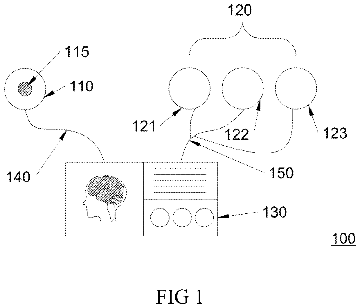
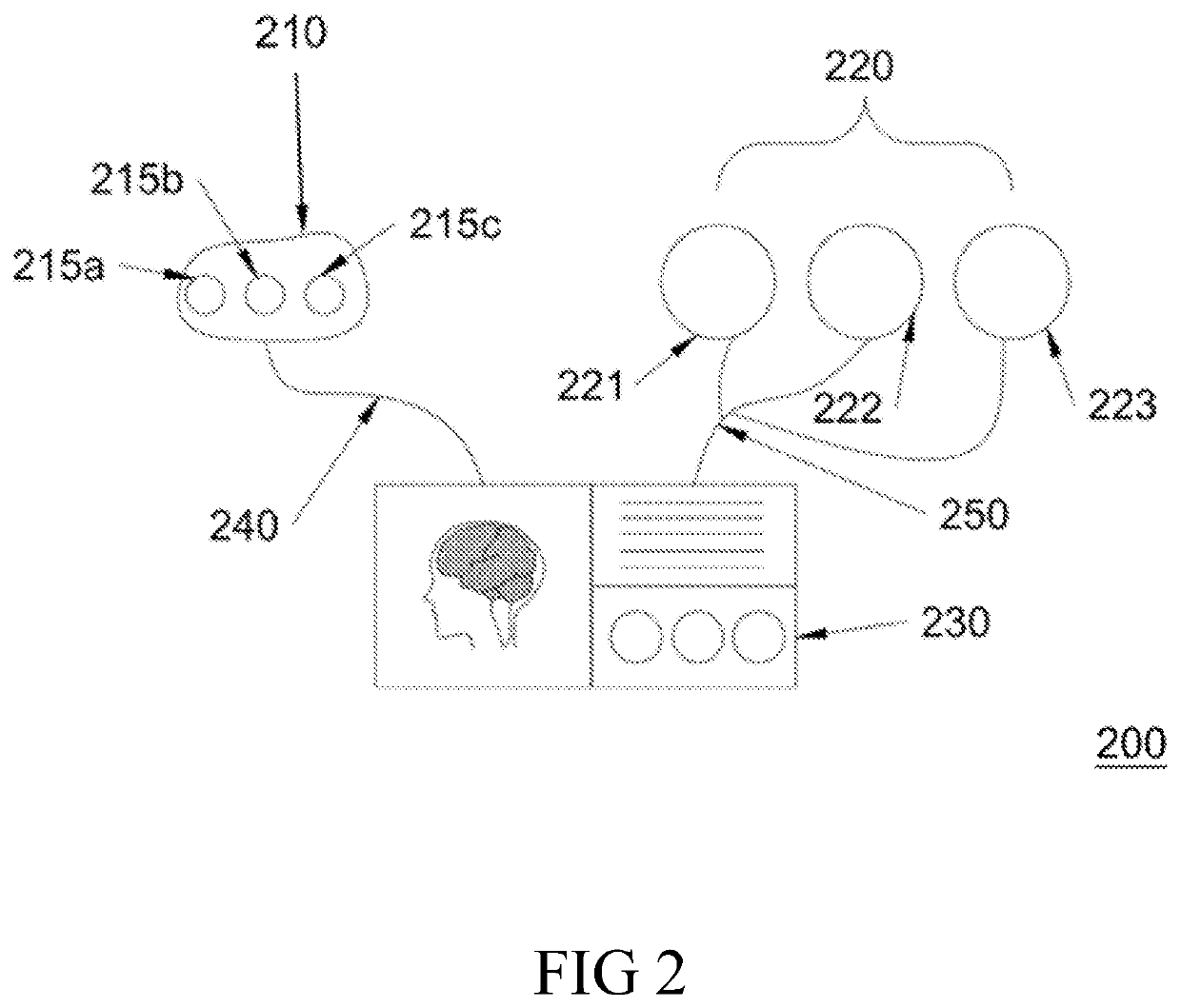
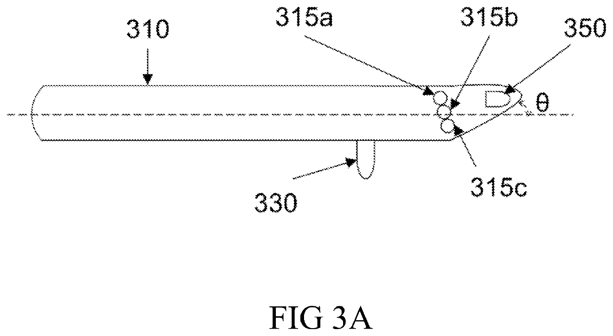
A device for measuring brain oxygen level of a subject, including a probe (210) and a detecting means (220), which are respectively coupled to a processor (230). According to the example, the probe (210) includes three light sources (215a, 215b, 215c) that simultaneously emit the first, second, and third NIR wavelengths across the brain of the subject. The first NIR wavelength is the isosbestic wavelength for oxy-hemoglobin (HbO2) and deoxy-hemoglobin (Hb), the second NIR wavelength is shorter than the first NIR wavelength, and the third NIR wavelength is longer than the first NIR wavelength. The detecting means (220) includes a first, second and third detectors (221, 222, 223) for respectively detecting the NIR intensities of the first, second and third NIR wavelengths traveled across the brain. The processor (230) is configured to determine blood oxygen level based on the measured NIR intensities of the first, second and third NIR wavelengths by use of build-in algorithm derived from Beer-Lambert Law. And method of detecting brain hematoma of a subject by use of the present device.

Description

CROSS REFERENCE[0001]This application claims priority from U.S. provisional patent application 62 / 080,038, filed on November 14, the content of which is incorporated herein by reference.BACKGROUND OF THE INVENTION1. Field of the Invention[0002]The present disclosure in general, relates to the determination of brain oxygen level and the detection of brain hematoma. More particularly, the present disclosure relates to devices and methods for measuring the brain oxygen level and / or detecting the brain hematoma based on relative hemoglobin saturation measured at multiple sites of a subject using near infrared (NIR) wavelengths.2. Description of Related Art[0003]One of the most important principles for the initial resuscitation of a head trauma patient is to promptly identify and surgically evacuate traumatic intracranial hematomas. Hematoma is a condition by which blood accumulates outside blood vessel generally as a result of a hemorrhage or trauma. During the resuscitation, time is cr...

Claims


Application Information

Patent Timeline
Patent Type & Authority Patents(United States)
IPC IPC(8): A61B5/1455A61B5/00
CPCA61B5/14551A61B5/0059A61B5/14553A61B5/6803A61B5/4064A61B5/7235
Inventor CHANG, WEN-HANLU, CHIH-WEI
Owner MACKAY MEMORIAL HOSPITAL
Why Eureka
Try Eureka