Biomaterial scaffold for regenerating the oral mucosa

a biomaterial and oral mucosa technology, applied in the field of tissue engineering, can solve the problems of oral mucosa loss, material biocompatibility, and difficult and necessary oral mucosa repair, and achieve the effect of high strength and convenient handling

Active Publication Date: 2022-02-08
HISTOCELL SL
View PDF12 Cites 2 Cited by
  • Summary
  • Abstract
  • Description
  • Claims
  • Application Information

AI Technical Summary

Benefits of technology

[0008]The authors of the present invention have developed a new biomaterial characterized by having a macroporous scaffold structure which provides said material with a high strength, being easily handled in its swollen state and having stability, elasticity, flexibility and plasticity, as it can recover its shape after applying a deformation force. Furthermore, the biomaterial of the invention has shown to withstand a suture process in its swollen state without breaking or destroying, which assures correct fixing to the damaged tissue.

Problems solved by technology

Among other characteristics, these materials are biocompatible due to their permanent contact with living tissues.
Among soft tissues deficiencies, oral mucosa repair represents a challenging and necessary mission due to the complex structure of this tissue.
The loss of oral mucosa as a result of various surgical procedures, injuries or other clinical conditions is a real problem in view of the need to obtain a suitable coverage that effectively repairs the created defect.
However, these grafts present complications such as rejection (allografts, xenografts), donor site morbidity (autografts), maintenance of original tissue characteristics, provide enough structural properties to allow new tissue development while preventing collapse by surrounding tissues, limited functions and are aesthetically unappealing.

Method used


Image

  • Biomaterial scaffold for regenerating the oral mucosa
  • Biomaterial scaffold for regenerating the oral mucosa
  • Biomaterial scaffold for regenerating the oral mucosa

Examples


example 1

on of the Biomaterial Scaffold

[0153]A solution containing a mixture of human blood plasma (76 ml), PBS 1× (16.5 ml) and Amchafibrin (1.5 ml) was prepared. Agarose 4% (5 ml) and calcium chloride 10% (1 ml) were added to said solution at the same time. The proportions are measured in v / v with respect to the total volume of the resulting mixture.

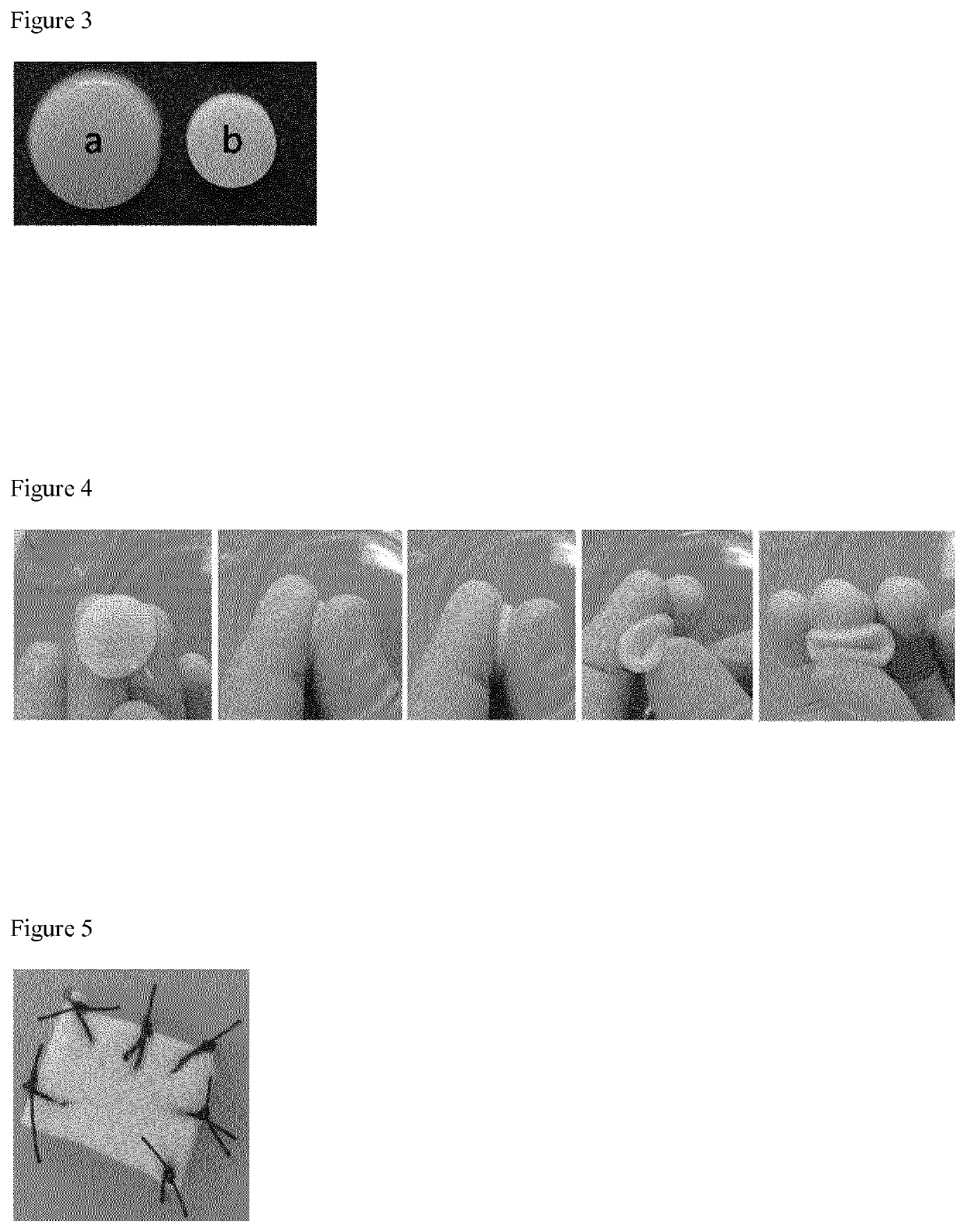
[0154]The resulting mixture was mixed thoroughly and added to a mold. The mixture was left to gel. FIG. 3(a) shows the resulting hydrogel prior to be subjected to lyophilization.

[0155]The hydrogel formed was washed gently with water and lyophilized. In that purpose hydrogels were frozen at −80° C. and water was sublimated afterwards at 0.024 mbar an −48° C.

example 2

on of the Biomaterial Scaffold and Physical Characterization Thereof

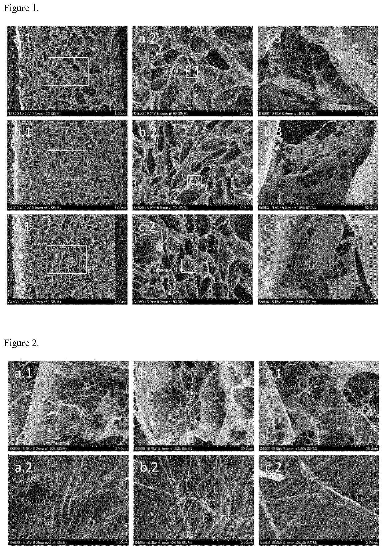
[0156]A solution containing a mixture of human blood plasma (76 ml), PBS 1× (16.5 ml) and Amchafibrin (1.5 ml) was prepared. Agarose 4% (5 ml) and calcium chloride 10% (1 ml) were added to said solution at the same time. The proportions are measured in v / v with respect to the total volume of the resulting mixture.

[0157]The resulting mixture was mixed thoroughly and added to a mold. The mixture was left to gel.

[0158]The hydrogel formed was then subjected to a freezing-thawing process by freezing the hydrogel at −20° C. during 12 hours and subsequently the frozen hydrogel was thawed at room temperature during 3 hours.

[0159]FIG. 3 (b) shows the resulting hydrogel prior to be subjected to lyophilization.

[0160]After that, the resulting hydrogel was washed gently with water and lyophilized. In that purpose hydrogels were frozen at −80° C. and water was sublimated afterwards at 0.024 mbar an −48° C.

[0161]The same procedure...

example 3

roof-of-Principle

[0183]In a first approach, a biomaterial synthesized according to example 2 was subcutaneously implanted in a rat model in order to evaluate the response of the surrounding tissue to the biomaterial implanted in terms of potential toxicity events and degradability. Wistar AG rats more than 8 weeks of age were used in this study following housing and management according to Ethical Committee rules.

[0184]In each animal, two incisions at both sides of the lower back were performed and 1 cm2 of biomaterial was introduced subcutaneously through one of the incisions (see FIG. 10), leaving the other as control (physiological saline solution). At 7, 14, 30 and 60 days after biomaterial implanted, rats were sacrificed and biomaterial and surrounding tissues obtained for histological evaluation.

[0185]No signs of macroscopic external inflammation, erythema or edema were observed at any time of the experience in the adjacent tissues. Signs of neovascularization were observed bo...


PUM

PropertyMeasurementUnit
timeaaaaaaaaaa
concentrationaaaaaaaaaa
concentrationsaaaaaaaaaa

Abstract

The present invention refers to a process for preparing a biomaterial scaffold said method comprising, (a) providing a hydrogel comprising a fibrin network and a polysaccharide network; (b) subjecting the hydrogel of step a) to a freeze-thawing process to physically crosslink the hydrogel; and (c) subjecting the physically cross-linked hydrogel obtained after conducting the step b), to a lyophilization. The invention also relates to the biomaterial scaffold obtainable by the process as defined above, as well said biomaterial scaffold for its use to partially or completely increase, restore or replace the functional activity of a diseased or damaged oral mucosa.

Description

FIELD OF THE INVENTION[0001]The present invention belongs to the field of tissue engineering, and more particularly to biomaterials suitable for regenerating, repairing, and / or replacing soft tissues.BACKGROUND[0002]A new, highly promising field of knowledge known as “Tissue Engineering” has been developed in medical and dental research over recent years. The main objectives of this innovative field are aimed at bioartificial regeneration, repair or replacement of the actual tissues and organs of the human body that were damaged by various factors, such as injuries, burn, by acquired diseases such as cancer or certain congenital abnormalities. Tissue engineering is based primarily on three fundamental components: 1) Cells, 2) Biomaterials, and 3) Biomolecules, inducers or growth factors.[0003]In the case of biomaterials, they are used in the production of biological systems and applied in various branches of medicine. Among other characteristics, these materials are biocompatible du...

Claims


Application Information

Patent Timeline
Patent Type & Authority Patents(United States)
IPC IPC(8): A61L27/52A61L27/22A61L27/26A61L27/56A61L27/20A61L27/38
CPCA61L27/225A61L27/20A61L27/26A61L27/38A61L27/52A61L27/56A61L2430/34C08L89/00C08L5/12A61P1/02A61P1/04A61P17/02A61P31/00A61P35/00A61P43/00A61L27/3616
Inventor CASTRO FEO, MARÍA BEGOÑABAIGET ORTS, AMPARO
Owner HISTOCELL SL
Why Eureka
Try Eureka