Wireless endoscopic surgical device

a surgical device and endoscope technology, applied in the field of minimally invasive surgical and interventional endoscope devices and methods, can solve the problems of tool breakage, decreased popularity of techniques, compounding challenges, etc., and achieve the effect of reducing bending stress

Active Publication Date: 2022-07-12
SCOPERNICUS LLC
View PDF40 Cites 0 Cited by
  • Summary
  • Abstract
  • Description
  • Claims
  • Application Information

AI Technical Summary

Benefits of technology

[0020]In the first embodiment of the invention, the endoscope incorporates folded optics to maintain a 0° angle between the imaging axis and the tool bore access. The device uses a folded path to maintain a line of sight (0°) with the offset imaging axis providing space for an axial tool bore, such as an axial tool bore parallel offset from the imaging axis or an imaging axis parallel offset from an axial tool bore, allowing the tool bore and the imaging axis to coexist without interfering with each other. See FIG. 7. The tool bore, parallel to the line of sight, alleviates bending stresses on electrosurgical devices (cauterizing and transection tool) or other mechanical cutting, clamping, holding, or specimen removal tools, while in the bore.

Problems solved by technology

For single-port surgery, the challenges are compounded by the necessity to share the single incision with both an endoscope and one or more instruments.
This technique has declined in popularity because of issues related to the ‘sword fighting’ of instruments going through a common entry point.
The disadvantage of this approach is that during the harvesting procedure, side entry creates increased torque and excessive stiction forces during manipulation of the tool, which can cause tool breakage.
These unwarranted forces occur because the tool is not parallel to the endoscope along its entire length.
The plate can cause the top of the tool to hang or catch on the plate.
So much equipment requires so many equipment boxes that the suite must have a storage rack, further using up valuable space in overcrowded operating rooms.
Broken bundles can lead to degraded image quality and require its replacement.
Moreover, while the disclosed scopes have bent imaging paths, they do not have any degree of parallelism between the tool bore and the imaging path along the scope length.
But as far as patient safety is concerned, reusable devices frequently pose a higher infection risk than disposable medical devices.
Traditional endoscopes can have problems: first is the failure of a component of a system, especially if it is a reprocessed scope, and second is the bulk or unwieldy nature of a system.
Endoscopes are delicate instruments and can become damaged with repeated use, cleaning, or resterilization.
Owing to the cost, most operating rooms (ORs) do not have many back-up scopes.
Fiber-optic bundles can be cost-effective, but they can display optical artifacts from packing density that can worsen with length.
Current technology limits the length that GRIN lenses can be economically produced.
Ergonomic or logistic problems frequently seen in the OR suite stem from having many cords.
As the wired devices are used during the procedures, the cords inevitably entangle.
Frequently, such tangling breaks surgical components during the procedure, causing an FDA-reportable incident.
In some surgery cases, the fiber-optic light cable and camera power cord stretching from the equipment-laden tower to the patient table causes clutter and becomes a tripping or other safety hazard, especially with many operators and technicians working in a small OR.
Lack of reasonable solutions to draping cables and cords within the OR calls for wireless connectivity within the OR.
Cord clutter also interferes with the ability of the operator to manipulate the tools inside or alongside an endoscope.
In addition to endoscope use, set-up also carries inherent safety issues.
The external light source box can get hot and cause burns if mishandled.
Additionally, damage or failure in a scope discovered during system set-up could trigger not only repair work, but if no back-up scopes were immediately available, it could also force conversion of a minimally invasive surgical procedure to an open surgical procedure with associated costs, complications, and patient discomfort.
Their maintenance adds time and resource costs to hospital operations, as well.
Thus, while any medical device could be discarded after a single use, in some cases doing so would be unreasonable to those of ordinary skill in the art.
Even with functioning components, device assembly still takes time.
Specimen removal can be especially risky for cancerous tissue.
If the specimen removal procedure is compromised, cancerous tissue can be spread throughout the site and perhaps further.

Method used


Image

  • Wireless endoscopic surgical device
  • Wireless endoscopic surgical device
  • Wireless endoscopic surgical device

Examples


Embodiment Construction

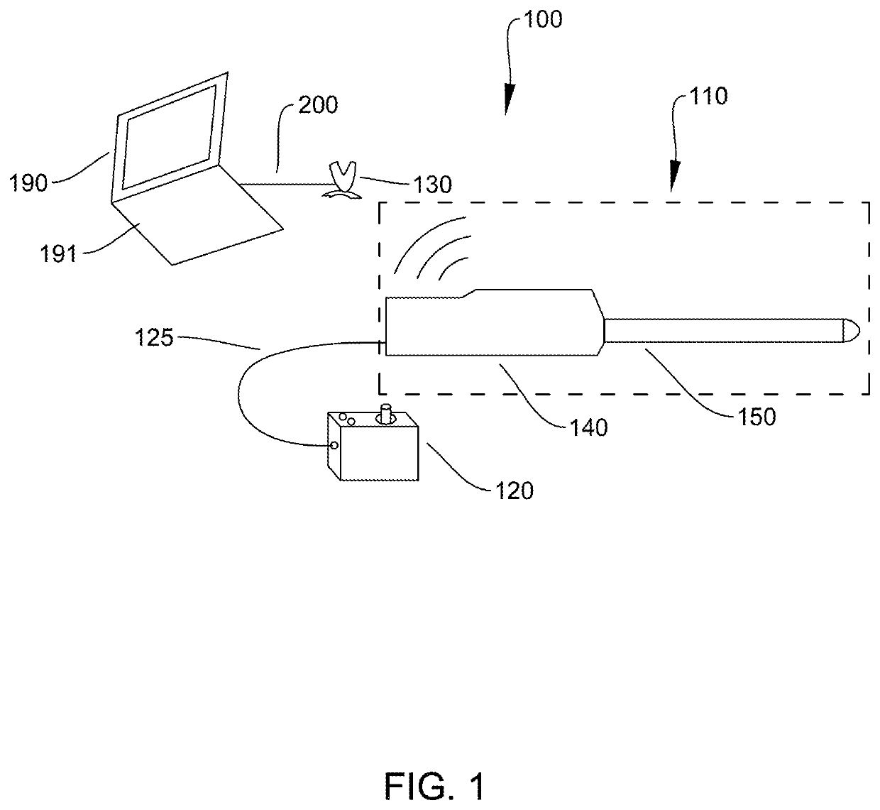
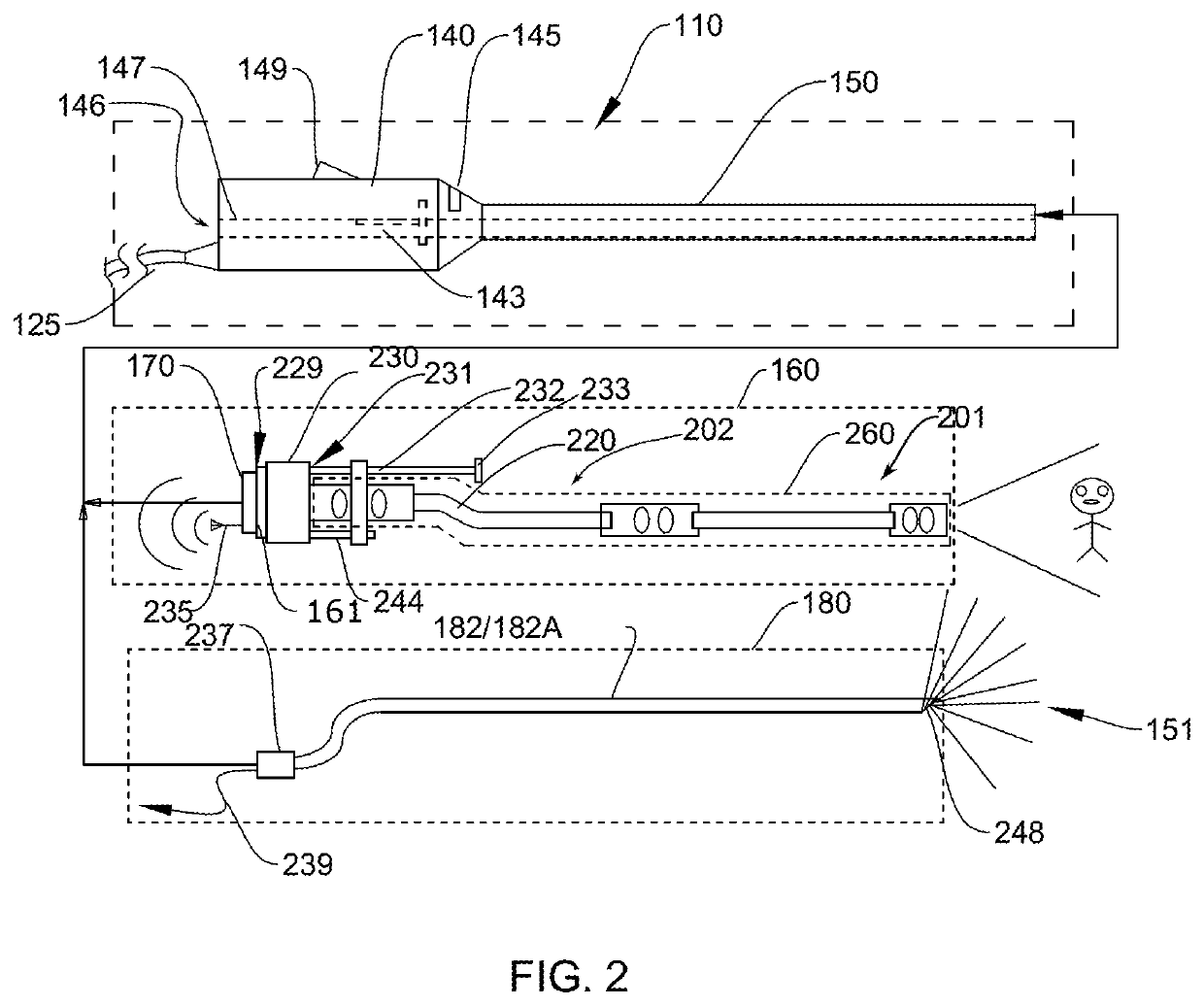
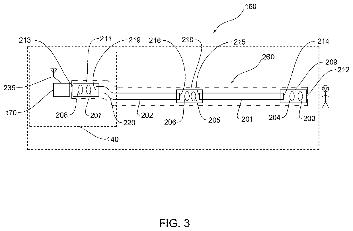
[0035]100 EN system[0036]110 EN device[0037]120 PCM[0038]125 Cable[0039]130 Receiver[0040]140 Handle[0041]143 T-slot[0042]145 Cutout[0043]146 Tool port[0044]147 Tool bore[0045]149 Ventilation openings[0046]150 Conduit[0047]151 Tip[0048]160 Imaging module[0049]161 lens[0050]170 camera[0051]180 Light module[0052]182 waveguide pipe[0053]182A optical fiber bundle[0054]182B Circumferential single fiber bundle[0055]190 Monitor[0056]191 Base[0057]200 Data cable or data line[0058]201 Distal CF bundle[0059]202 Proximal CF bundle[0060]203-208 Optic elements[0061]209-211 Dual-lens housings[0062]212 Distal Imaging assembly (IA) end[0063]213 Proximal IA end[0064]214 Distal CF bundle distal end[0065]215 Distal CF bundle proximal end[0066]218 Proximal CF bundle distal end[0067]219 Proximal CF bundle proximal end[0068]220 S-curve[0069]229 Coupler[0070]230 Focus system[0071]231 Imaging Plate[0072]232 Wheel shaft[0073]233 Focus wheel[0074]235 Antenna[0075]237 LED[0076]238 Heat sink[0077]239 wiring[00...


PUM


Abstract

An endoscopic surgical device used for minimally invasive procedures comprises a handheld component and a power control module. The handheld component consisting of a handle, and a conduit houses a wireless imaging module and a single LED light source. The imaging module comprises a wired or wireless camera coupled to an optically folded assembly or imaging assembly. A battery-operated power module can control both the intensity of the LED and the camera action. The handle and conduit are designed to accommodate surgical tools, wherein the imaging model is parallel offset to the tool, or the tool are parallel offset to the imaging. In alternative embodiments, the handheld component is self-contained.

Description

REFERENCE TO RELATED REFERENCES[0001]This application is a Continuation-in-Part of U.S. patent application Ser. No. 15 / 094,992, filed on Apr. 8, 2016, abandoned, which is a Continuation-in-Part of U.S. patent application Ser. No. 13 / 759,920, filed on Feb. 5, 2013, abandoned; the contents of both applications are incorporated by reference in their entirety.FIELD OF THE INVENTION[0002]The field of the invention generally relates to minimally invasive surgical and interventional endoscopic devices and methods. More particularly, the field of the invention relates to tool-bearing endoscopes for endoscopic or robotic surgical procedures in general, cardiac, thoracic, endovascular, urologic, gynecologic, colorectal, oncologic, endocrine, pediatric, head and neck, hand, metabolic, and bariatric laparoscopic surgery; and minimally invasive orthopedic and neurological surgery, as well as for vascular access surgical procedures and methods of vessel harvesting during cardiac surgery.BACKGROUN...

Claims


Application Information

Patent Timeline
Patent Type & Authority Patents(United States)
IPC IPC(8): A61B1/06A61B1/04A61B1/018A61B1/00A61B1/07
CPCA61B1/00108A61B1/00006A61B1/00016A61B1/00066A61B1/018A61B1/042A61B1/0669A61B1/0684A61B1/07
Inventor KAWULA, PAUL JOHNMCAVINN, JAMESWALSH, PATRICK T.
Owner SCOPERNICUS LLC
Why Eureka
Try Eureka