Vitro repair of bone and/or cartilage defects

a technology of cartilage and bone, applied in the direction of skeletal/connective tissue cells, prostheses, drug compositions, etc., can solve the problems of no beneficial effect on clinical parameters, no improvement in clinical parameters, gross appearance or microscopic appearance of defects when compared to saline controls, and 35% of horses cannot return to previous use within racing or less possibilities of obtaining previous levels of racing

Inactive Publication Date: 2002-06-27
INTERFACE BIOTECH
View PDF7 Cites 49 Cited by
  • Summary
  • Abstract
  • Description
  • Claims
  • Application Information

AI Technical Summary

Problems solved by technology

Approximately 35% of the horses cannot return to their previous use within racing or with less possibilities of obtaining previous levels of racing.
Surgery of the shoulder is difficult due to the depth of the joint below the muscles in the area.
Results of this study indicated that no beneficial effect on clinical parameters, the gross appearance, or the microscopic appearance of defects when compared to saline controls.
Some evidence from this study suggests that intraarticular PSGAG may have a detrimental effect on the healing of articular cartilage.
Controversy exists as to whether these lesions are a manifestation osteochondritis secondary to a joint trauma, or a combination of stress and trauma.
Human articular cartilage is incapable of undergoing self-repair since chondrocytes lose their mitotic ability during the first year of life.
Defects in articular cartilage, especially in weight-bearing joints, will predictably deteriorate toward osteoarthritis.
No conventional method may prevent this deterioration.
Drilling of the subchotidral bone can lead to fibrocartilage formation, which is non-resilient and can only be considered a temporary repair that slowly degrades.
The period of cultivation is dependent on the stage and conditions of the harvested mesenchymal and / or precursor cells from mammals and it is therefore difficult to define the time period used for the propagation of the mesenchymal and / or precursor cells in the growth and selection medium to be differentiated into chondroblast / chondrocytes or osteoblasts / osteocytes.

Method used


Image

  • Vitro repair of bone and/or cartilage defects
  • Vitro repair of bone and/or cartilage defects

Examples


example 2

[0107] Stimulation Molecule

[0108] Collagen II was purified from large scale culturing of porcine chondrocytes as well as proteoglycans derived from the same porcine cartilage source. Collagen II was obtained from large scale culturing of mammalian chondrocytes (preferably species specific to the recipient of the cells), cultured as explants whereby the adequate amount of Collagen II is present.

example 3

[0109] Protocol for Cultivating Cartilage Explants to a Monolayer Culture:

[0110] A cartilage biopsy is harvested from the patients knee and immediately transferred into aseptic growth medium, supplemented with L-ascorbic acid [50 .mu.g / ml (300 .mu.mol / l)] and gentaricin sulfate [50 .mu.g / ml (10 mmol / l)], Fungizone [2 .mu.g / ml (2.2 .mu.mol / l)] in tissue culture flasks. The cartilage biopsy is then washed carefully with new growth medium and dissected in growth medium into 2-4 mm cartilage pieces (explants). When initially placed in the culture, it takes the cartilage explants approximately several days depending on donor material, to reach a constant metabolic state. Having reached such a steady state (steady state is a balance between synthesis and catabolism), the pre chondroblasts / chondroblasts are now stimulated by growth factors present in the growth medium, which diffundate through the cartilage matrix and bind to various binding proteins present in the matrix as well as bindi...

example 4

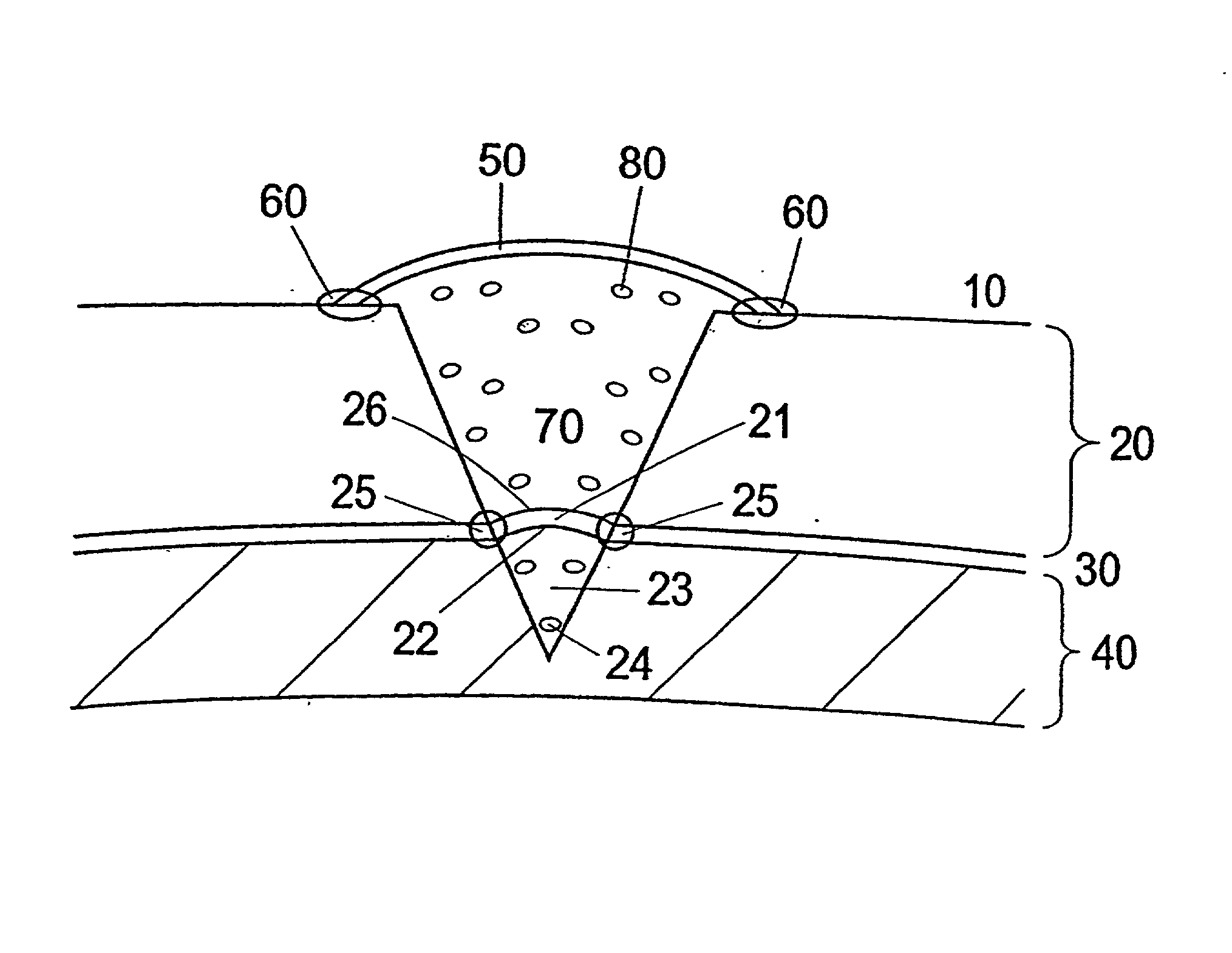
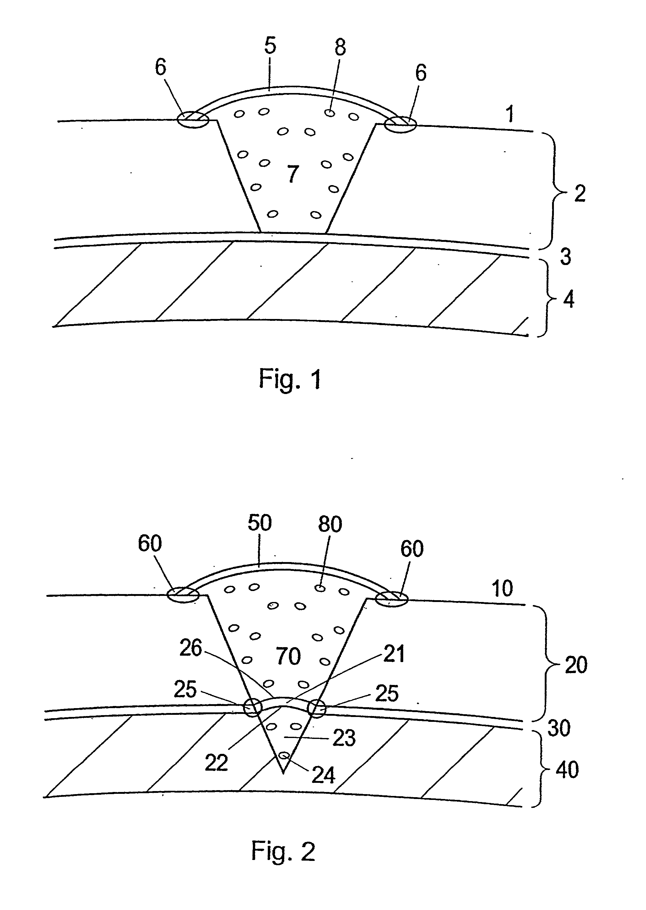
[0114] Membrane to be Used for the Treatment and Use of the Membrane

[0115] During open knee surgery, a membrane collagen type I / (III) is used, purchased from Geistlich Biomaterials, Wolhusen, Switzerland or from Baxter (Denmark). The membrane is cut into a size, which is somewhat larger than the cartilage defect, so that the membrane can cover the cartilage defect and extend around 1 / 2 cm beyond the rim of the defect. The membrane is sutured together with the cartilage rim. The border between the membrane and the cartilage is sealed using Tisseel Due Quick (Baxter). The collagen type I membrane is pretreated with a 1-5 ml collagen type II solution (3-10 mg collagen II / ml (Dep. Material Engineering, Drexel University, Philadelphia, Pa.) for 5-15 minutes at room temperature prior to use for implantation.

[0116] a) When performing arthroscopic surgery, the surface of the collagen I( / III) membrane, which is facing the cartilage defect is pretreated with component II a thrombin solution w...


PUM

PropertyMeasurementUnit
sizeaaaaaaaaaa
depthaaaaaaaaaa
stressaaaaaaaaaa

Abstract

The present invention relates to methods and materials for in vivo repair of cartilage or bone and cartilage defects in mammals. The invention relates to membranes carrying a composition comprising at least one stimulation molecule, which is capable of inducing signal transduction in chondroblasts/chondrocytes and/or osteoblasts/osteocytes. Furthermore the invention relates to a method for the preparation of chondroblasts/chondrocytes and/or osteoblasts/osteocytes suspensions.

Description

[0001] The present invention relates to method and materials for in vivo repair of cartilage defects or bone and cartilage defects in joints in mammals, such as human and horses.BACKGROUND OF INVENTION[0002] More than one million human arthroscopic procedures and total joint replacements are performed each year in the U.S. and Europe together. Included in these numbers are in the U.S., about 90,000 total knee replacements, and around 50,000 procedures for repairing defects in the knee alone per year (In: Praemer, A., Furner, S., Rice, D. P., Musculoskeletal Conditions in the United States, Park Ridge, Ill.: American Academy of Orthopaedic Surgeons, 1992, 125).[0003] Among different breeds of horses in U.S. and in Europe there are around five million registered "expensive" Thoroughbred Racehorses used in horse races, whereof an estimated 60% is directly or indirectly owned by U.S. horse-owners. Of all breeds Thoroughbreds is the most frequent sufferer of degenerative joint disease, m...

Claims


Application Information

Patent Timeline
Patent Type & Authority Applications(United States)
IPC IPC(8): A61B17/00A61B17/06A61F2/00A61F2/02A61F2/28A61F2/30A61F2/46A61K35/12A61L27/00A61L27/22A61L27/24A61L27/38C12N5/00C12N5/077
CPCA61B17/06166C12N2533/90A61F2/28A61F2/2846A61F2/30756A61F2002/0086A61F2002/2817A61F2002/2835A61F2002/30062A61F2002/30064A61F2002/30322A61F2002/30448A61F2002/30589A61F2002/30761A61F2002/30762A61F2002/3093A61F2002/4635A61F2210/0004A61F2220/005A61F2250/0026A61F2310/00365A61K35/12A61L27/227A61L27/24A61L27/3817A61L27/3821A61L27/3826A61L27/383A61L27/3847A61L27/3852A61L27/3891A61L27/3895A61L2430/06C12N5/0068C12N5/0654C12N5/0655C12N2500/38C12N2533/54A61B2017/00495A61P19/00
Inventor OSTHER, KURTSTORGAARD, PETER
Owner INTERFACE BIOTECH
Why Eureka
Try Eureka