
Method of regenerating neurons

a technology of regenerating neurons and axons, which is applied in the field of regenerating neurons, can solve the problems of failure of neurons to regenerate axons in the damaged brain and spinal cord, the consequences of brain and spinal cord injury are often severe and prolonged, and the reasons why regeneration normally fails are still poorly understood

Inactive Publication Date: 2003-11-20
UNIV COLLEGE OF LONDON
View PDF0 Cites 4 Cited by
  • Summary
  • Abstract
  • Description
  • Claims
  • Application Information

AI Technical Summary

Problems solved by technology

The failure of neurons to regenerate axons in the damaged brain and spinal cord is a major cause of human suffering in many common neurological disorders such as brain and spinal cord trauma, stroke, cerebral palsy and multiple sclerosis.
In contrast, axonal regeneration in the central nervous system (CNS) of adult mammals is normally minimal and the consequences of injury to the brain and spinal cord are often severe and prolonged.
Although various ways have been discovered to bring about the limited regeneration of some classes of axons in the CNS, the reasons why regeneration normally fails are still poorly understood.
Typically, restoration of neural functions may result in an increase in the length of the longest neurite per cell relative to control cultures, increases in the number of neurites per cell, increases in axonal outgrowth and regeneration; hypinnervation and the formation of inappropriate connections.
The neurons to be regenerated may be damaged due to a disease ie. any anatomical abnormality or impairment of the normal functioning of an organism or any parts thereof including those arising directly from physical injury.
However, in adeno-associated virus vectors this characteristic integration is lost due to deletion of rep proteins in an attempt to decrease the risk of the emergence of replication competent adeno-associated viruses.
Although adenovirus vectors are known to have a low capacity for integration into genomic DNA, this feature is counterbalanced by the high efficiency of gene transfer afforded by these vectors.
Although this is a very simple and consistent method, it fails to take into consideration that, for example, in an otherwise identical pair of sequences, one insertion or deletion will cause the following amino acid residues to be put out of alignment, thus potentially resulting in a large reduction in % homology when a global alignment is performed.
However, these more complex methods assign "gap penalties" to each gap that occurs in the alignment so that, for the same number of identical amino acids, a sequence alignment with as few gaps as possible--reflecting higher relatedness between the two compared sequences--will achieve a higher score than one with many gaps.

Method used


Image

  • Method of regenerating neurons
  • Method of regenerating neurons
  • Method of regenerating neurons

Examples


example 1

Structure of FLRT-3

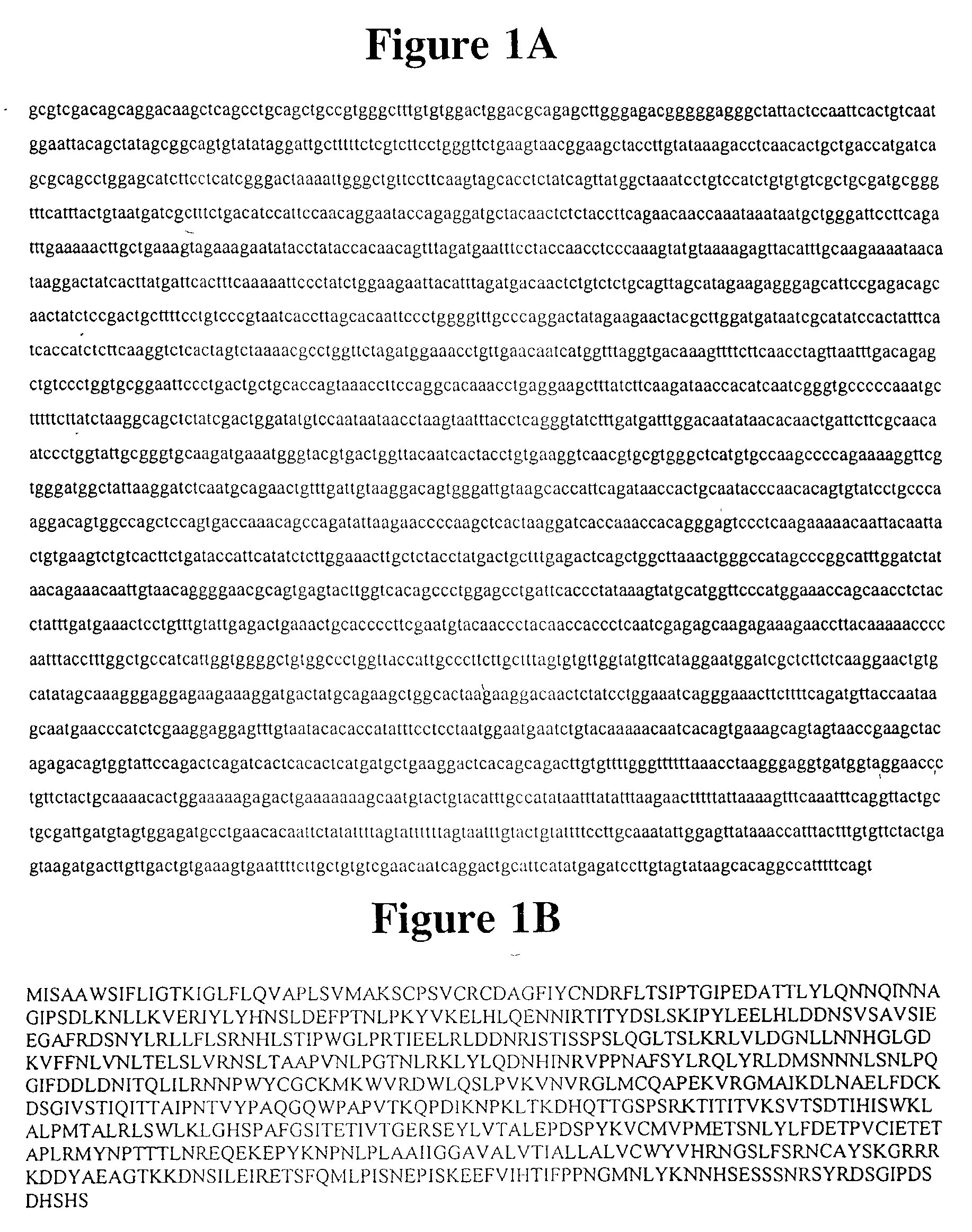
[0339] We have used differential display of mRNA and cDNA library screening to isolate a gene encoding a protein, previously identified as FLRT-3 in human muscle samples (FIG. 1; Lacy S E et al. (1999) Genomics 15;62:417-26). It is demonstrated herein that FLRT-3 is up-regulated in adult rat DRG 3 days after sciatic nerve crush.

[0340] Structure prediction analysis (FIG. 7) suggests that FLRT-3 is a single pass transmembrane protein comprising a 530 amino acid N-terminal extracellular domain, a 20 amino acid transmembrane domain, and a 99 amino acid C-terminal intracellular domain. Much of the extracellular face consists of a 320 amino acid leucine rich region, containing 10 leucine rich repeats, which is flanked by cysteine rich regions. A fibronectin type III domain follows this. The intracellular region contains no recognisable motifs. This indicates that FLRT-3 may be a cell surface receptor or cell adhesion molecule. It is similar in some respects to NOGO A re...

example 2

Expression of FLRT-3

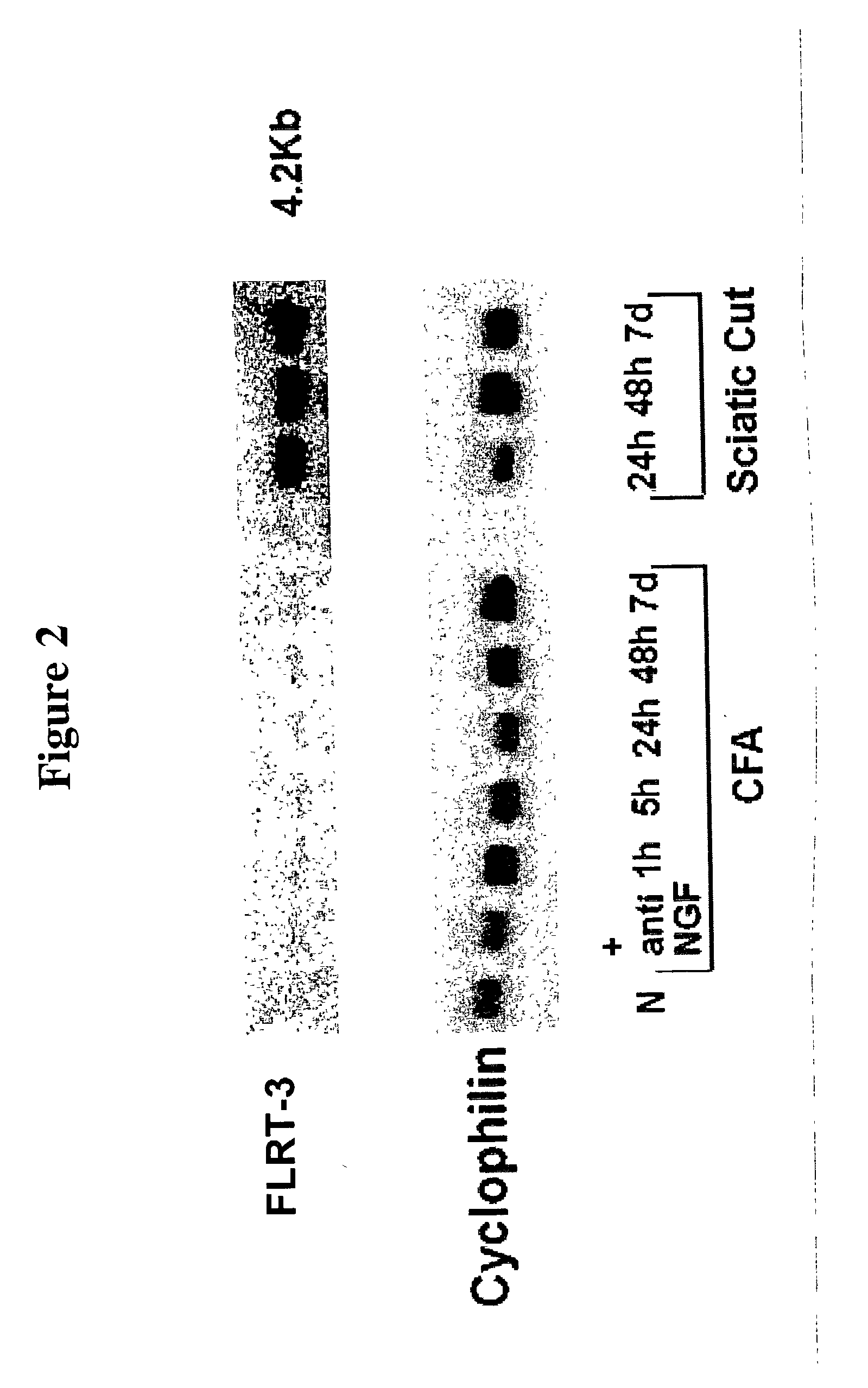
[0341] As shown in FIG. 2, FLRT-3 mRNA is up-regulated in adult rat lumbar DRG after sciatic cut. Up-regulation is consistent between 24 hours and 7 days post cut. Peripheral inflammation induced by injection of complete Freund's adjuvant (CFA) into the rat hind paw has no effect on FLRT-3 expression. The northern membrane reprobed with constitutively expressed cyclophilin shows equivalent loading of total RNA. (N, naive; NGF, nerve growth factor; h, hours; d, day.)

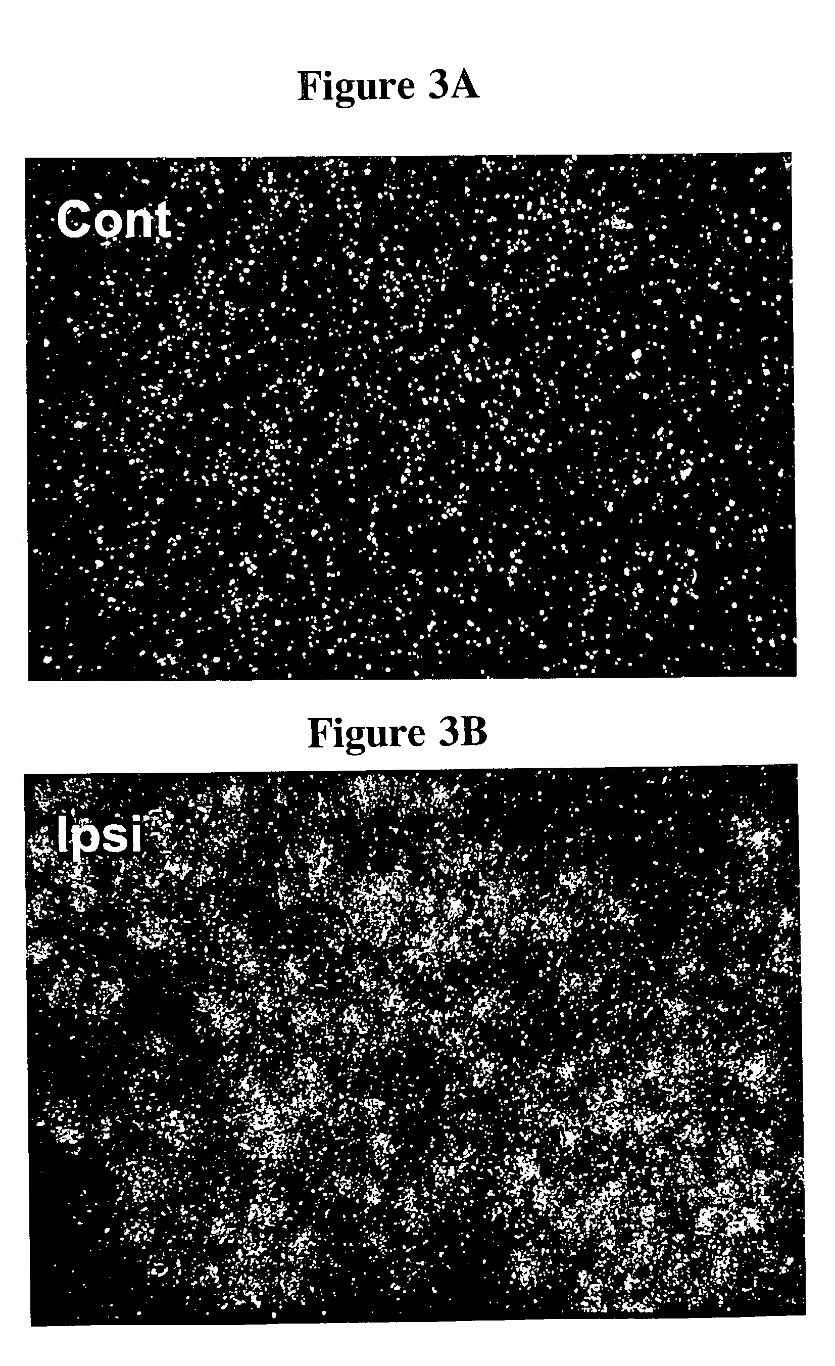
[0342] Using radio-isotopic ISH, FIG. 3 demonstrates that FLRT-3 mRNA is up-regulated in most neurons of ipsilateral (Ipsi) adult rat lumbar DRG 3 days after sciatic cut. The DRG from the contralateral (uncut) (Cont) side is shown for comparison.

[0343] Radio-isotopic ISH analysis of adult rat lumbar spinal cord (FIG. 4) demonstrates the up-regulation of FLRT-3 mRNA in the ipsilateral (Ipsi) large motor neurons 3 days after sciatic cut. Spinal cord from the contralateral (uncut) (Cont) side is shown for...

example 3

Knockdown of FLRT-3 Protein

[0348] Knockdown of FLRT-3 protein in cultured dissociated adult rat DRG by the application of antisense oligonucleotide produces a decrease in neurite length of approximately 44% and a decrease in neurite number per cell of approximately 43%, relative to control cultures with sense oligonucleotide added (FIG. 10). These decreases are significant (P<0.001, 2-way ANOVA). Prior to statistical analysis, data for neurite length were normalised under a logarithmic function, while those for neurite number were normalised under a square-root function. Data were accumulated from 3 separate experiments.

[0349] The up-regulation of FLRT-3 expression in DRG after peripheral nerve injury suggests that it may play a role in the promotion of neurite outgrowth during regeneration. The effects on neurite outgrowth of knocking down and overexpressing FLRT-3 in vitro in dissociated adult rat DRG are strong evidence for this. Knockdown of FLRT-3 in these cultured neuronal cel...


PUM

PropertyMeasurementUnit
Tmaaaaaaaaaa
Tmaaaaaaaaaa
diameteraaaaaaaaaa

Abstract

The invention relates to a method of regenerating neurons comprising administering to a subject in need thereof FLRT-3 to cause neuronal regeneration.

Description

[0001] The present invention relates to methods for regenerating neurons, assay methods and products derived therefrom.[0002] In particular, the present invention relates to a method of regenerating neurons comprising administering to a subject in need thereof FLRT-3 to cause neuronal regeneration.BACKGROUND TO THE INVENTION[0003] The failure of neurons to regenerate axons in the damaged brain and spinal cord is a major cause of human suffering in many common neurological disorders such as brain and spinal cord trauma, stroke, cerebral palsy and multiple sclerosis.[0004] Axonal regeneration in injured peripheral nerves is vigorous and often leads to functional recovery. In contrast, axonal regeneration in the central nervous system (CNS) of adult mammals is normally minimal and the consequences of injury to the brain and spinal cord are often severe and prolonged. Although various ways have been discovered to bring about the limited regeneration of some classes of axons in the CNS, ...

Claims


Application Information

Patent Timeline
Patent Type & Authority Applications(United States)
IPC IPC(8): A61K38/17G01N33/566
CPCG01N33/566A61K38/1709
Inventor HUNT, STEPHEN P.ROBINSON, MICHELLELIVESEY, FREDERICK
Owner UNIV COLLEGE OF LONDON
Learn More