

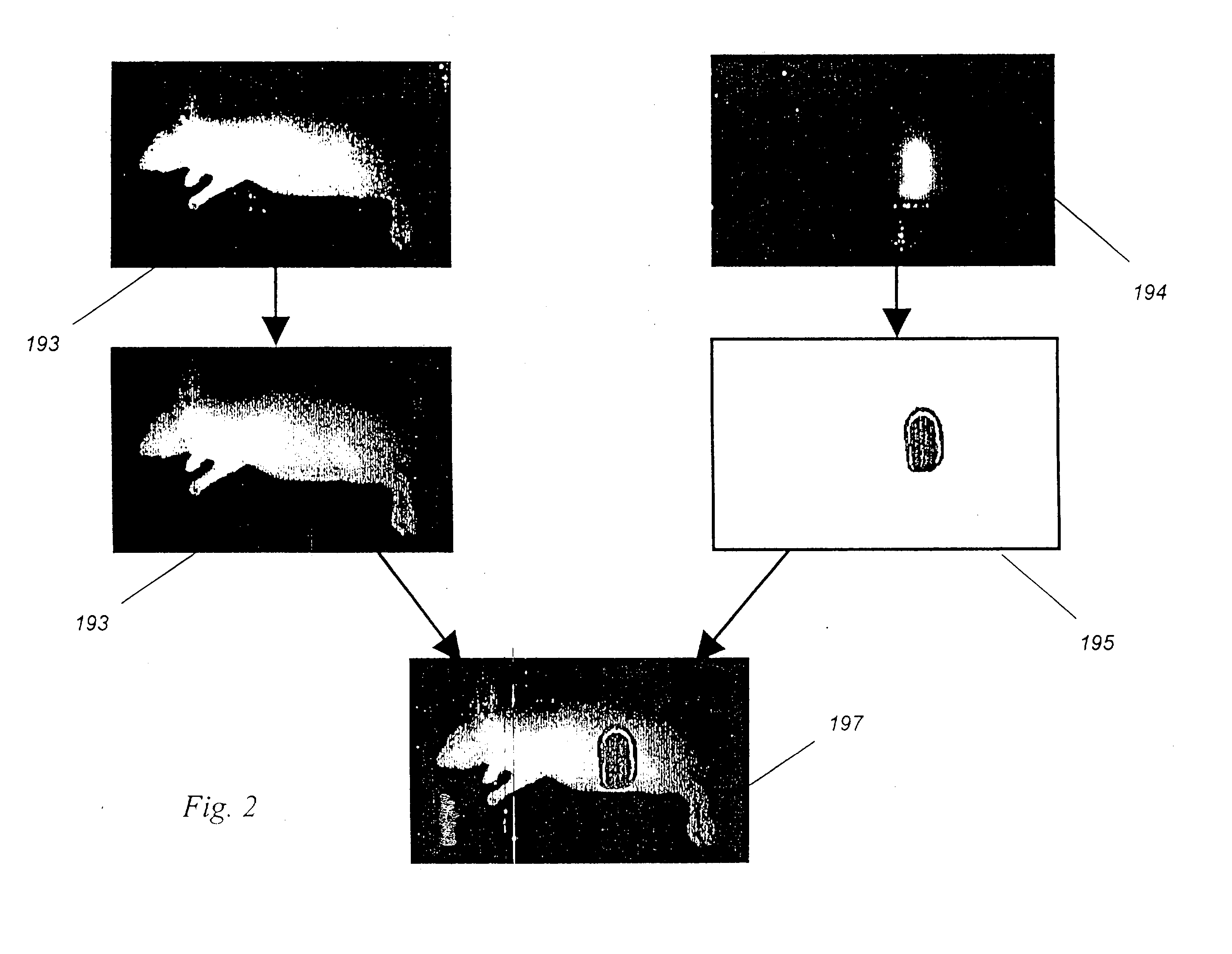
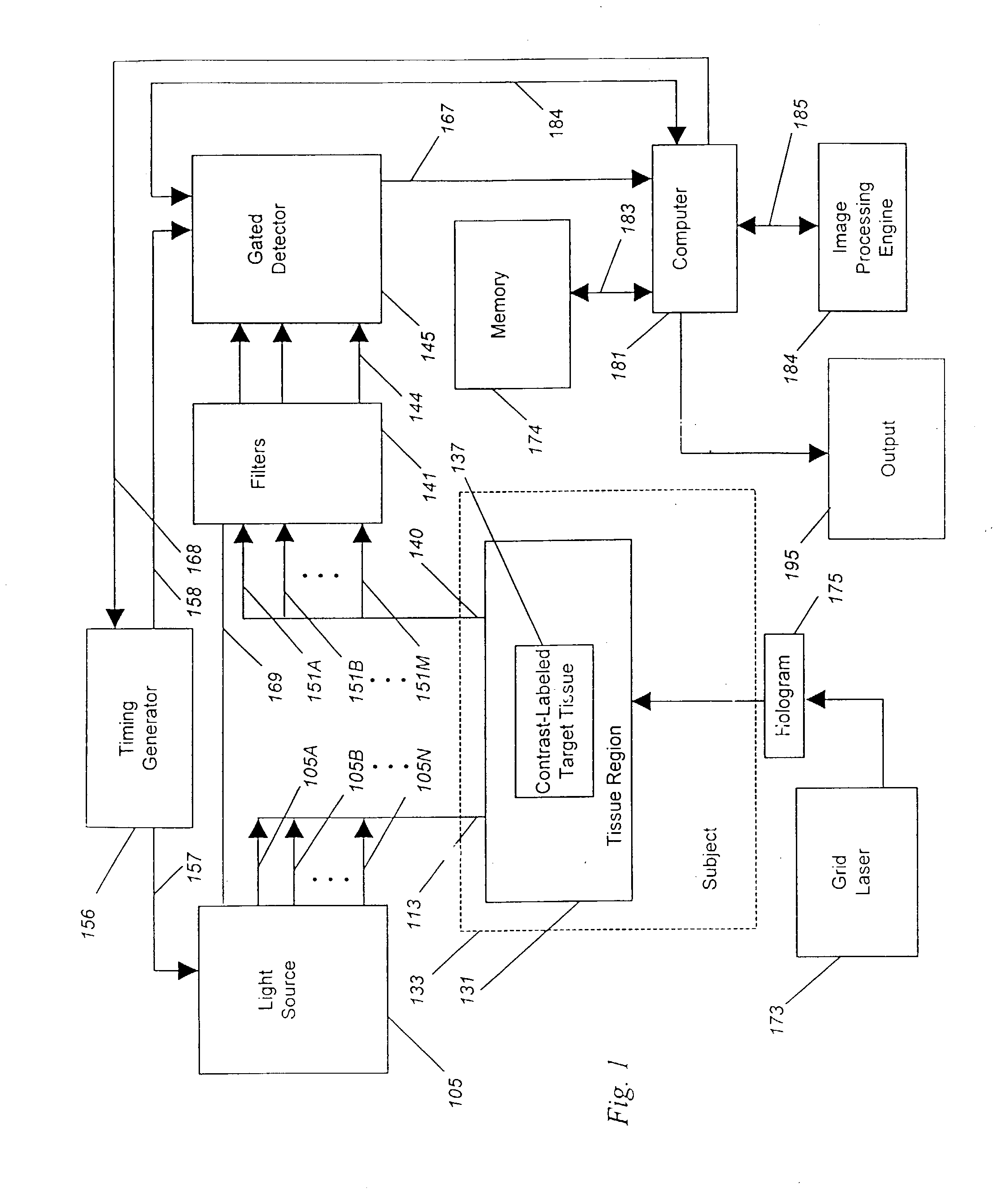
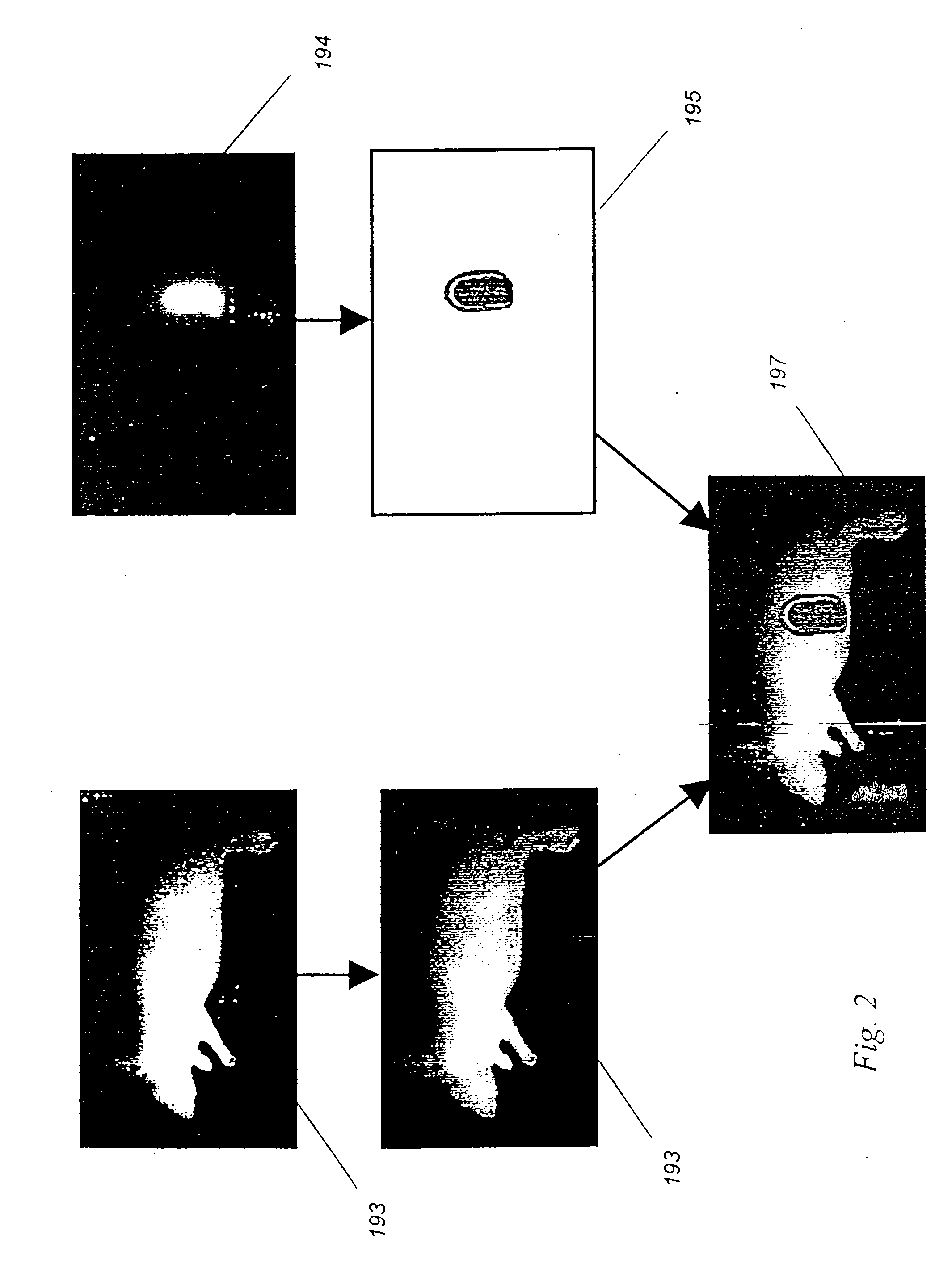
Optical imaging of induced signals in vivo under ambient light conditions
an optical imaging and induced signal technology, applied in the direction of catheters, fluorescence/phosphorescence, spectroscopy, etc., can solve the problems of poor signal to noise ratio, and limited application of such systems in vivo during real-time medical or surgical procedures
- Summary
- Abstract
- Description
- Claims
- Application Information
AI Technical Summary
Benefits of technology
Problems solved by technology
Method used
Image

Examples
example 1
Mathematical Simulations
[0099] To model the effect of gating an optical camera system, we simulated the optics of tissue and cancer using a computer model developed for this purpose. This model incorporates data regarding measured light levels in an operating room, measured reflectance of light from exposed human tissues, optical contrast and filter characteristics, and both known and measured collection losses.
[0100] There is significant background light in an operating room. We measured human skin in an operating room to have a typical reflected irradiance of up to 3 mW / cm.sup.2, or about 10.sup.14 photons / sec / mm.sup.2. The standard deviation, or noise, in this background reflected light is related to the square root of the photon count, or about 10.sup.7 photons for a one second measurement.
[0101] Now consider a 1 mm wide tumor, in the shape of a sphere, which has been marked using the binding of a contrast agent targeted to a particular cell surface receptor. Such a tumor likely...
example 2
Working Imaging System Validates Computer Model Projections
[0107] In order to test the validity of the images generated using the computer model shown in Example 1:, we constructed a working gated camera and laser system as described under the preferred embodiment, and tested this system on a tumor model.
[0108] In this experiment, the reflected background light in a dimmed room was adjusted to 0.375 nW / cm.sup.2, or 75 million photons / sec / pixel. Although this level of light is well below that which is found in the operating room, a low level of ambient light was required as we wished to test the camera in both gated and ungated modes to validate the improvement achieved through the use of gating. In a more brightly lit room, the ambient light would have overflowed the camera, making it difficult to compare the signal to noise both with and without gating in ambient light.
[0109] For a tumor model, we constructed a fiber-based light emitting diode (LED) system that emits a known number...
example 3
Imaging Of A Dye Coupled to Antibody in Ambient Light Ex Vivo
[0121] Using the camera system described in Example 2, we added a synchronized laser light source and demonstrated the ability of the system to image trace quantities of an antibody-coupled dye, present in the same concentration and volume as expected in vivo for similarly sized tumors labeled using antibody-targeted dyes.
[0122] As noted in Example 1, the concentration of dye in a tumor is about 3.2 nM dye. In this example, we diluted Cy 7 dye (Nycomed Amersham, Buckinghamshire, England) in a free-acid form using serial dilutions to provide the same number of dye molecules per unit volume as expected to be found within an antibody-labeled tumor. Intermediate dilutions of dye were verified using a spectrophotometer measuring at 760 nm, a wavelength for which the extinction coefficient of Cy 7 is about 200,000 per cm per molar solution.
[0123] Next, we placed measured volumes of dye, equivalent in size and dye content to tumo...
PUM
 Abstract
Description
Claims
Application Information