
Device and method for the photodynamic diagnosis of tumor tissue

a tumor tissue and photodynamic diagnosis technology, applied in the field of tumor tissue photodynamic diagnosis devices and methods, to achieve the effect of convenient identification and retrieval

Inactive Publication Date: 2004-04-29
UNIV OF UTAH RES FOUND
View PDF44 Cites 26 Cited by
  • Summary
  • Abstract
  • Description
  • Claims
  • Application Information

AI Technical Summary

Benefits of technology

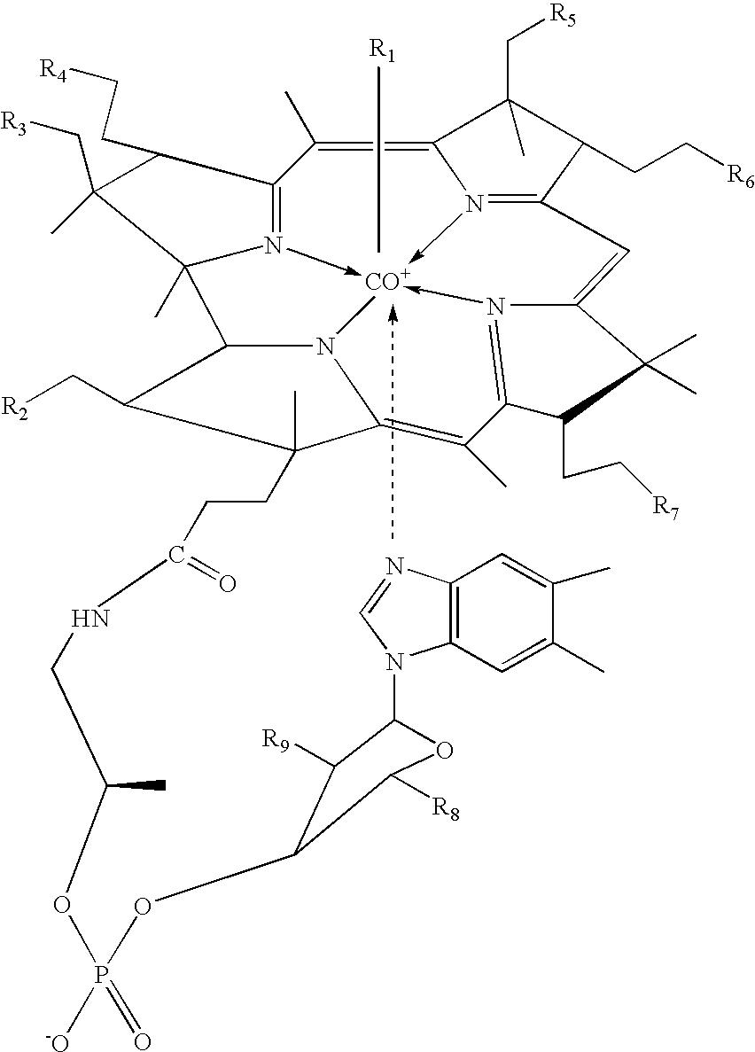
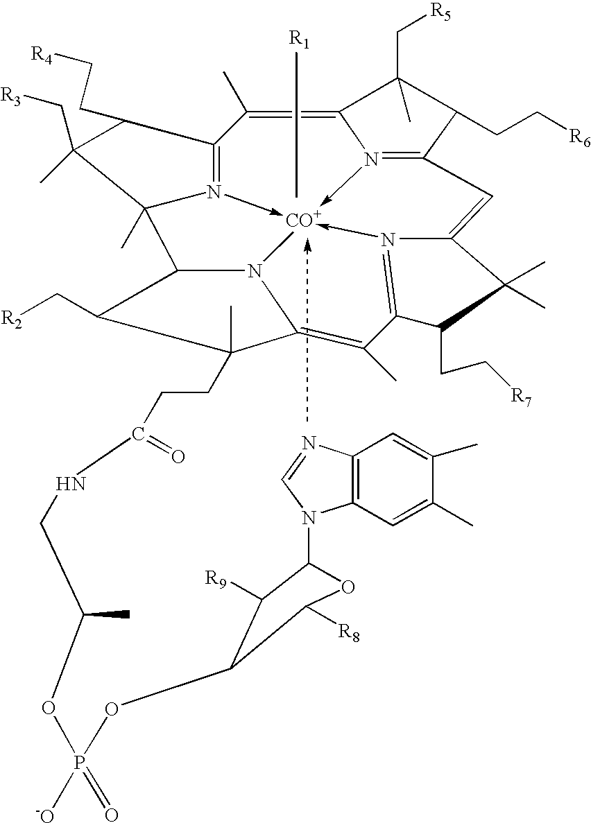
[0011] According to one embodiment, fluorescent cobalamins comprised of a fluorescent, phosphorescent, luminescent or light-producing compound that is covalently linked to cobalamin can be used in conjunction with the above-described apparatuses. These fluorescent cobalamins can be used as a diagnostic and prognostic marker (a) to distinguish cancer cells and tissues from healthy cells and tissues, including identifying lymph nodes containing cancer cells, and (b) to determine if an individual will respond positively to chemotherapy using cobalamin-therapeutic bioconjugates. The fluorescent cobalamins offer the properties of (1) rapid transport and storage by cancer cells (maximum uptake occurs at 4-6 hours), (2) a bright fluorophore that can be visually detected at very low concentrations, and (3) nontoxic components.
[0013] In a second aspect, the fluorescent cobalamins are used to identify atypical cells such as neoplastic cells, dysplastic cells, or hyperplastic cells. More particularly, the fluorescent cobalamins are used to distinguish cancer cells from healthy cells. In one embodiment, a fluorescent cobalamin is administered to a patient prior to surgery. The presence of fluorescence, phosphorescence, luminescence or emitted light in cancer cells is used by the surgeon to define the tissue to be removed, whether in a primary tumor or in a metastatic site. In a second embodiment, a fluorescent cobalamin is administered to a patient in a manner suitable for uptake by lymph nodes draining the situs of the tumor. The presence of fluorescence, phosphorescence, luminescence or emitted light identifies those lymph nodes that should be removed during surgery. In this latter embodiment, laparoscopic, endoscopic and microscopic techniques can be utilized to identify lymph nodes with cancer cells. The use of these techniques facilitates the identification and retrieval of positive lymph nodes.

Problems solved by technology

This procedure is controversial, as it fails to detect metastatic disease in about 12% of patients (McMasters et al., 1999).

Method used


Image

  • Device and method for the photodynamic diagnosis of tumor tissue
  • Device and method for the photodynamic diagnosis of tumor tissue
  • Device and method for the photodynamic diagnosis of tumor tissue

Examples


example 1

Synthesis of Fluorescent Cobalamin by Attachment of the Fluorophore to Cobalt

[0098] As a visual indicator of cobalamin localization, five fluorescent analogues of cobalamin were prepared by covalently attaching fluorescein to cobalamin. Under green light illumination, the fluorescein molecule emits yellow light that can be detected by the dark-adapted eye to concentrations lower than 0.1 ppm. This emission enables the sensitive detection of cancer cells via epifluorescence microscopy, as well as by visual inspection. Each of the five fluorescent cobalamins exhibited intrinsic fluorescence. All of these compounds were synthesized by reacting aminopropyl chloride with cob(I)alamin to produce aminopropylcob(III)alamin in accordance with published techniques. In a subsequent step, aminopropylcob(III)alamin was reacted with a variety of fluorophore isothiocyanates (i.e. fluorescein isothiocyanate, "FITC") to produce the corresponding fluorophore that is linked to cobalamin through an ami...

example 2

Uptake of Cobalamin Bioconjugates by Cancer Cells

[0100] A leukemic myeloblast preparation was made from a bone marrow aspirate of a 61-year old patient having acute myelogenous leukemia (AML) M1 (minimally mature myeloblasts in the FAB classification). Cells were treated three days post-harvest with a fluorescent cobalamin prepared as described in Example 1. Differential uptake of fluorescent cobalamin analogues, as determined by fluorescence microscopy or fluorescence flow cytometry, in normal and leukemic human bone marrow cells was found. The difference between normal marrow cells and leukemic myeloblasts (cancer cells) is particularly noteworthy, with no detectable cobalamin being taken up by normal cells. A bone marrow sample from a healthy individual showed no fluorescent labeling. Uptake of a doxorubicin-cobalamin conjugate, originally synthesized as a potential chemotherapeutic compound, was seen in P-388 marine leukemia cells and in HCT-116 human colon tumor cells. These re...

example 3

Preparation of Cyanocobalamin Monocarboxylic Acids

[0101] The b-, d-, and e-monocarboxylic acids were prepared by acid-catalyzed hydrolysis of cyanocobalamin. See FIG. 2. Briefly, cyanocobalamin (527.0 mg, 0.389 mmol) was placed into a 100 ml round bottom flask and dissolved in 40 ml of 0.5 M HCl. The flask was placed in a water bath at 50.degree. C. and stirred for 4 hours. The reaction was monitored via HPLC (Waters, Inc. 3.9.times.300 mm DeltaPak 100 C-18 column) using the gradient tabulated in Table 1.

1TABLE 1 0.5 M H.sub.3PO.sub.4 (pH Time (min) Flow Rate (ml / min) 3.0 w / NH.sub.3OH) 9:1 CH.sub.3CN:H.sub.20 0.0 20 90.0 10.0 2.0 2.0 90.0 10.0 18.0 2.0 83.7 16.3 23.0 2.0 30.0 70.0 25.0 2.0 30.0 70.0 30.0 2.0 90.0 10.0

[0102] After 4 hours the reaction was cooled to room temperature. The pH was adjusted to 7.0 with NaOH (10%) using a pH meter. The crude material was desalted using a C-18 SepPak column (Waters, Inc. PIN WATO23635) by first rinsing the column with 10 ml methanol followe...


PUM


Abstract

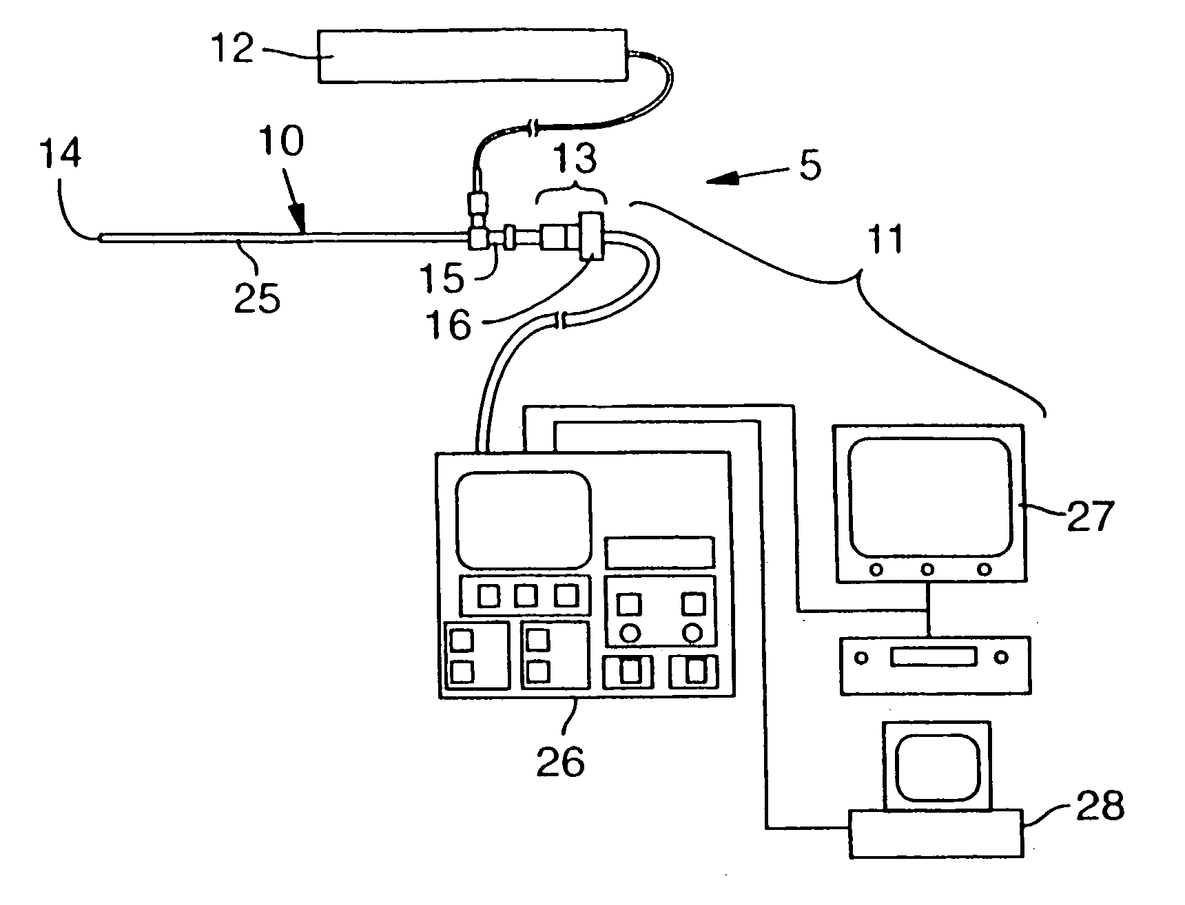
An apparatus that includes a surgical telescopic device having a distal end and a proximal end; a camera coupled to the proximal end of the surgical telescopic device; and a holographic notch filter interposed between the camera and the proximal end of the surgical telescopic device. The camera preferably is a charge-coupled-device camera ("CCD camera"). The apparatus may also include a focusing lens or an alternative type of filter such as a long-pass filter or an infrared filter. The surgical telescopic device may be used by illuminating the material with non-white light and detecting the emitted fluorescence, phosphorescence or luminescence.

Description

[0002] The present disclosure relates to devices and methods for exciting a biologically-targeted fluorescent molecule with light from a light source (e.g., a laser). According to certain embodiments, fluorescent cobalamins (sometimes referred to herein as CobalaFluors) can be used with the disclosed devices and methods. The fluorescent cobalamins comprise a fluorescent, phosphorescent, luminescent or light producing compound that is covalently linked to cobalamin. These fluorescent cobalamins can be used as diagnostic and prognostic markers (a) to distinguish cancer cells and tissues from healthy cells and tissues, including identifying lymph nodes containing cancer cells, and (b) to determine if an individual will respond positively to chemotherapy using cobalamin-based therapeutic bioconjugates.[0003] The publications and other materials cited herein are referenced in the following text by author and date and are listed alphabetically by author in the appended bibliography.[0004]...

Claims


Application Information

Patent Timeline
IPC IPC(8): A61B1/313
CPCA61B1/313
Inventor MCGREEVY, JAMESGRISSOM, CHARLES B.CANNON, MICHELLE
Owner UNIV OF UTAH RES FOUND
Learn More