Method for determining molecular affinities for human serum albumin
a human serum albumin and molecular affinity technology, applied in the field of new fluorescence-based assays, can solve the problem of reducing the concentration of free drugs available for physiological action
- Summary
- Abstract
- Description
- Claims
- Application Information
AI Technical Summary
Benefits of technology
Problems solved by technology
Method used
Image

Examples
example 1
General Procedure
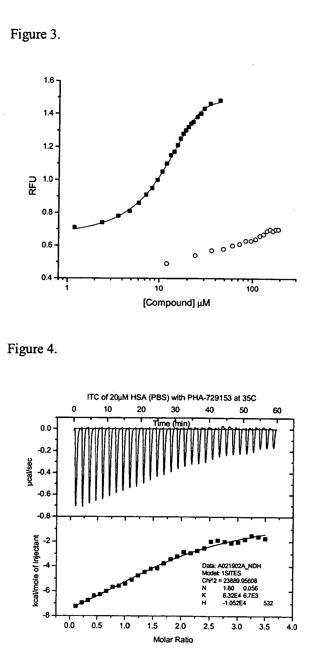
Buffered probe solution (200 μL of 0.5 μM probe in phosphate buffered saline) was added to each well of a 96 well plate using the automated liquid injection device on the instrument. To each well, except wells in the last row, was added 15 μL of 20 μM HSA. Test compounds were added to wells, except the last two rows of the plate, equilibrated for 60 minutes at room temperature and then the fluorescence was read for the test compound wells (f), the row of wells containing probe, buffer and HSA (f0), and the last row of wells containing only probe and buffer (fb). Precision in the fluorescence measurements was determined to be within 3-6% and assay reproducibility over an extended period of one month was within 10% using multiple preparations of HSA, probe solution and stock test compound.
example 2
Chemicals, Equipment and Assay Stock Solutions
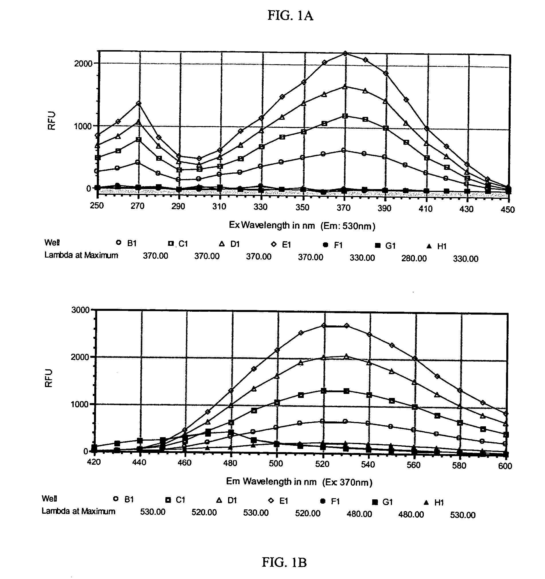
Human serum albumin, fraction V, with a purity ≧95% as indicated by cellulose acetate electrophoresis was purchased from Calbiochem (catalog no. 12666). Dulbecco's phosphate buffered saline (PBS) without magnesium or calcium was purchased from Gibco. Dimethylsulfoxide was purchased from EM Science. HSA stock solution was prepared in Dulbecco's PBS at a nominal concentration of 5 mg / mL (77 μM). Accurate concentration of the HSA stock solution was determined by measuring the absorbance of 500 μl of stock solution at 280 run with a Perkin Elmer Lambda 40 UV Spectrophotometer, ε280=35,600 M−1cm−1. HSA stock solutions were stored at 5° C. A stock solution of THE PROBE, FW 302.3 g / M, was prepared at 10 mM in DMSO, 0.45 mg of THE PROBE in 150 μL DMSO. This solution was stored in an amber bottle at 5° C.
A SPECTRAmax GEMINI dual-scanning microplate spectrofluorometer (Molecular Devices, Sunnyvale, Calif.) was used to collect the fluorescence ...
example 3
The Probe Displacement Assay Procedure
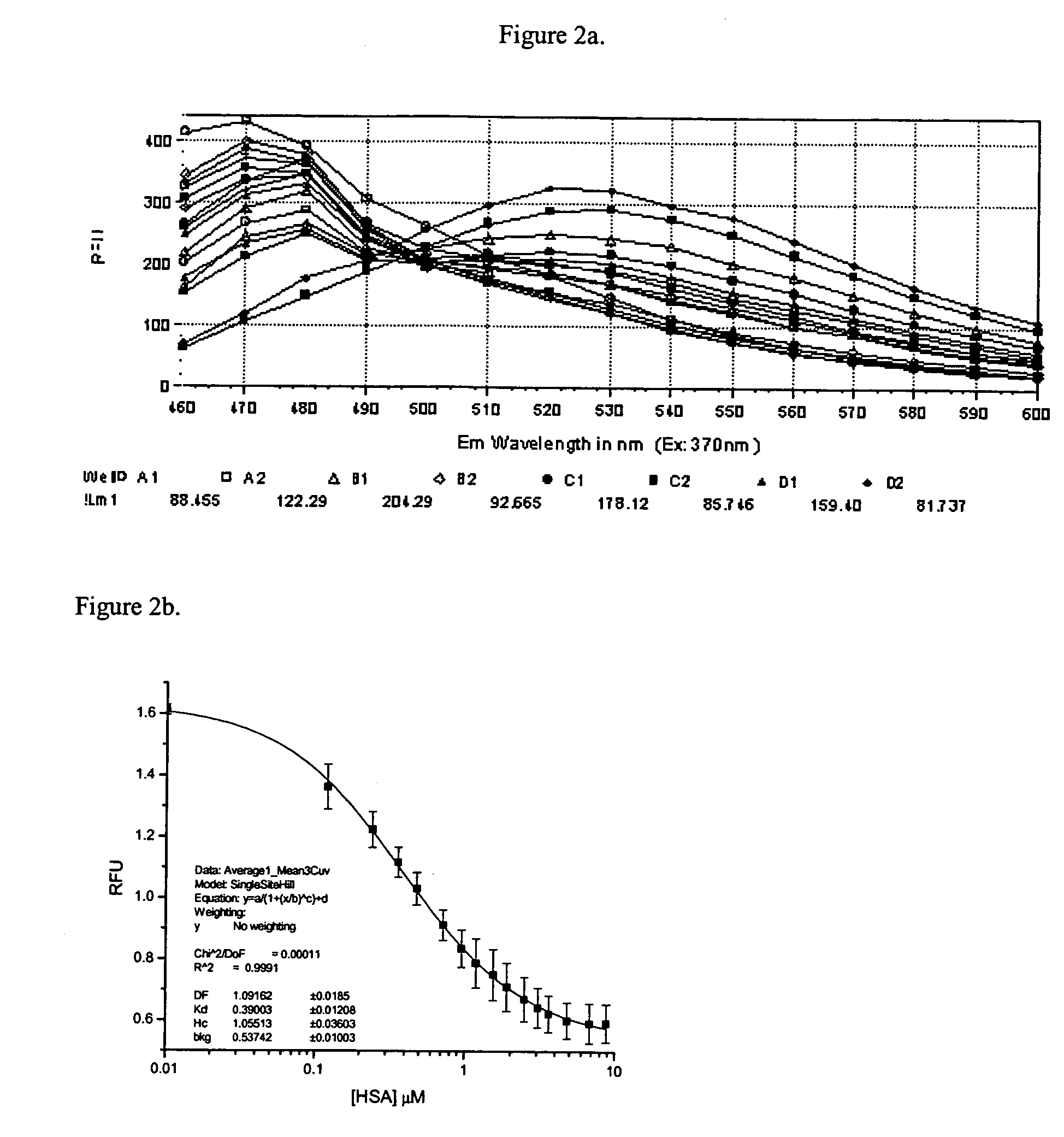
Analyte solutions were prepared at 5 mM in DMSO. Aliquots, 0.5 μL, were dispensed into assay wells in triplicate starting with wells A1, B1 and C1. This resulted in a final analyte concentration of 12 μM after addition of 200 μL of PBS plus THE PROBE and 15 μL of HSA at 20 μM. The next analyte was added to wells D1, E1 and F1. In this manner, 2 analytes per column or 21 compounds in all could be assayed on one 96 well plate. The last two columns of the assay plate were reserved for assay of the standards and the last two rows were reserved for replicate assay of controls and blanks. A typical plate layout is shown in Table 1.
A 5 μl aliquot of THE PROBE was diluted into 100 mL of PBS for a final probe concentration of 0.5 μM. Injector number one of the Polarstar was filled with this solution and 200 μl of the probe solution was added to each well of the assay plate. The plate was transferred to a SpectraMax Gemini reader for data collection. ...
PUM
| Property | Measurement | Unit |
|---|---|---|
| binding affinity | aaaaa | aaaaa |
| fluorescence | aaaaa | aaaaa |
| molecular affinities | aaaaa | aaaaa |
Abstract
Description
Claims
Application Information
 - R&D
- Intellectual Property
- Life Sciences
- Materials
- Tech Scout
- Unparalleled Data Quality
- Higher Quality Content
- 60% Fewer Hallucinations
Browse by: Latest US Patents, China's latest patents, Technical Efficacy Thesaurus, Application Domain, Technology Topic, Popular Technical Reports.
© 2025 PatSnap. All rights reserved.Legal|Privacy policy|Modern Slavery Act Transparency Statement|Sitemap|About US| Contact US: help@patsnap.com