Biological laser printing via indirect photon-biomaterial interactions

a biomaterial and laser printing technology, applied in the field of laser and/or photon transfer, can solve the problems of limited application or inherent limitations of the maple dw, low reproducibility of the technique, and low absorption of certain matrix materials

Inactive Publication Date: 2005-01-27
THE UNITED STATES OF AMERICA AS REPRESENTED BY THE SECRETARY OF THE NAVY
View PDF28 Cites 32 Cited by
  • Summary
  • Abstract
  • Description
  • Claims
  • Application Information

AI Technical Summary

Problems solved by technology

MAPLE DW has limited applications or inherent limitations due to the physical properties of the biological materials and surrounding media needed to ensure accurate pattern formation.
In addition, the reproducibility of the technique can be low due to inconsistencies in the parameters necessary for ablation of the matrix material.
Also, because the absorptivity of certain matrix materials is quite low, there is the potential for damage to biological materials from direct and indirect interaction with the incident laser radiation.
This leads to difficulty in patterning multiple biological material types and difficulty in rapid design of micro-scale constructs.
It is possible to obtain sub-micron features using MCP, but there is little control over the amount of material transferred.
It is possible that different materials could be patterned using multiple stampings, but this could lead to possible contamination of stamps and has inherent limitations in the proximity of the different material types.
Also, MCP is dependent on biomaterial-substrate adhesion, and therefore not universal to all substrate materials.
This technique allows for manipulation and placement of individual cells, but has limitations in the fact that it can take hours to write a moderate number of cells.
In addition there is the potential for DNA damage from the extended time that cells or other biological materials are exposed to the laser beam.
Laser transfer of biological materials presents a challenge due to the fragility of many biological materials.
They can be harmed by shear stress when they are removed from the target substrate and by impact stress when they land on the receiving substrate.
UV damage can also result when a UV laser is used.
A disadvantage of these methods is that they can still expose the material to be deposited to high amounts of energy that may not be suitable for biological materials.

Method used


Image

  • Biological laser printing via indirect photon-biomaterial interactions
  • Biological laser printing via indirect photon-biomaterial interactions
  • Biological laser printing via indirect photon-biomaterial interactions

Examples


example 1

[0057] Deposition of Eukaryotic Cells in a Large-Scale Array Format

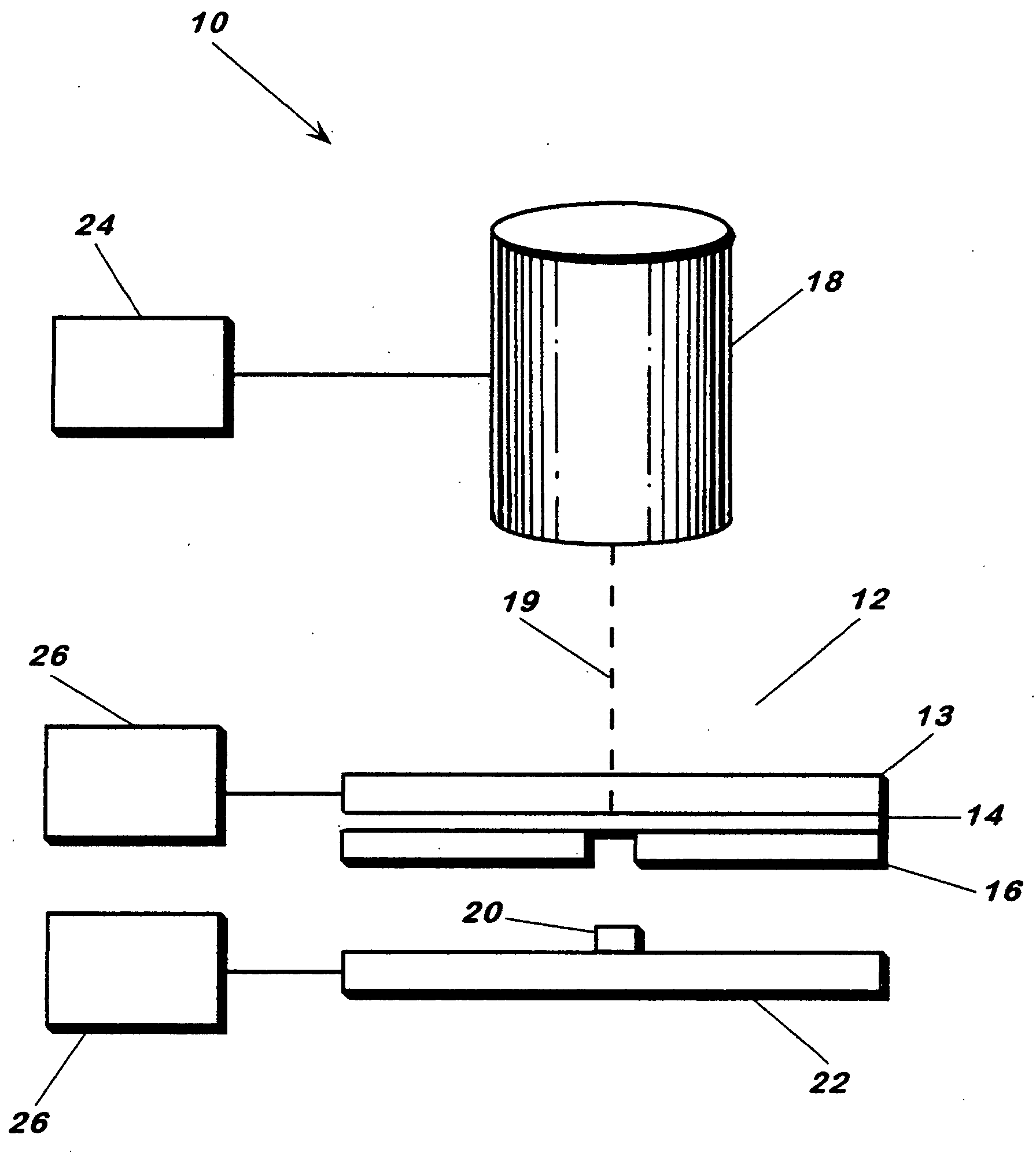
[0058] Human osteosarcoma cells (ATCC #: CRL-1427, designation: MG-63) were cultured as per literature, trypsinized and placed in a deposition media composed of 50% (v / v) DMEM, 45% (v / v) Fetal Bovine Serum and 5% glycerol (v / v). Cell concentration in the deposition media was found to be 106 cells / mL. 12 μL of the deposition solution was spread across a 4 cm2 area of the target and then placed in the apparatus for transfer. Arrays of cell were deposited onto 1×3″ glass slides coated with a 200 μm thick Matrigel™ basement membrane matrix, immersed in DMEM and placed in an incubator 5 minutes after transfer. Twenty hours post-transfer, the cells were washed with 1×PBS and assayed via a live / dead viability / cytotoxicity kit (Molecular Probes, L-3224) (FIG. 7).

[0059] In this deposition, the laser spot was 100 μm in diameter at the laser absorption interface and laser fluence was 160 mJ / cm2. Deposition spots were approxim...

example 2

[0061] ATCC Designation MG 63 Human Osteosarcoma Cells and C2C12 Mouse Myoblast Cells

[0062] MG63 cells were initially obtained from ATCC (USA) and cultured in 5% humidified CO2 in air at 37° C. in Dulbecco's Modified Eagle Medium (DMEM) with high glucose, 10% (v / v) fetal bovine serum, and 1% (v / v) streptomycin. FIG. 4 shows a patterned row of cells transferred from a titanium-coated quartz support and deposited onto a quartz substrate coated with matrigel (50 μm). The apparatus was as described above. By varying laser spot size, the number of cells deposited could be controlled. At ˜100 μm spot size and an energy of 0.35 μJ / pulse, near-single cell transfers were obtained, at cell transfer rate of 1 cell / shot (±0.5). Laser fluence was 4.4 mJ / cm2.

example 3

[0063] ATCC Designation MG 63 Human Osteosarcoma Cells

[0064] Human osteosarcoma cells along with 10 μm fluorescent beads were transferred from a titanium-coated quartz support using the energy conversion method. The cells and beads were in a 25% glycerol, 25% cell media (DMEM), and 50% aqueous bead (0.15 M NaCl, 0.05% Tween 20, 0.02% thimerosal) solution. Laser fluence was 160 mJ / cm2 and spot sizes were 100 μm in diameter.


PUM

PropertyMeasurementUnit
thickaaaaaaaaaa
diameteraaaaaaaaaa
widthaaaaaaaaaa

Abstract

A method of laser forward transfer is disclosed. Photo energy is directed through a photon-transparent support and absorbed by an interlayer coated thereon. The energized interlayer causes the transfer of a biological material coated thereon across a gap and onto a receiving substrate.

Description

[0001] This application claims the benefit of U.S. Provisional Patent Application No. 60 / 476,377, filed on Jun. 6, 2003 and U.S. Provisional Patent Application No. 60 / 542,841, filed on Feb. 10, 2004. All applications named are incorporated by reference. The U.S. patent application to Ringeisen et al., titled “Biological Laser Printing from Tissue via Indirect Photon-Biomaterial Interactions,” designated NC 96,075, filed on the same day as the present application is also incorporated by reference.BACKGROUND OF THE INVENTION [0002] 1. Field of the Invention [0003] The invention is generally related to methods of laser and / or photon transfer. [0004] 2. Description of the Prior Art [0005] Direct write technologies have gained popularity with the increased interest in biological sensors and microarrays, and the push for engineered tissues to replace organ transplants. These techniques allow for increased ability to manipulate biological materials in very small volumes with much better ac...

Claims


Application Information

Patent Timeline
Patent Type & Authority Applications(United States)
IPC IPC(8): A61B18/20B05D1/00B05D3/00B05D3/06B23K26/00B32B7/02B32B7/04B32B9/02B32B9/04B41J2/435B41M3/00B41M5/382B41M5/385B41M5/41B41M5/46C12MC12N13/00C23C14/04C23C14/28C23C14/32H05B6/64
CPCA61B18/20G01N2001/2886B01J2219/00603B01J2219/00641B01J2219/00659B01J2219/00725B01J2219/00743B41M5/38207B41M5/385B41M5/41B41M5/465C12N13/00C23C14/048C23C14/28G01N1/2813B01J2219/00527
Inventor BARRON, JASONRINGEISEN, BRADLEY R.KIM, HEUNGSOOWU, PETER
Owner THE UNITED STATES OF AMERICA AS REPRESENTED BY THE SECRETARY OF THE NAVY
Why Eureka
Try Eureka