
Materials and methods for near-infrared and infrared intravascular imaging

a technology of infrared and intravascular imaging, applied in the field of imaging tissue and organs, can solve the problem that small fluorophores only provide short time windows for imaging, and achieve the effects of short time windows, prolonged imaging of the vasculature, and rapid leakag

Inactive Publication Date: 2005-01-27
BETH ISRAEL DEACONESS MEDICAL CENT INC +1
View PDF41 Cites 71 Cited by
  • Summary
  • Abstract
  • Description
  • Claims
  • Application Information

AI Technical Summary

Benefits of technology

Traditionally, in vivo near-infrared (NIR) fluorescence imaging of the vasculature is performed with low-molecular weight organic dyes such as indocyanine green (ICG) or the carboxylic acid of IRDye78 (IRDye78-CA). Unfortunately, these small fluorophores only provide short time windows for imaging (i.e., immediately after intravenous injection) since they rapid leak out of the vasculature and into tissue. Advantageously, emissive semiconductor nanocrystals of the appropriate size and coating permit provide prolonged imaging of the vasculature with high sensitivity. The nanocrystals are excited by light, and emit light, thereby replacing the need to produce images using X-ray technology. The size of NIR nanocrystals keeps particles in vasculature for prolonged imaging. The method allows for sensitive detection of normal tissue and tumor vascular during surgery, sensitive detection of sites of bleeding during surgery, and sensitive measurement of tissue perfusion during surgery and surgical repairs.

Problems solved by technology

Unfortunately, these small fluorophores only provide short time windows for imaging (i.e., immediately after intravenous injection) since they rapid leak out of the vasculature and into tissue.

Method used


Image

  • Materials and methods for near-infrared and infrared intravascular imaging
  • Materials and methods for near-infrared and infrared intravascular imaging
  • Materials and methods for near-infrared and infrared intravascular imaging

Examples


examples

Animals. Animals were used in accordance with an approved institutional protocol. Male Sprague-Dawley rats were from Charles River Laboratories (Wilmington, Mass.). Hairless athymic nu / nu mice were from Taconic (Germantown, N.Y.). Rats and mice were anesthetized with 65 mg / kg and 50 mg / kg intraperitoneal pentobarbital, respectively.

Reagents. Sterile Intralipid™ (20%) was purchased from Baxter (Deerfield, Ill.). Water was purified on a Milli-Q system (Millipore, Bedford, Mass.). Olive oil was from Filippo Berio (Viareggio, Italy). Oxyhemoglobin (OxyHb) was prepared from normal human donors as described in Drabkin, J. Biol. Chem. 164:703-723 (1946), which is incorporated by reference in its entirety. Deoxyhemoglobin (DeoxyHb) was prepared by treatment of OxyHb with 1% sodium dithionite (Sigma, St. Louis, Mo.). Albumin, Cohn Fraction V was also from Sigma. All solutions except Intralipid were filtered through 0.2 μm filters (Millipore) prior to use to eliminate scatter. Trioctylphos...


PUM


Abstract

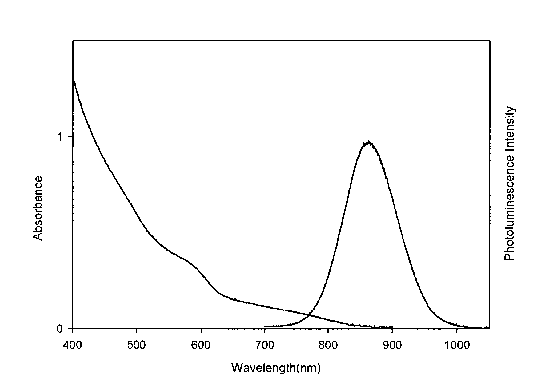
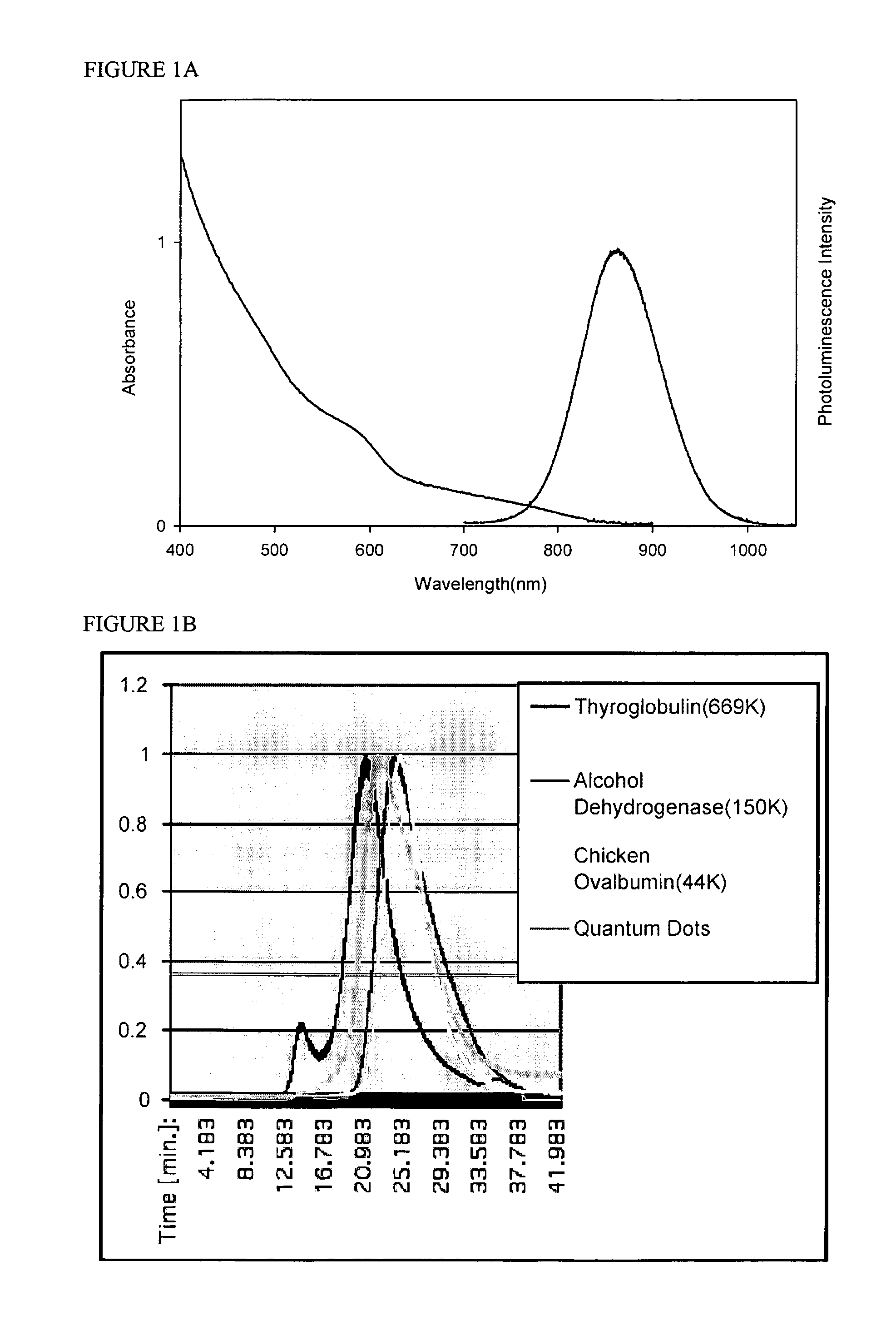
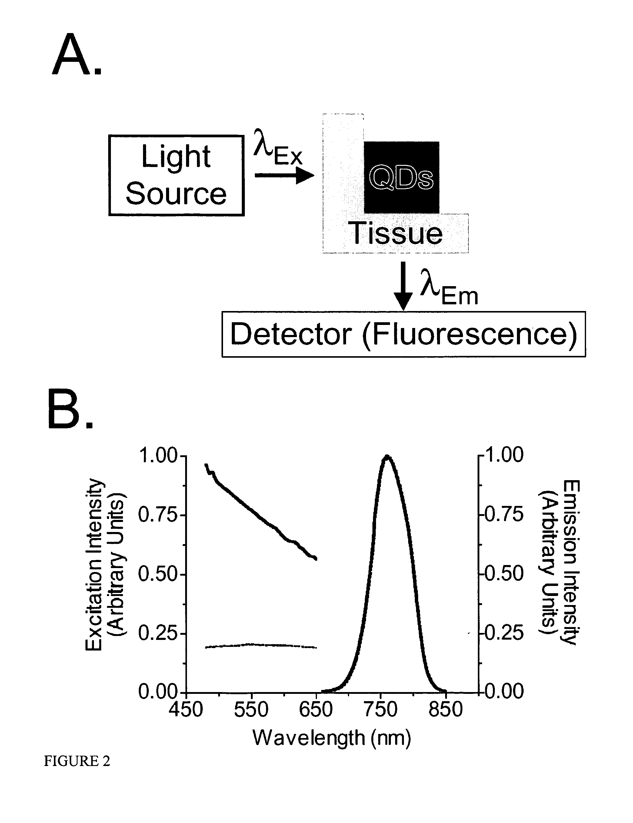
Vasculature can be imaged with emissive semiconductor nanocrystals, for example, in the near infrared.

Description

TECHNICAL FIELD The invention relates to imaging tissue and organs. BACKGROUND Semiconductor nanocrystals (QDs) having small diameters can have properties intermediate between molecular and bulk forms of matter. For example, nanocrystals based on semiconductor materials having small diameters can exhibit quantum confinement of both the electron and hole in all three dimensions, which leads to an increase in the effective band gap of the material with decreasing crystallite size. Consequently, both the optical absorption and emission of nanocrystals shift to the blue (i.e., to higher energies) as the size of the crystallites decreases. Semiconductor nanocrystals can have a narrow fluorescence band whose emission wavelength is tunable with the size and material of the nanocrystals. SUMMARY In general, imaging of vasculature can be performed by introducing emissive semiconductor nanocrystals into tissue. The emissive nanocrystals can emit irradiation in the near-infrared (NIR) or in...

Claims


Application Information

Patent Timeline
IPC IPC(8): A61BA61B5/00A61B5/05A61B6/00A61K49/00
CPCA61B5/0059A61K49/0067A61B5/418A61B5/415
Inventor FRANGIONI, JOHN V.BAWENDI, MOUNGI G.KIM, SUNGJEELIM, YONG TAIK
Owner BETH ISRAEL DEACONESS MEDICAL CENT INC
Learn More