
Secretion of neublastin
a technology of neublastin and secretion, which is applied in the direction of peptide/protein ingredients, virus, dna/rna fragmentation, etc., to achieve the effects of reducing the likelihood of immune attack, reducing the risk of infection, and increasing the safety and control of therapy
- Summary
- Abstract
- Description
- Claims
- Application Information
AI Technical Summary
Benefits of technology
Problems solved by technology
Method used
Image

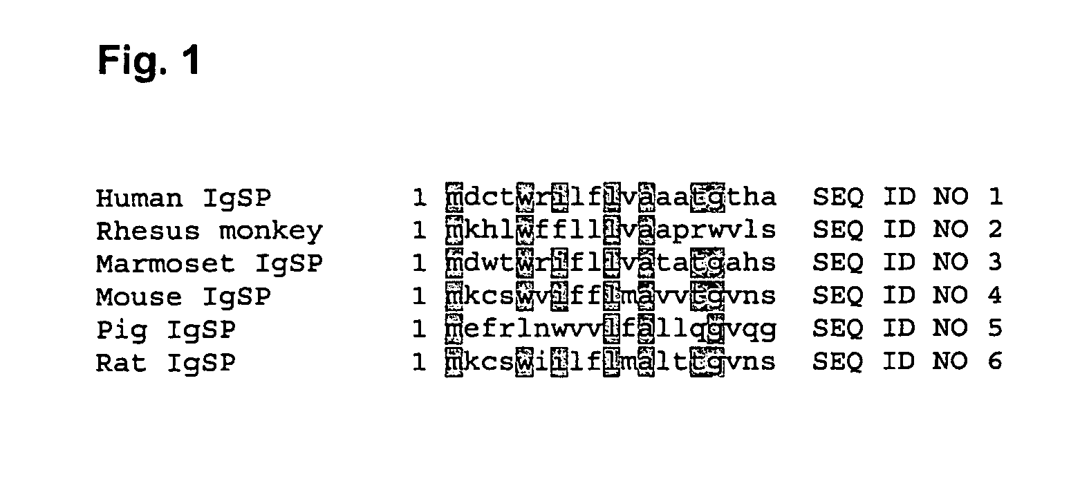
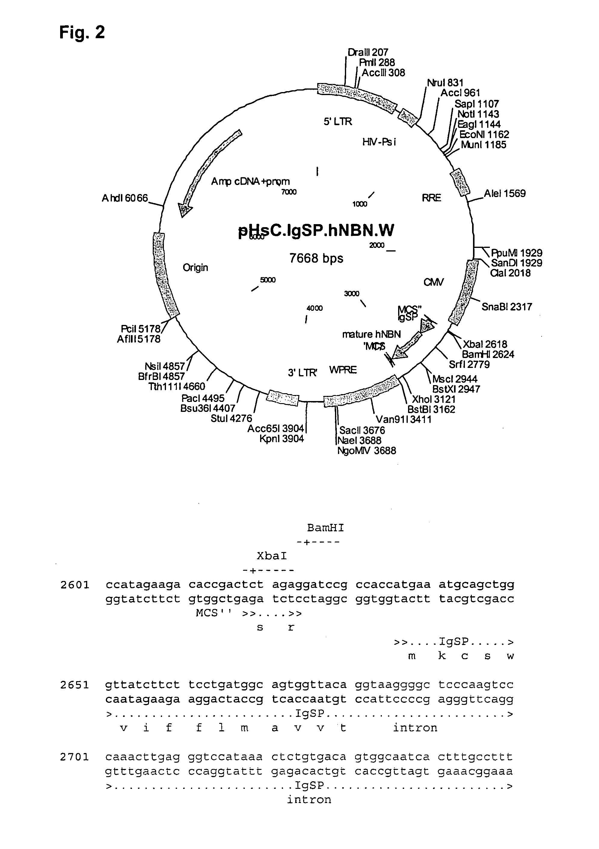
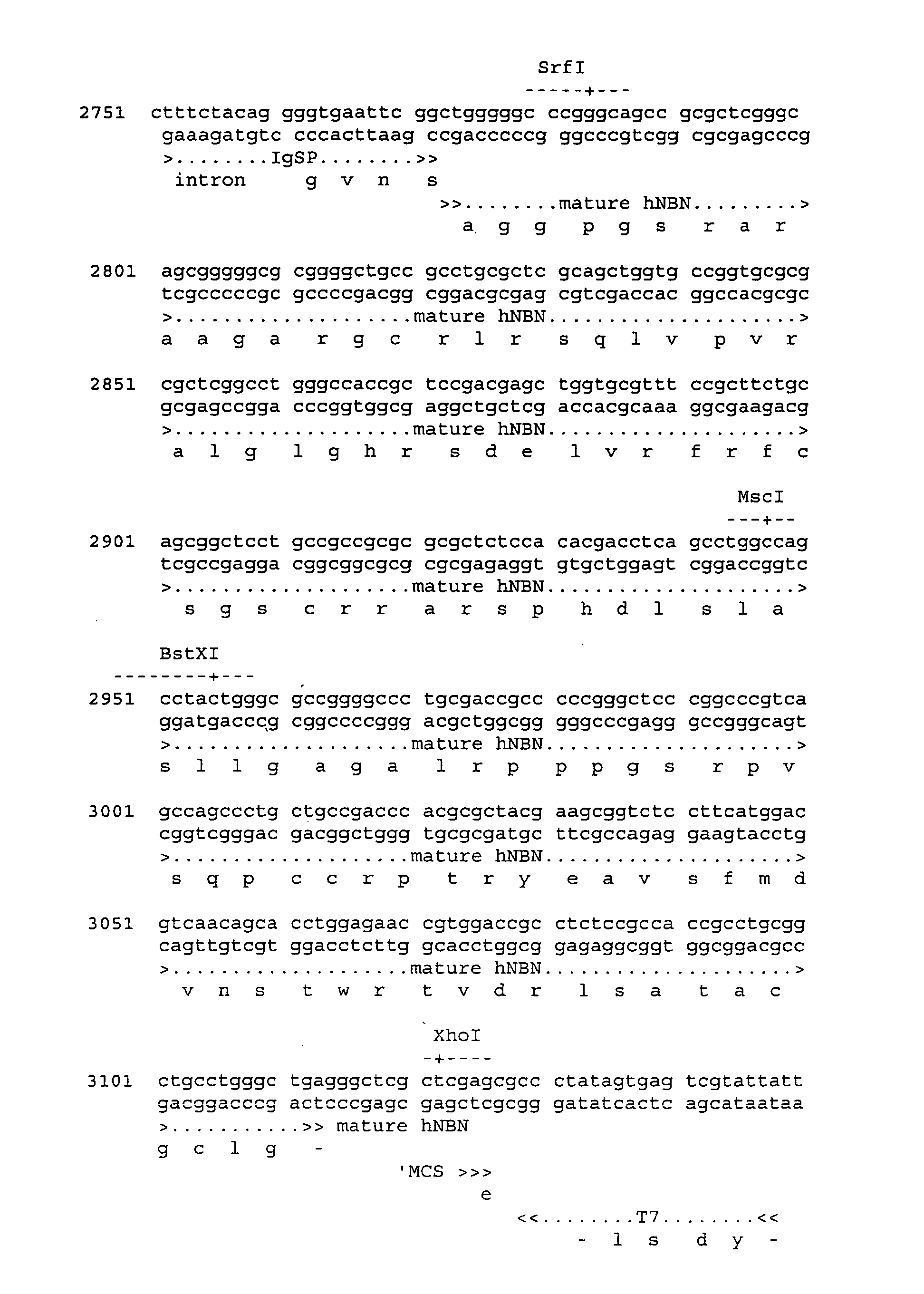
Examples
example 1
In Vitro Transfection with IgSP-NBN Construct
[0239] Construction of IgSP-NBN Constructs
[0240] IgSP.NBN was generated by overlap PCR. In the first amplification step, the mature fragment of NBN was amplified by PCR from the pUbi1z.NBN.BamHI vector using the primers NBNs-IgSP.Flap (5′-GGTGAATTCGGCTGGGGGCCCGGGCAGCC-3′) and NBNas+XhoI(5′-TATACTCGAGCGAGCCCTCAGCCCAGGCA-3′). In a second PCR reaction, the IgSP sequence was amplified from the pNUT-IgSP-CNTF vector (ref. U.S. Pat. No. 6,361,741) using the primers IgSPKozak1s+BamHI (5′-TATAGGATCCGCCACCATGAAATGCAGCTGGGTTATC-3′) and IgSPas-NBN.Flap (5′-GGCCCCCAGCCGMTTCACCCCTGTAGAAAG-3′). In the third step the products of step 1 and 2 were combined in a final PCR reaction that generates IgSP-NBN by using equal amounts of the two products as template with the primers IgSPKozak1s+BamHI and NBN-as+XhoI.
[0241] To generate a plasmid-based expression vector the resulting fragment was cloned in pNS1n digested with BamHI / XhoI. In this vector, the IgSP...
example 2
In Vitro Transduction With IgSP-NBN Construct
[0249] Generation of a Lentiviral IgSP-NBN Construct and Virus Stocks
[0250] To generate a lentiviral construct, pNS1n-IgSP-NBN was digested with BamHI-XhoI and the IgSP-hNBN PCR fragment (as described in example 1) was ligated into BamHI / XhoI-digested pHsCXW resulting in pHsCXW.IgSP.NBNw (FIG. 3). pHsCXW is a derivative of a self-inactivating lentiviral transfer construct, pHR′-SIN−18 including a WPRE element (Dull et al., J.Virol., 72(11):8463-71(1998); Zufferey et al., J.Virol., 72(12):9873-80(1998): Zufferey et al. J.virol., 73 (4):2886-92 (1999)) generated by replacing the large non-viral part of the transfer construct with the pUC19 backbone. The sequence of pHsCXW can be accessed through GenGank ID: AY468486.
[0251] Replication-defective LV-sC.IgSP.NBN.W virus particles are generated by co-transfection of pHsC.IgSP.NBN.W with pMD.G (VSV-G pseudo-typing vector) and pBR8.91 (packaging vector) (Zufferey et al., Nat. Biotech., 15:871-...
example 3
Analysis of NBN Protein Expressed from IgSP-NBN Constructs
[0257] Western Blot Analysis of Cell Supernatants
[0258] NBN present in cell supernatants from transfected or transduced cell cultures was concentrated by affinity binding to GFRα3-Ig prior to Western blot analysis. Briefly, 4 wells of a Nunc MaxiSorp plate were coated with 300 μl Goat anti human Fc 1 μg / ml (Jackson Immunoresearch Laboratories, TriChem, Denmark) in 50 mM NaHCO3 (pH=9.6) for 16 h at 4° C. The wells were blocked with 400 μl 1% BSA in PBS for 1 h at room temperature, followed by 3 washes in PBST. 300 μl / well GFRα3 / Fc fusion protein 1 μg / ml (R&D Systems, UK) was then added and the plate was incubated for 1 h at room temperature to maximise binding. The wells were then emptied and washed again 3 times with PBST. 300 μl supernatant collected from transfected or transduced cell cultures was then added to each of the 4 wells and incubated for at least 3 h with gently shaking at room temperature. The wells were then ...
PUM
| Property | Measurement | Unit |
|---|---|---|
| Mass | aaaaa | aaaaa |
| Area | aaaaa | aaaaa |
Abstract
Description
Claims
Application Information