Nucleic acid sequencing by Raman monitoring of uptake of nucleotides during molecular replication

a technology of nucleotide uptake and nucleic acid sequencing, applied in the field of molecular biology and genomics, can solve the problems of high cost, inefficiency and time-consuming, laborious process,

Inactive Publication Date: 2005-07-07
INTEL CORP
View PDF16 Cites 35 Cited by
  • Summary
  • Abstract
  • Description
  • Claims
  • Application Information

AI Technical Summary

Problems solved by technology

This process is laborious, expensive, inefficient and time-consuming.
It also typically requires the use of fluorescent or radioactive labels, which can potentially pose safety and waste disposal problems.

Method used


Image

  • Nucleic acid sequencing by Raman monitoring of uptake of nucleotides during molecular replication
  • Nucleic acid sequencing by Raman monitoring of uptake of nucleotides during molecular replication
  • Nucleic acid sequencing by Raman monitoring of uptake of nucleotides during molecular replication

Examples


example 1

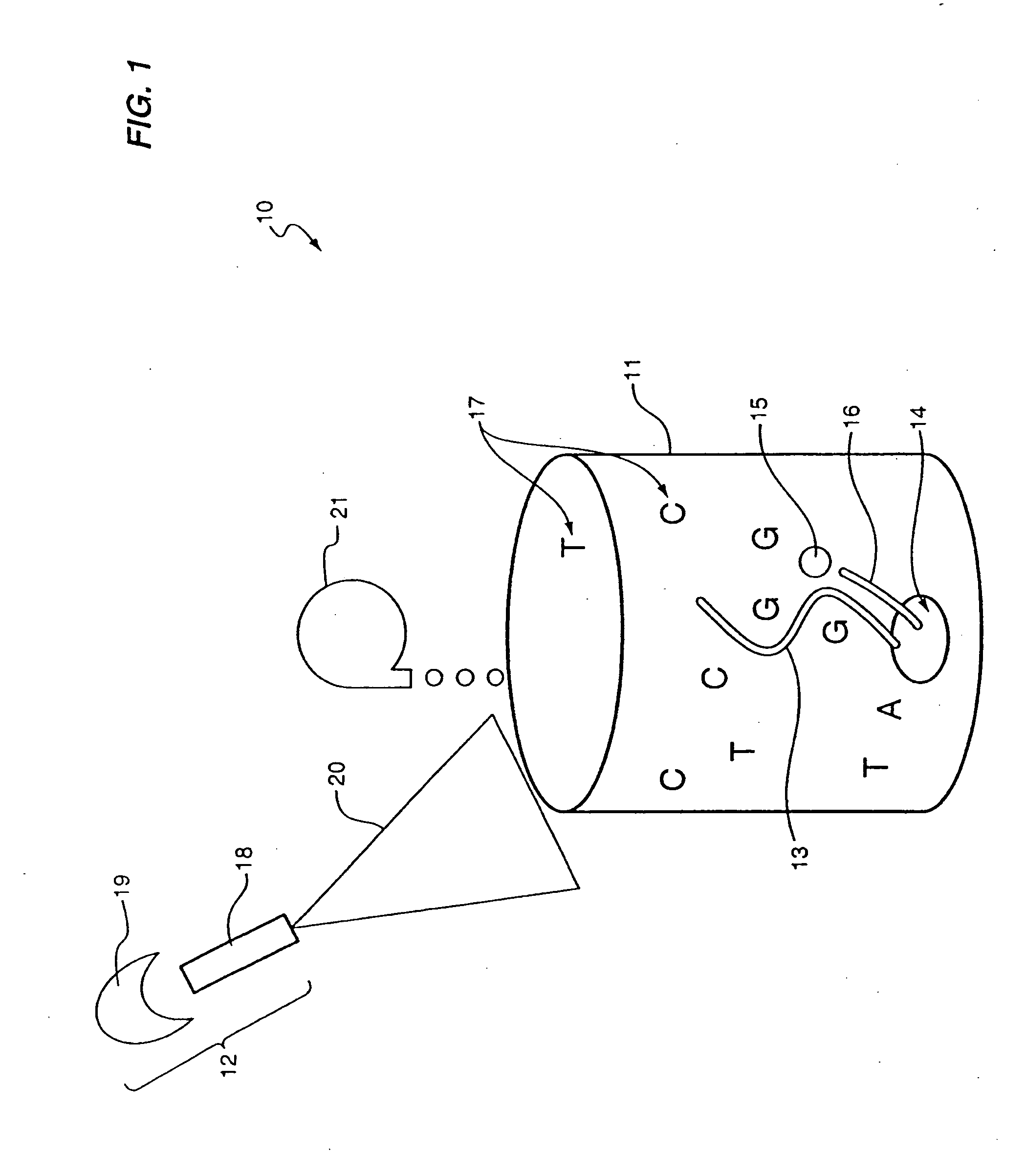
Raman Detection of Nucleotides

[0108] Methods and Apparatus

[0109] In a non-limiting example, the excitation beam of a Raman detection unit was generated by a titanium:sapphire laser (Mira by Coherent) at a near-infrared wavelength (750˜950 nm) or a gallium aluminum arsenide diode laser (PI-ECL series by Process Instruments) at 785 nm or 830 nm. Pulsed laser beams or continuous beams were used. The excitation beam was passed through a dichroic mirror (holographic notch filter by Kaiser Optical or a dichromatic interference filter by Chroma or Omega Optical) into a collinear geometry with the collected beam. The transmitted beam passed through a microscope objective (Nikon LU series), and was focused onto the Raman active substrate where target analytes (nucleotides or purine or pyrimidine bases) were located.

[0110] The Raman scattered light from the analytes was collected by the same microscope objective, and passed the dichroic mirror to the Raman detector. The Raman detector incl...

example 2

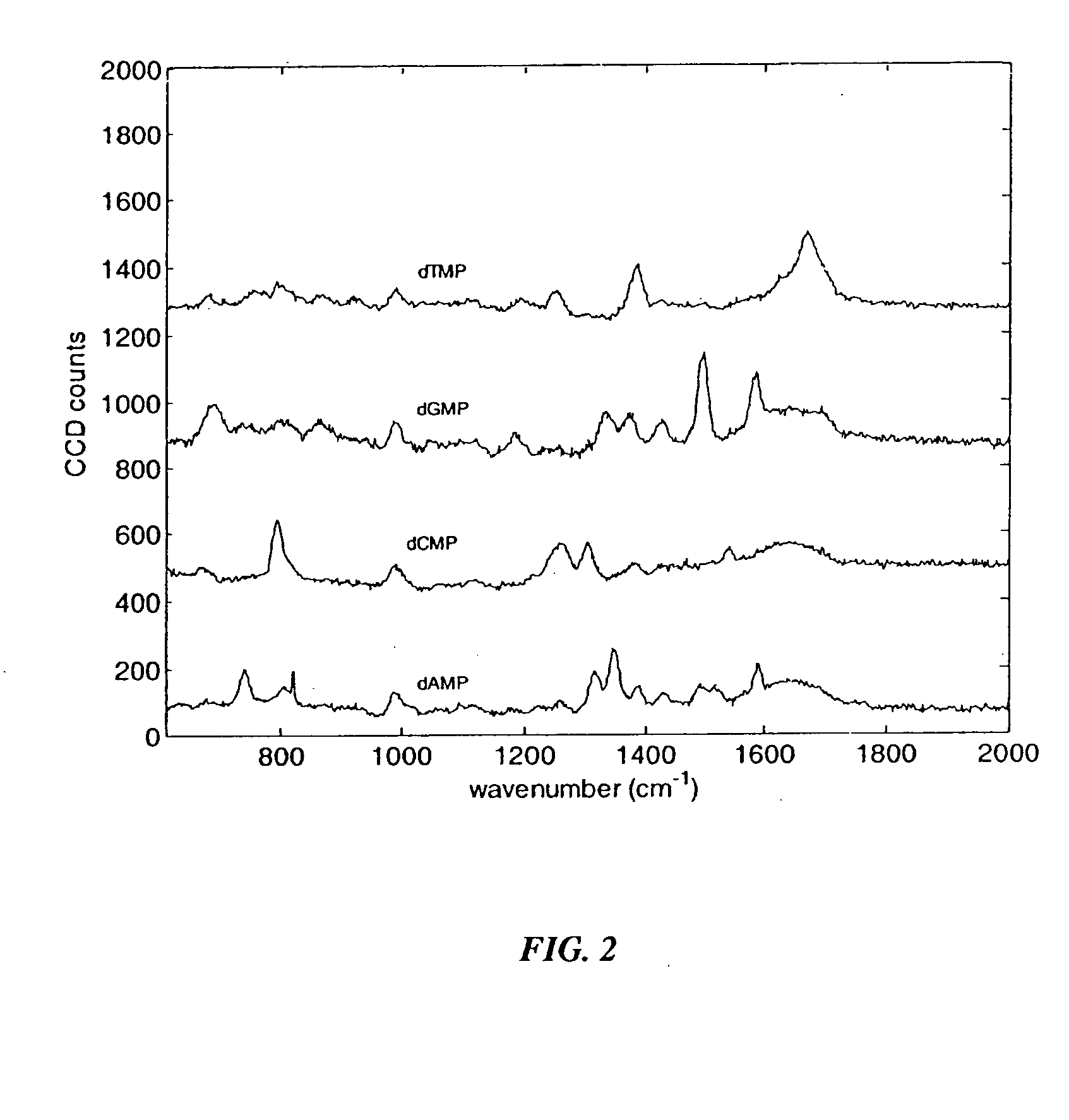
Raman Emission Spectra of Nucleotides, Purines and Pyrimidines

[0116] The Raman emission spectra of various analytes of interest were obtained using the protocol of Example 1, with the indicated modifications. FIG. 2 shows the Raman emission spectra of a 100 mM solution of each of the four nucleoside monophosphates, in the absence of surface enhancement and without Raman labels. No LiCl was added to the solution. A 10 second data collection time was used. Excitation occurred at 514 nm. Lower concentrations of nucleotides can be detected with longer collection times, added electrolytes and / or surface enhancement. For each of the following figures, a 785 nm excitation wavelength was used. As shown in FIG. 2, the unenhanced Raman spectra showed characteristic emission peaks for each of the four unlabeled nucleoside monophosphates.

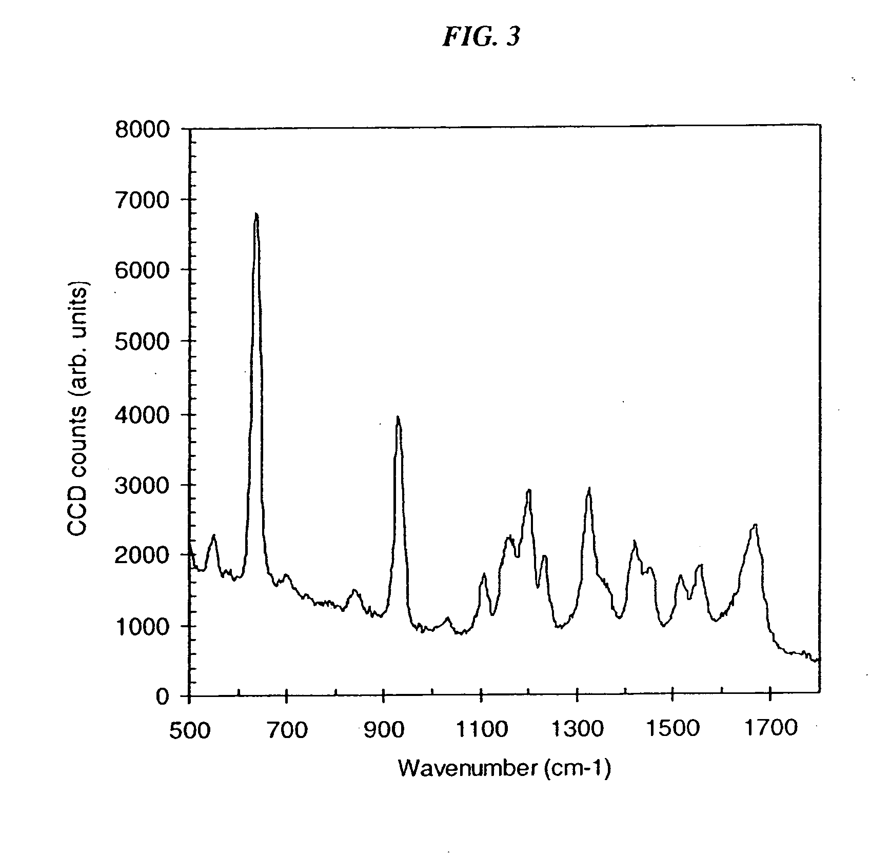
[0117]FIG. 3 shows the SERS spectrum of a 1 nm solution of guanine, in the presence of LiCl and silver nanoparticles. Guanine was obtained from dGMP by acid ...

example 3

SERS Detection of Nucleotides and Amplification Products

[0123] Silver Nanoparticle Formation

[0124] Silver nanoparticles used for SERS detection were produced according to Lee and Meisel (1982). Eighteen milligrams of AgNO3 were dissolved in 100 mL (milliliters) of distilled water and heated to boiling. Ten mL of a 1% sodium citrate solution was added drop-wise to the AgNO3 solution over a 10 min period. The solution was kept boiling for another hour. The resulting silver colloid solution was cooled and stored.

[0125] SERS Detection of Adenine

[0126] The Raman detection system was as disclosed in Example 1. One mL of silver colloid solution was diluted with 2 mL of distilled water. The diluted silver colloid solution (160 μL) (microliters) was mixed with 20 μL of a 10 nM (nanomolar) adenine solution and 40 μL of LiCl (0.5 molar) on an aluminum tray. The LiCl acted as a Raman enhancing agent for adenine. The final concentration of adenine in the sample was 0.9 nM, in a detection vol...


PUM

PropertyMeasurementUnit
diameteraaaaaaaaaa
diameteraaaaaaaaaa
diameteraaaaaaaaaa

Abstract

The methods and apparatus disclosed herein are useful for detecting nucleotides, nucleosides, and bases and for nucleic acid sequence determination. The methods involve detection of a nucleotide, nucleoside, or base using surface enhanced Raman spectroscopy (SERS). The detection can be part of a nucleic acid sequencing reaction to detect uptake of a deoxynucleotide triphosphate during a nucleic acid polymerization reaction, such as a nucleic acid sequencing reaction. The nucleic acid sequence of a synthesized nascent strand, and the complementary sequence of the template strand, can be determined by tracking the order of incorporation of nucleotides during the polymerization reaction.

Description

BACKGROUND OF THE INVENTION [0001] 1. Field of the Invention [0002] The present methods and apparatus relate to the fields of molecular biology and genomics. More particularly, the disclosed methods and apparatus concern nucleic acid sequencing. [0003] 2. Background Information [0004] Genetic information is stored in the form of very long molecules of deoxyribonucleic acid (DNA), organized into chromosomes. The human genome contains approximately three billion bases of DNA sequence. This DNA sequence information determines multiple characteristics of each individual. Many common diseases are based at least in part on variations in DNA sequence. [0005] Determination of the entire sequence of the human genome has provided a foundation for identifying the genetic basis of such diseases. However, a great deal of experimentation remains to be done to identify the genetic variations associated with each disease. This experimentation requires DNA sequencing of portions of chromosomes in in...

Claims


Application Information

Patent Timeline
Patent Type & Authority Applications(United States)
IPC IPC(8): B01L3/00C12Q1/68G01N21/05G01N21/65
CPCB01L3/5027G01N2021/0346B01L2200/0647B01L2300/0896B01L2400/0454B01L2400/0463B82Y5/00B82Y15/00B82Y30/00C12Q1/6869G01N21/05G01N21/658G01N2021/651G01N2021/653G01N2021/655G01N2021/656B01L3/502761C12Q2565/628
Inventor BERLIN, ANDREWKIRCH, STEVEN J.NEUBAUER, GABIRAO, VALLURIYAMAKAWA, MINEO
Owner INTEL CORP
Why Eureka
Try Eureka