Methods of diagnosing and treating pre-eclampsia or eclampsia

a technology of eclampsia and edema, which is applied in the field of detection and treatment of subjects having preeclampsia or eclampsia, can solve the problems of cerebral edema and seizures seen in eclampsia, and achieve the effect of reducing the level of free plgf and gaining molecular weigh

Inactive Publication Date: 2005-08-04
BETH ISRAEL DEACONESS MEDICAL CENT INC
View PDF26 Cites 56 Cited by
  • Summary
  • Abstract
  • Description
  • Claims
  • Application Information

AI Technical Summary

Benefits of technology

[0009] We have also discovered that PlGF levels in the urine can be used as a diagnostic tool to detect pre-eclampsia or eclampsia, or a predisposition thereto. The free form of PlGF has an average molecular weight of about 30 kDa and is small enough to be filtered by the kidney and released into the urine. PlGF, when complexed to sFlt-1, has a much greater molecular weight and would therefore not be released into the urine. When the levels of sFlt-1 are increased, sFlt-1 can complex to PlGF, thereby reducing the levels of free PlGF released into the urine. As a result, urine analysis for free PlGF levels can be used to diagnose pre-eclampsia or eclampsia or a patient at risk for having the same.

Problems solved by technology

Excess sFlt-1 may also lead to eclampsia by disrupting the endothelial cells that maintain the blood-brain barrier and / or endothelial cells lining the choroids plexus of the brain thus leading to cerebral edema and the seizures seen in eclampsia.

Method used


Image

  • Methods of diagnosing and treating pre-eclampsia or eclampsia
  • Methods of diagnosing and treating pre-eclampsia or eclampsia
  • Methods of diagnosing and treating pre-eclampsia or eclampsia

Examples


example 1

Increased Levels of sFlt-1 mRNA and Protein in Pregnant Women With Pre-Eclampsia

[0110] In an attempt to identify novel secreted factors playing a pathologic role in pre-eclampsia, we performed gene expression profiling of placental tissue from women with and without pre-eclampsia using Affymetrix U95A microarray chips. We found that the gene for sFlt-1 was upregulated in women with pre-eclampsia.

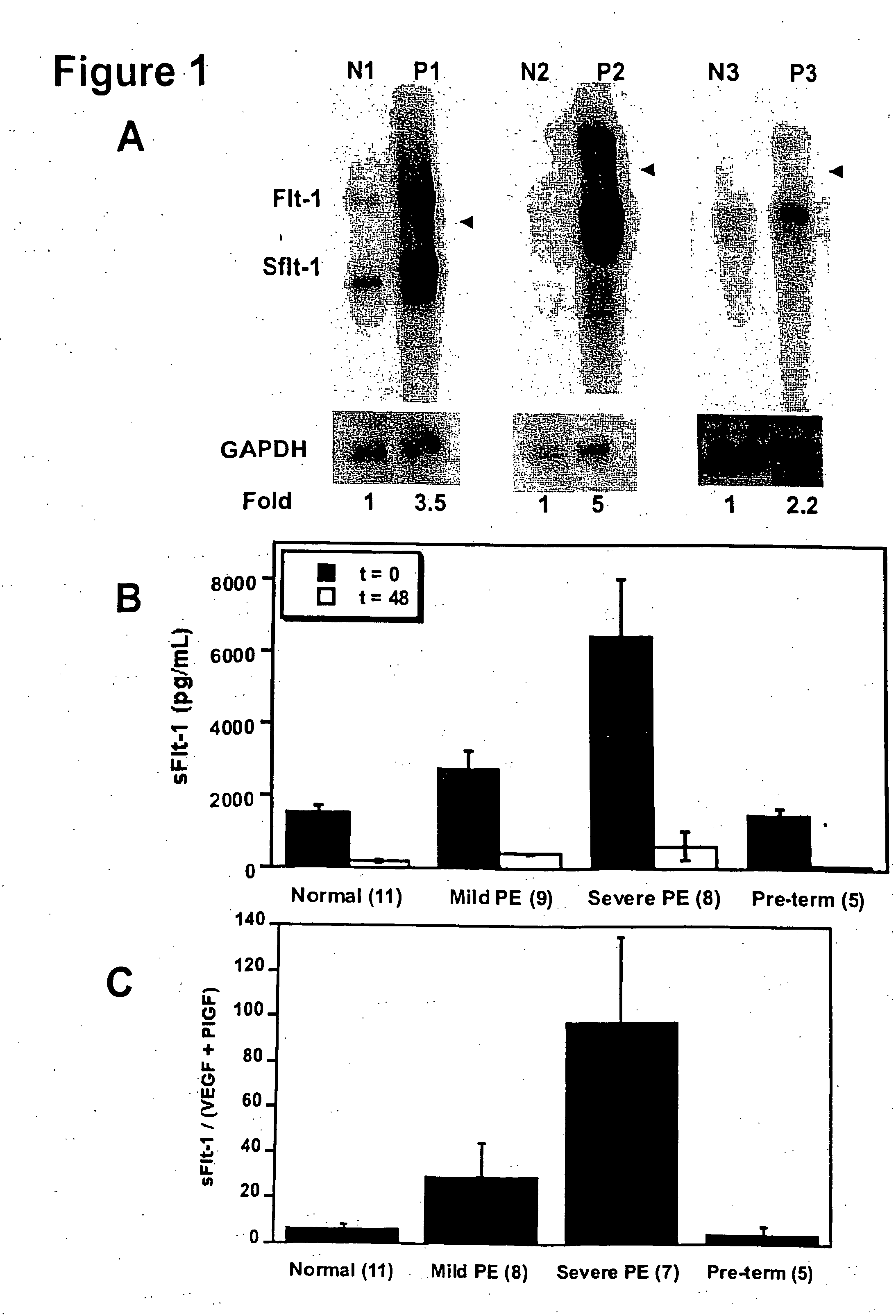
[0111] In order to confirm the upregulation of sFlt-1 in pre-eclampsia, we performed Northern blots to analyze the placental sFlt-1 mRNA levels (FIG. 1A) and ELISA assays to measure serum protein levels of sFlt-1 (FIG. 1B) in pre-eclamptic pregnant women as compared with normotensive pregnant women. Pre-eclampsia was defined as (1) a systolic blood pressure (BP)>140 mmHg and a diastolic BP>90 mmHg after 20 weeks gestation, (2) new onset proteinuria (1+by dipstik on urinanalysis, >300 mg of protein in a 24 hour urine collection, or random urine protein / creatinine ratio >0.3, and (3) resolut...

example 2

Serum from Women with Pre-Eclampsia Inhibits Angiogenesis in an In Vitro Endothelial Tube Assay

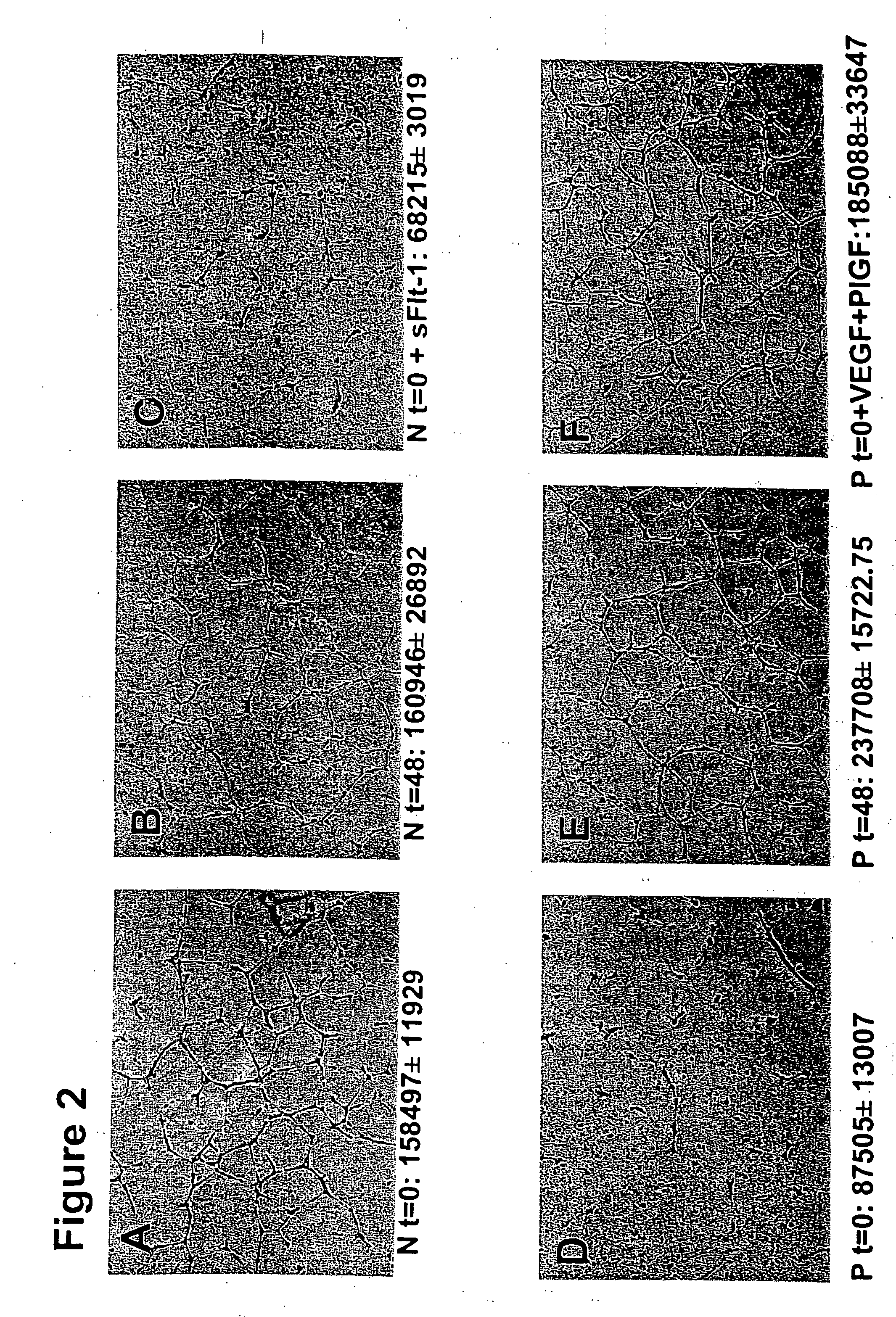
[0114] We hypothesized that excess circulating sFlt-1 in patients with pre-eclampsia causes endothelial dysfunction and leads to an anti-angiogenic state. To address this, we used an endothelial tube assay as an in vitro model of angiogenesis. Growth factor reduced Matrigel (7 mg / mL, Collaborative Biomedical Products, Bedford, Mass.) was placed in wells (100 μl / well) of a pre-chilled 48-well cell culture plate and incubated at 37° C. for 25-30 minutes to allow polymerization. Human umbilical vein endothelial cells (30,000+in 300 μl of endothelial basal medium with no serum, Clonetics, Walkersville, Md.) at passages 3-5 were treated with 10% patient serum, plated onto the Matrigel coated wells, and incubated at 37° C. for 12-16 hours. Tube formation was then assessed through an inverted phase contrast microscope at 4× (Nikon Corporation, Tokyo, Japan) and quantitatively analyzed (tube area...

example 3

sFlt-1 Inhibits VEGF and PlGF Induced Vasodilation of Renal Microvessels

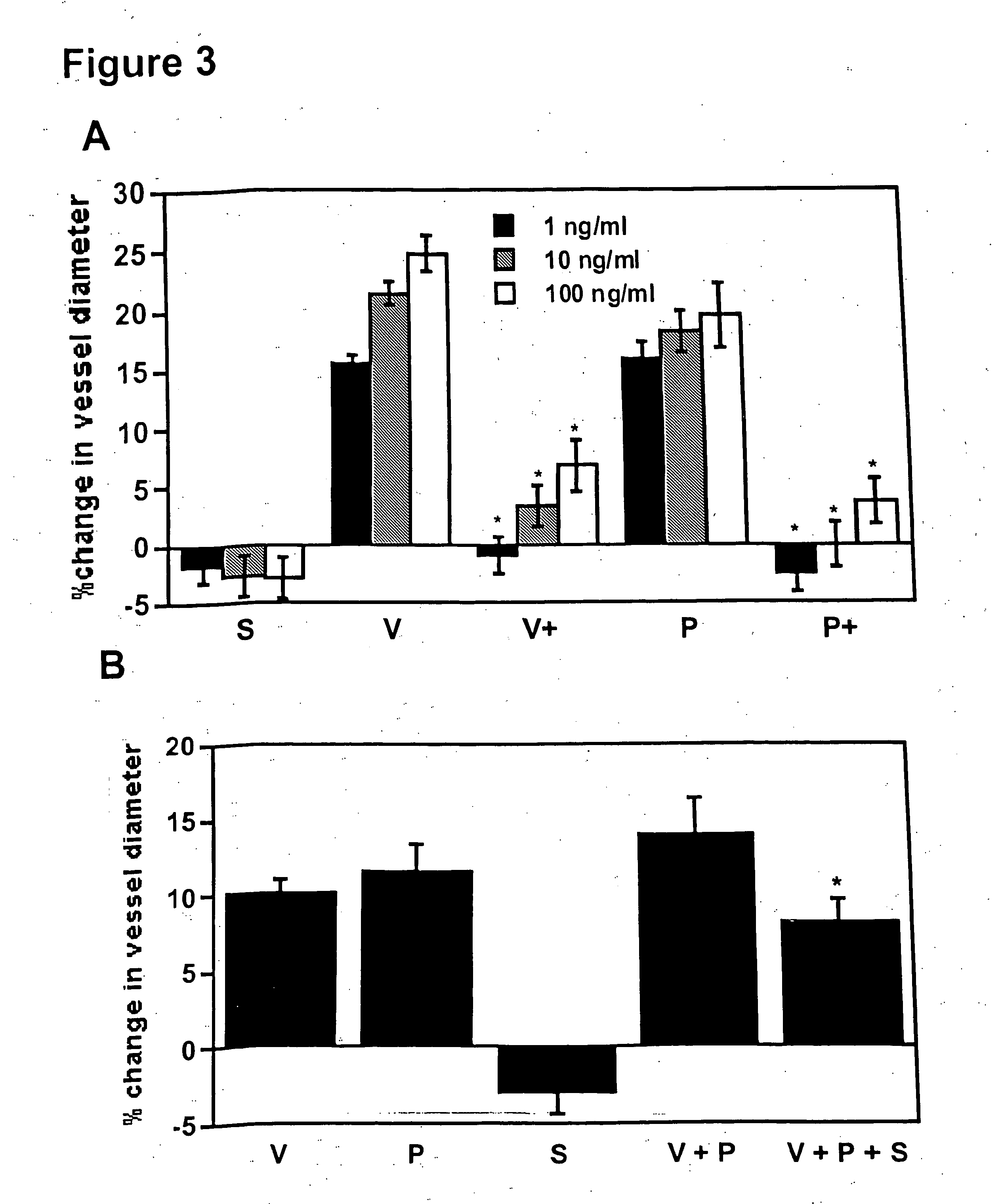
[0116] The causative role of sFlt-1 in vasoconstriction was determined using an in vitro microvascular reactivity experiment. Microvascular reactivity experiments were done as described previously using rat renal microvessels (Sato et al., J. Surg. Res., 90: 138-143, 2000). Kidney artery microvessels (70-1701 m internal diameter) were dissected from rat kidneys using a 10× to 60× dissecting microscope (Olympus Optical, Tokyo, Japan). Microvessels were placed in an isolated microvessel chamber, cannulated with dual glass micropipettes measuring 30-60 μm in diameter, and secured with a 10-0 nylon monofilament suture (Ethicon, Somerville, N.J.). Oxygenated (95% oxygen and 5% carbon dioxide) Krebs' buffer solution warmed to 37° C. was continuously circulated through the vessel chamber and a reservoir containing a total of 100 ml of the solution. The vessels were pressurized to 40 mmHg in a no-flow state using a bur...


PUM

PropertyMeasurementUnit
concentrationaaaaaaaaaa
average molecular weightaaaaaaaaaa
concentrationaaaaaaaaaa

Abstract

Disclosed herein are methods for diagnosing pre-eclampsia and eclampsia or a propensity to develop pre-eclampsia or eclampsia by detecting the levels of placental growth factor in a subject.

Description

CROSS REFERENCE TO RELATED APPLICATIONS [0001] This application is a continuation-in-part of U.S. patent application Ser. No. 10 / 771,518, filed Feb. 4, 2004, which is a continuation-in-part of U.S. patent application Ser. No. 10 / 624,809, filed Jul. 21, 2003, which claims the benefit of U.S. Provisional Application Nos. 60 / 467,390 filed on May 2, 2003, 60 / 451,796, filed on Mar. 3, 2003, and 60 / 397,481, filed on Jul. 19, 2002, each of which is herein incorporated by reference in their entirety.FIELD OF THE INVENTION [0002] In general, this invention relates to the detection and treatment of subjects having pre-eclampsia or eclampsia. BACKGROUND OF THE INVENTION [0003] Pre-eclampsia is a syndrome of hypertension, edema, and proteinuria that affects 5 to 10% of pregnancies and results in substantial maternal and fetal morbidity and mortality. Pre-eclampsia accounts for at least 200,000 maternal deaths worldwide per year. The symptoms of pre-eclampsia typically appear after the 20th week...

Claims


Application Information

Patent Timeline
Patent Type & Authority Applications(United States)
IPC IPC(8): A61K31/522C07K16/12C07K16/28C12Q1/48G01N33/68G01N33/74
CPCA61K31/522A61K2039/505C07K16/1217C07K16/2863G01N2800/368G01N33/689G01N33/6893G01N33/74G01N2500/00C12Q1/485A61P15/00A61K38/18
Inventor KARUMANCHI, S. ANANTHSUKHATME, VIKAS P.
Owner BETH ISRAEL DEACONESS MEDICAL CENT INC
Why Eureka
Try Eureka