
Stent grafts with bioactive coatings

a technology of stent grafts and coatings, applied in the field of pharmaceutical compositions, can solve the problems of reducing pressure, stent grafts however have a number, and their tendency to burst, so as to reduce prevent the risk of rupture, and reduce the risk of aneurysm ruptur

Inactive Publication Date: 2005-08-04
ANGIOTECH INT AG (CH) +1
View PDF79 Cites 99 Cited by
  • Summary
  • Abstract
  • Description
  • Claims
  • Application Information

AI Technical Summary

Benefits of technology

[0019] Also provided by the present invention are methods for treating patients having aneurysms (e.g., abdominal, thoracic, or iliac aortic aneurysms), for bypassing a diseased portion of a vessel, or for creating communication or a passageway between one vessel and another (e.g., artery to vein or vice versa, or artery to artery or vein to vein), such that risk of rupture of the aneurysm is reduced. As utilized herein, it should be understood that ‘reduction in the risk of rupture’ or ‘prevention of the risk of rupture’ refers to a statistically significant reduction in the, number, timing, or, rate of rupture, and not to a permanent prohibition of any rupture.

Problems solved by technology

Blood within this excluded sac thromboses and the aneurysm thus has no flow within it, presumably reducing the pressure and thus its tendency to burst.
Presently available stent grafts however have a number of problems.
For example, current stent grafts are prone to persistent leakage around the area of the stent graft.
Hence, pressure within the sac stays at or near arterial pressure and there is still a risk of rupture.
This can be persistent from the time of insertion because of poor sealing between the stent graft and vessel wall, or can develop later because the seal is lost.
In addition, this problem can develop due to changes in the position or orientation of the stent graft in relation to the aneurysm as the aneurysm grows, shrinks, elongates or shortens with time after treatment.
The second type of perigraft leak can occur because there are side arteries extending out the treated segment of blood vessel.
The third type of perigraft leak can occur because of disarticulation of the device (in the case of modular devices) or because of the development of holes within the graft material because continuous pulsation of the vessel causes the graft material to rub against a metallic stent tyne eventually causing graft failure.
Disarticulation of the device can develop due to changes in shape of the aneurysm as it grows, shrinks, elongates or shortens with time after treatment.
Stent grafts are also limited in their application to only selected patients with aneurysms.
However, endovascular technology is only applicable to certain patients with AAA because (a) lack of a suitable route of access via the blood vessels to the intended site of deployment which prevents insertion of the device and (b) anatomy.
This requires surgical exposure of the insertion site, usually a common femoral artery and limits the application of the technology as a larger delivery device is more difficult to manipulate through the iliac artery to the intended site of delivery.
If the iliac arteries or aorta are very tortuous, (frequently the case in AAA), or heavily calcified and diseased (another frequent association with AAA), this may be a contraindication to treatment or cause of failure of attempted treatment because of inability to advance a device to the site of deployment or potential for iliac artery rupture.
Shorter “necks” at either end of the diseased segment, necks which are sloping rather than cylindrical, or necks which are smaller than the aneurysm but still dilated in comparison to the normal diameter for a vessel in this location predispose to failure of sealing around the stent graft or delayed perigraft leaks.
One further difficulty with present stent grafts is that over time certain devices have a tendency to migrate distally within the abdominal aorta.
Such migration results in device failure, perigraft leak and vessel occlusion.
Finally, there is long term uncertainty about the entire stent graft technology as a treatment for AAA.
Uncertainties about endovascular stent grafts include whether they will lower the aneurysm rupture rate, rate of perigraft leak, device migration, ability to effectively exclude aneurysms over a long term, and device rupture or disarticulation.

Method used


Image

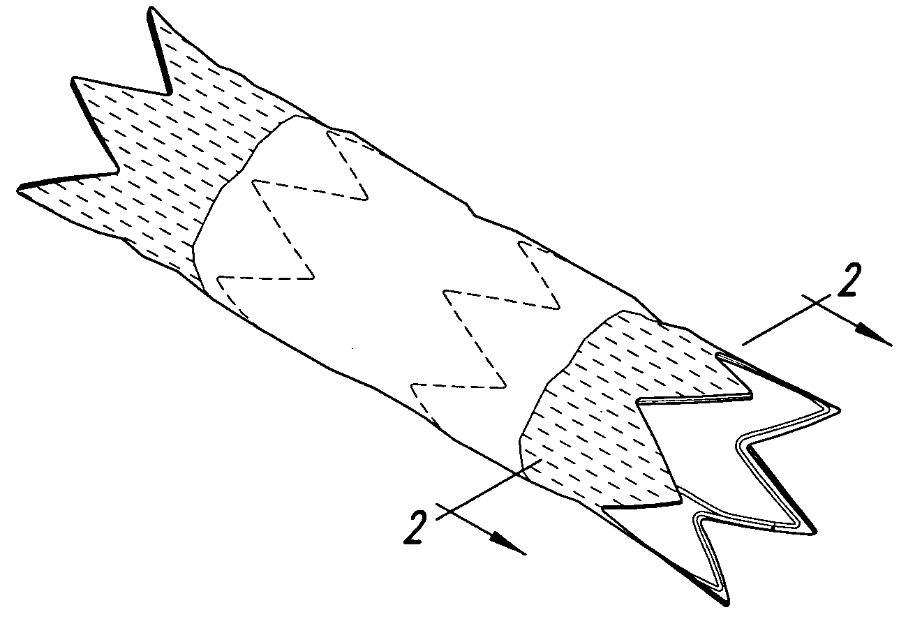
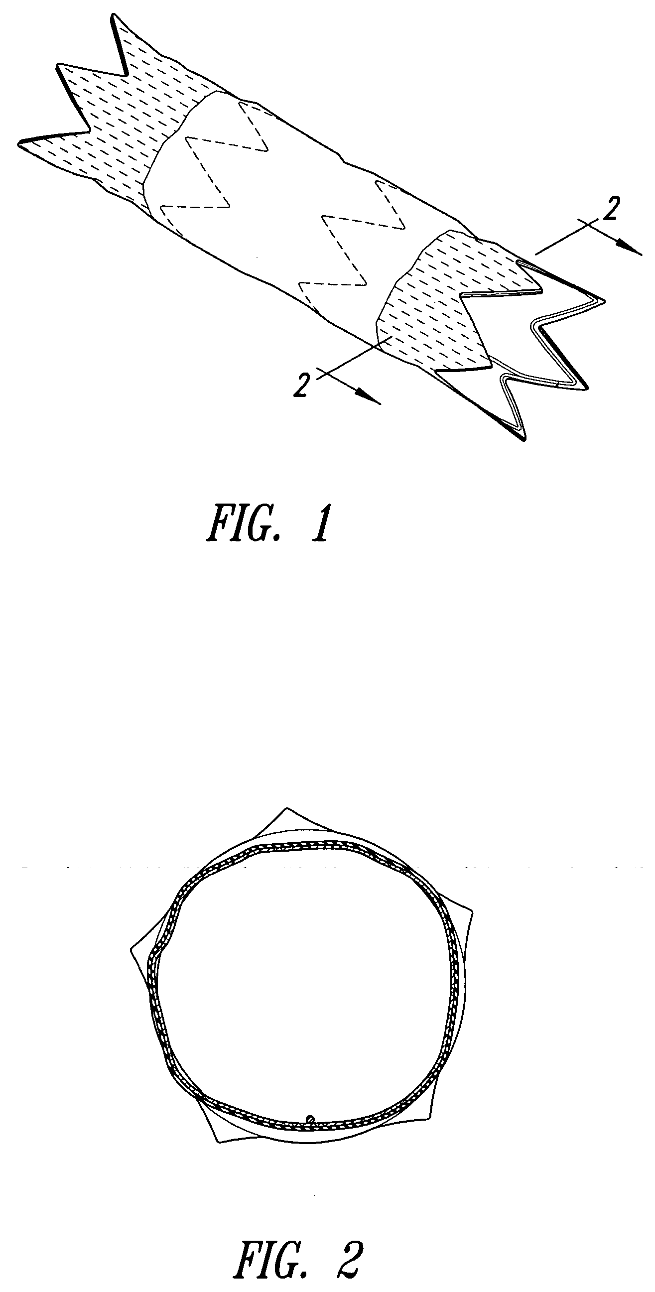
  • Stent grafts with bioactive coatings
  • Stent grafts with bioactive coatings

Examples


example 1

Coating of Intra-Anatomic Aortic Grafts with Fibronectin

[0088] The coating apparatus consisted of an overhead stirrer (Fisher Scientific) orientated horizontally. A conical stainless steel head was attached to the revolving chuck of the stirrer. One end of the intra-anatomic aortic graft was pulled up onto the conical head until held firmly. The other end was attached to a clip-swivel device that held the graft in a horizontal position, but allowed the graft to rotate along its axis. The stirrer was then set to rotate at 30 rpm so that the whole graft rotated along the horizontal axis at this speed. A 1% (w / w) fibronectin (Calbiochem, San Diego, Calif.) solution in sterile water was prepared. Two hundred microlitres of this solution was slowly pipetted as a 3 mm wide ring located 5 mm from the end of the graft fixed in the conical steel head over a period of 2 minutes as the graft rotated. The fibronectin was then dried under a stream of nitrogen as the graft continued to rotate. W...

example 2

Coating of Intra-Anatomic Aortic Grafts with Poly-L-Lisine

[0089] The coating apparatus consisted of a Fisher overhead stirrer orientated horizontally. A conical stainless steel head was attached to the revolving chuck of the stirrer. One end of the intra-anatomic aortic graft was pulled up onto the conical head until held firmly. The other end was attached to a clip-swivel device that held the graft in a horizontal position, but allowed the graft to rotate along its axis. The stirrer was set to rotate at 30 rpm so that the whole graft rotated along the horizontal axis at this speed. A 1% (w / w) poly-L-Lysine (Sigma, St. Louis, Mo.) solution in sterile water was prepared. Two hundred microliters of this solution was slowly pipetted as a 3 mm wide ring located 5 mm from the end of the graft fixed in the conical steel head over a period of 2 minutes as the graft rotated. The poly-L-Lysine was then dried under a stream of nitrogen as the graft continued to rotate. When dry, the graft wa...

example 3

Coating of Intra-Anatomic Aortic Grafts with N-Carboxybutyl Chitosan

[0090] The coating apparatus consists of a Fisher overhead stirrer orientated horizontally. A conical stainless steel head is attached to the revolving chuck of the stirrer. One end of the intra-anatomic aortic graft is pulled up onto the conical head until held firmly. The other end is attached to a clip-swivel device that holds the graft in a horizontal position, but allows the graft to rotate along its axis. The stirrer is set to rotate at 30 rpm so that the whole graft rotates along the horizontal axis at this speed. A 1% (w / w) n-carboxybutyl chitosan (Carbomer, Westborough, Mass.) solution in sterile water is prepared. Two hundred microlitres of this solution is slowly pipetted as a 3 mm wide ring located 5 mm from the end of the graft fixed in the conical steel head over a period of 2 minutes as the graft rotates. The n-carboxybutyl chitosan is dried under a stream of nitrogen as the graft continues to rotate...


PUM

PropertyMeasurementUnit
Adhesion strengthaaaaaaaaaa
Adhesivityaaaaaaaaaa
Metallic bondaaaaaaaaaa

Abstract

Stent grafts are provided comprising an endoluminal stent and a graft, wherein the stent graft releases an agent which induces the in vivo adhesion of the stent graft to vessel walls, or, otherwise induces or accelerates an in vivo fibrotic reaction causing said stent graft to adhere to vessel wall. Also provided are methods for making and using such stent grafts.

Description

CROSS-REFERENCES TO RELATED APPLICATIONS [0001] This application is a continuation-in-part of U.S. application Ser. No. 10 / 862,019, filed Jun. 4, 2004, which is a continuation of U.S. application Ser. No. 09 / 859,899, filed May 16, 2001, which is continuation-in-part of U.S. application Ser. No. 09 / 476,490, filed Dec. 30, 1999, which claims priority to U.S. Provisional Application No. 60 / 114,731 filed Dec. 31, 1998, and U.S. Provisional Application No. 60 / 116,726 filed Jan. 20, 1999, which applications are incorporated by reference in their entirety.TECHNICAL FIELD [0002] The present invention relates generally to pharmaceutical compositions, methods and devices, and more specifically, to compositions and methods for preparing stent grafts to make them more adherent to, or, more readily incorporated within a vessel wall. BACKGROUND OF THE INVENTION [0003] Stent grafts have been developed in order to not only simply hold open a passageway, but also to bridge across diseased vasculatur...

Claims


Application Information

Patent Timeline
IPC IPC(8): A61F2/00A61F2/06A61L31/10A61L31/16
CPCA61F2/07A61F2002/065A61F2002/075A61F2250/0067A61L31/10A61F2/89A61L2300/00A61L2300/412A61L2300/418A61L2300/602A61L2300/606A61L31/16
Inventor MACHAN, LINDSAY S.JACKSON, JOHN K.HUNTER, WILLIAM L.
Owner ANGIOTECH INT AG (CH)
Learn More