
Apparatus for automated fresh tissue sectioning

a technology of fresh tissue and apparatus, applied in the direction of analytical using chemical indicators, laboratory glassware, instruments, etc., can solve the problems of poor histological quality of frozen sections, unsuitable for laser capture studies and 3-dimensional reconstruction of morphology or gene expression patterns, and many immunohistochemical target antigens and mrna target sequences, etc., to achieve rapid intra-operative evaluation of surgical margins

Inactive Publication Date: 2005-10-27
THE BOARD OF TRUSTEES OF THE UNIV OF ARKANSAS
View PDF28 Cites 1 Cited by
  • Summary
  • Abstract
  • Description
  • Claims
  • Application Information

AI Technical Summary

Benefits of technology

The present invention is a new technique for sectioning fresh tissue without prior processing, which allows for high-resolution volumric reconstruction of gene and protein expression patterns of large tissue specimens. This is done by using an electro-dissociation process that can produce very thin sections (4-10 μm) of fresh tissue with preserved tissue architecture and antigenicity. The technique can easily be automated, eliminating the requirement of a skilled technician. The device can be used to make serial sections of a complete tumor or lesion that can be stained and reconstructed on a computer to provide a virtual 3-D histological image of the lesion. The invention solves the problem of damage caused by preprocessing of the tissue, speeds up the analysis of samples taken from lesions, and allows for serial slices from lesions that can be used for 2-D and 3-D reconstructions.

Problems solved by technology

Unfortunately, traditional tissue fixation and processing prior to paraffin-embedding destroys many immunohistochemical target antigens and mRNA target sequences.
However, frozen sections are of poor histological quality due to ice-crystal artifacts, thus making them unsuitable for laser capture studies and 3-dimensional reconstruction of morphology or gene expression patterns.
Vibratome sectioning of frozen tissues is sometimes used in the research setting, but is not advantageous in the clinical setting.
Unfortunately, the vibratome cannot produce sections of soft tissues that are thin enough for high resolution work (4-10 μm) without rigidifying the specimen by freezing or fixing prior to sectioning.
Ice crystal formation cannot in practice be eliminated, because the extreme cooling rates needed to produce solid amorphous ice, or vitreous ice, cannot be realistically achieved.

Method used


Image

  • Apparatus for automated fresh tissue sectioning
  • Apparatus for automated fresh tissue sectioning
  • Apparatus for automated fresh tissue sectioning

Examples


Embodiment Construction

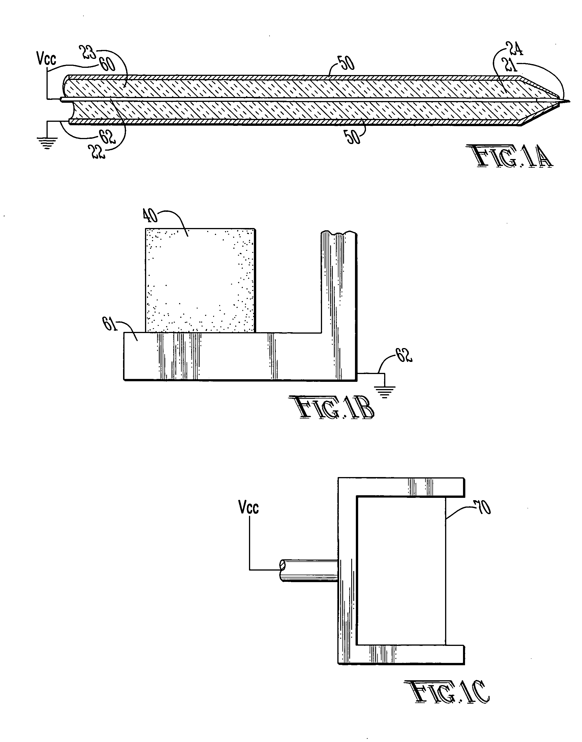
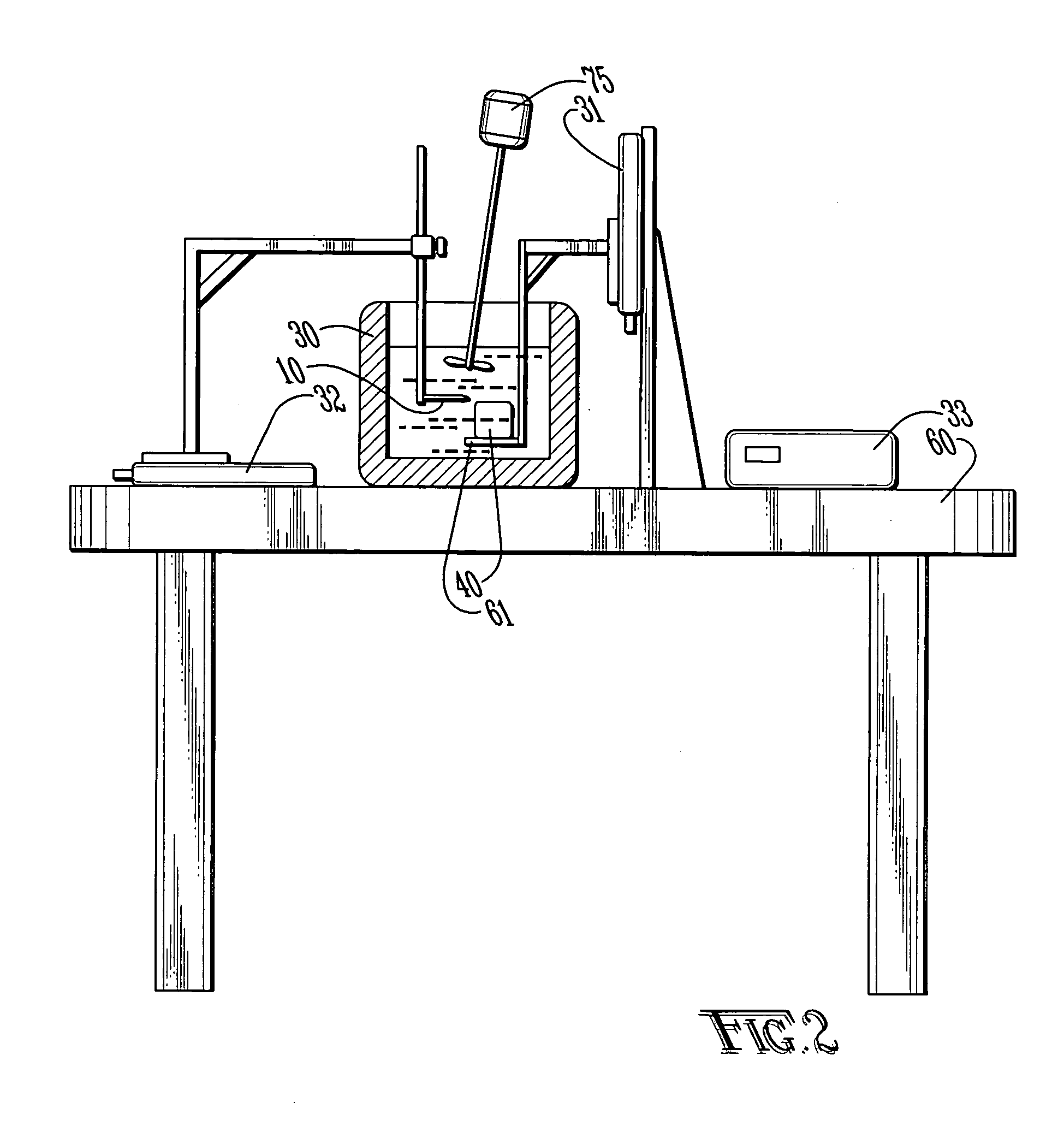
[0039] With reference to FIGS. 1A-C and 2, the preferred embodiments of the present invention may be described. The present invention is directed to satisfying the need to produce thin (4-10 μm) serial sections of large fresh tissue specimens that are suitable for high-resolution in situ protein / gene expression studies without ice artifacts or fixation-induced molecular damage.

[0040] Limitations of the existing sectioning techniques result from the fact that they rely on mechanical cutting, which in turn requires the tissue to be rigid. The present invention is a new approach to section tissue via an electro-dissociation process. The cutting tool is electrically biased with respect to the tissue sample which is submerged in a cooling bath. In one embodiment, the cutting tool may use focused radio frequency (RF) energy. The concept of electro-dissociation is known in devices known as electro-discharge machines (EDM) which are used to cut metals. Similar devices using this principle ...


PUM

PropertyMeasurementUnit
cryogenic temperaturesaaaaaaaaaa
thicknessaaaaaaaaaa
thicknessaaaaaaaaaa

Abstract

An apparatus for sectioning fresh unfixed tissue into very thin layers with preserved tissue architecture, antigenicity, mRNA content, and amenable to 3-D computer reconstruction. An electro-discharge machine (EDM) to accurately slice tissues through electro-dissociation of the tissue without mechanical or thermal damage. The tissue sample is placed on a holder submerged in a cooling bath comprising a liquid such as saline or water to minimize thermal effects and to provide a sink for dissociated ions. A cutting tool is electrically biased with respect to the tissue sample. A computer controlled EDM machine with x-y-z translation stage slices the tissue as defined by a predetermined program. The liquid in the cooling bath may be cooled to minimize tissue heating during cutting. In a preferred embodiment, the cutting tool may use focused RF energy to produce consecutive thin sections of fresh tissue for immunohistochemical and nucleic acids analyses by electro-dissociation without mechanical or thermal damage, ultimately allowing high-resolution volumetric reconstruction of gene and protein expression patterns of large tissue specimens.

Description

CROSS-REFERENCE TO RELATED APPLICATIONS [0001] Not applicable. STATEMENT REGARDING FEDERALLY SPONSORED RESEARCH OR DEVELOPMENT [0002] Not applicable. BACKGROUND OF THE INVENTION [0003] 1. Field of the Invention [0004] The present invention relates to the automated sectioning of consecutive thin sections of fresh tissues by electro-dissociation without mechanical force or thermal damage to the tissue. [0005] 2. Brief Description of the Related Art [0006] Routine histochemical analyses of thin tissue sections by light microscopy using chemical stains such as hematoxylin and eosin to highlight general nuclear and cytoplasmic features is the mainstay of surgical pathological diagnosis as well as morphological research. Another method, immunohistochemistry, provides more specific information about tissue sections by tagging a molecule of interest. Immunohistochemistry works on the principle of using an exogenous antibody raised against the molecule that is linked either to a fluorescent ...

Claims


Application Information

Patent Timeline
Patent Type & Authority Applications(United States)
IPC IPC(8): B01L3/00B23Q15/24C12M3/08G01N1/04G01N1/06
CPCC12M45/07G01N2001/045G01N1/06
Inventor SHAFIRSTEIN, GALFERGUSON, SCOTT L.FINK, LOUIS M.NORTH, PAULA E.WANER, MILTON
Owner THE BOARD OF TRUSTEES OF THE UNIV OF ARKANSAS
Learn More