Nanoparticle and microparticle based detection of cellular products

a technology of nanoparticles and cellular products, applied in the field of nanoparticles and microparticles based detection of cellular products, can solve the problems of inability to easily monitor the response of t-lymphocytes (“t-lymphocytes”) to antigens (including the antigen of pathogens), few sensitive, reliable and rapid techniques at present, and no reliable assay that can detect whether. , the effect of increasing the detection capability

Active Publication Date: 2005-12-15
CELLULAR TECH
View PDF7 Cites 56 Cited by
  • Summary
  • Abstract
  • Description
  • Claims
  • Application Information

AI Technical Summary

Benefits of technology

[0017] Because the markers used as part of this invention have clearly separate spectral features, one can detect multiple secreted soluble cell products simultaneously, thereby permitting increased speed of analysis and providing ability to compare differences between secreted products from the same cell culture. Further, because no enzyme reaction is needed, the assay devices and methods of this invention have improved reliability and accuracy.

Problems solved by technology

In contrast to the antibody response, the response of T-lymphocytes (“T-cells” or T Cells”) to antigen (including the antigen of pathogens) can not be easily monitored due to the fact that antigen reactive T cells occur in low frequencies and the fact that their secretory products are not typically stable (i.e. have a short half-life).
As a consequence of this, there are few sensitive, reliable and rapid techniques at present available that would reliably measure whether a patient has generated a T cell response to a particular pathogen, such as HIV.
There is no reliable assay that can detect whether a T cell response to HIV proteins has been generated, what proteins of the virus are primarily targeted, and which determinants within that protein are immunodominant.
There is also no reliable method available for testing the magnitude of the anti-viral T cell response (clonal sizes) and its quality (e.g. whether the response is pro- or antiinflammatory).
Heterogeneity of T cells, their products and the mode of function provide great challenges (particularly as compared to B cells).
Th1 cells are critical for the cellular defense and provide little help for antibody secretion.
It is detrimental to the host if an “inappropriate” type of T cell response is engaged (Th1 instead of Th2 or vice versa).
However, the conventional cytokine ELISA spot assay has not been a more sensitive tool than alternative assays (e.g. proliferation assays), displaying high background and generally a weak signal.
The quantification of the data is also problematic because of background problems and the subjective, visual evaluation.
Moreover, conventional 2 or 3-color systems used with conventional ELISA or ELISPOT methods cannot separately resolve individual spectral features of each colored detection reagent.
This procedure is time consuming and because of its complexity is difficult to carry out.
Additionally, substrates are not typically fully transparent, so a second substrate may mask an underlying substrate.
Moreover, as more substrates are used, the background becomes darker and less controlled.
The above problems with conventional ELISA methods therefore make assays of multiple cellular products difficult or impossible.

Method used


Image

  • Nanoparticle and microparticle based detection of cellular products
  • Nanoparticle and microparticle based detection of cellular products
  • Nanoparticle and microparticle based detection of cellular products

Examples


example 1

Use of Detection Surfaces in Detection Particle Based Assay

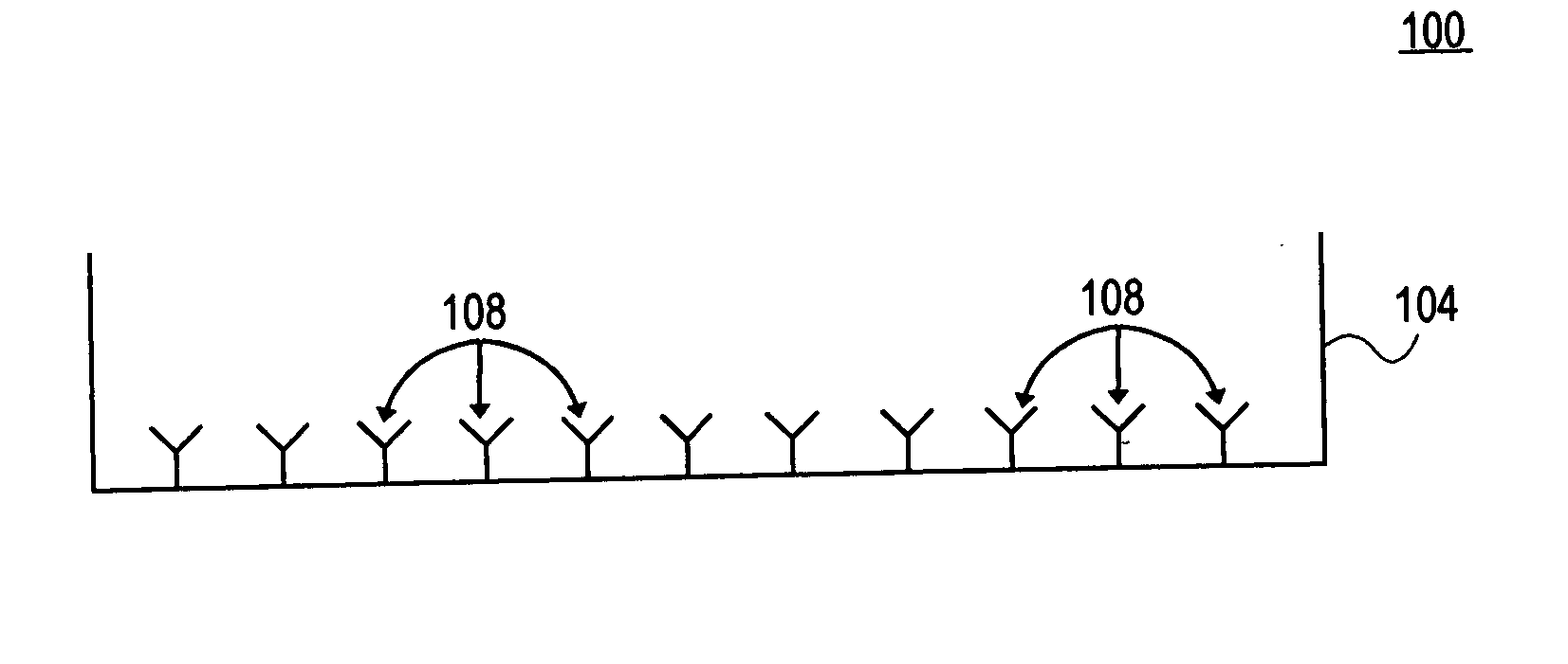
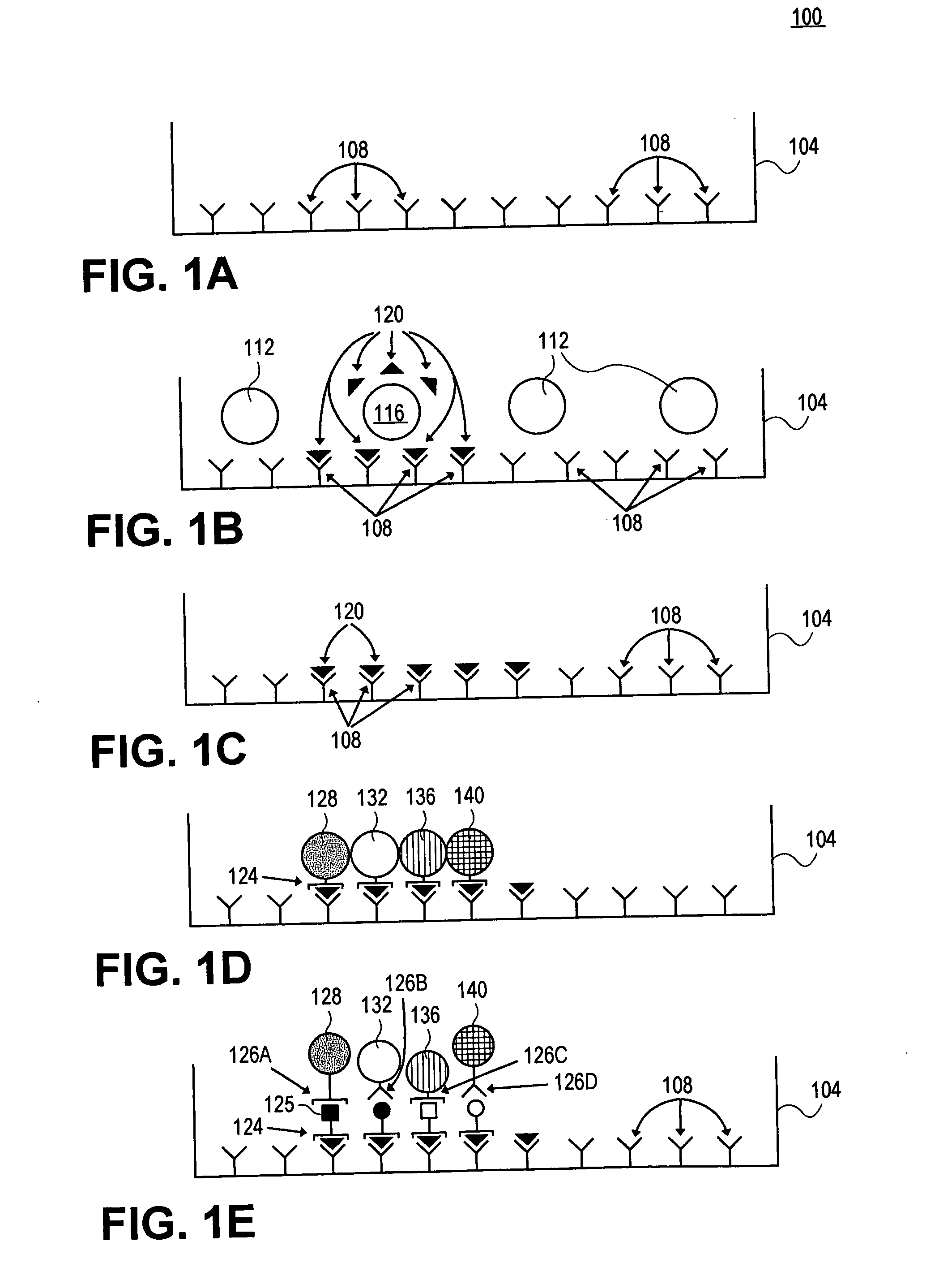
[0073] In this example, a detection surface is in a microtiter plate to detect cellular products. First, the detection surface is coated with anti-interferon gamma (IFN gamma antibody 1) as the capture reagent. After several hours of incubation (2 hrs to overnight), during which the capture reagent(s) bind(s) to the membrane, excess reagent is washed away. The unsaturated surfaces of the membrane are then blocked with irrelevant protein (bovine serum albumin or gelatin) to prevent subsequent nonspecific binding of proteins. Following the blocking step, the plates are washed to remove non-plate bound, excess blocking reagent.

[0074] At this point the detection substrate is properly prepared to test cells. In this case, human peripheral blood mononuclear lymphocytes (PBL) are added at a concentration of about 5×10⁵ per well. Specific antigen (e.g. HIV antigen) is added to the experimental wells, control wells contain no antig...

example 2

Peptide Screening

[0077] In this example, microwells are employed to screen and identify the antigenic determinants (i.e. MHC-bound peptides of the antigen generated by intracellular processing) that T cells recognize, using peptides. Usually, in studies that involved peptides, a few randomly chosen sequences of the antigen are tested as peptides. However, even overlapping sequences that walk down the molecule in steps of 5 to 10 amino acids do not necessarily detect all determinants. By contrast, in this example overlapping sets of peptides are employed that walk the molecule amino acid by amino acid. This peptide scan includes every possible determinant on an antigen and provides an exact mapping of the T cell repertoire.

[0078] While an assay system has been employed to test for antigen specific CD4 cells [G. Gammon et al., “T Cell Determinant Structure: Cores and Determinnant Envelopes in Three Mouse Major Histocompatibility Complex Haplotypes” J. Exp. Med. 173: 609-617 (1991)],...

example 3

Two Color Detection Particle Based Assay

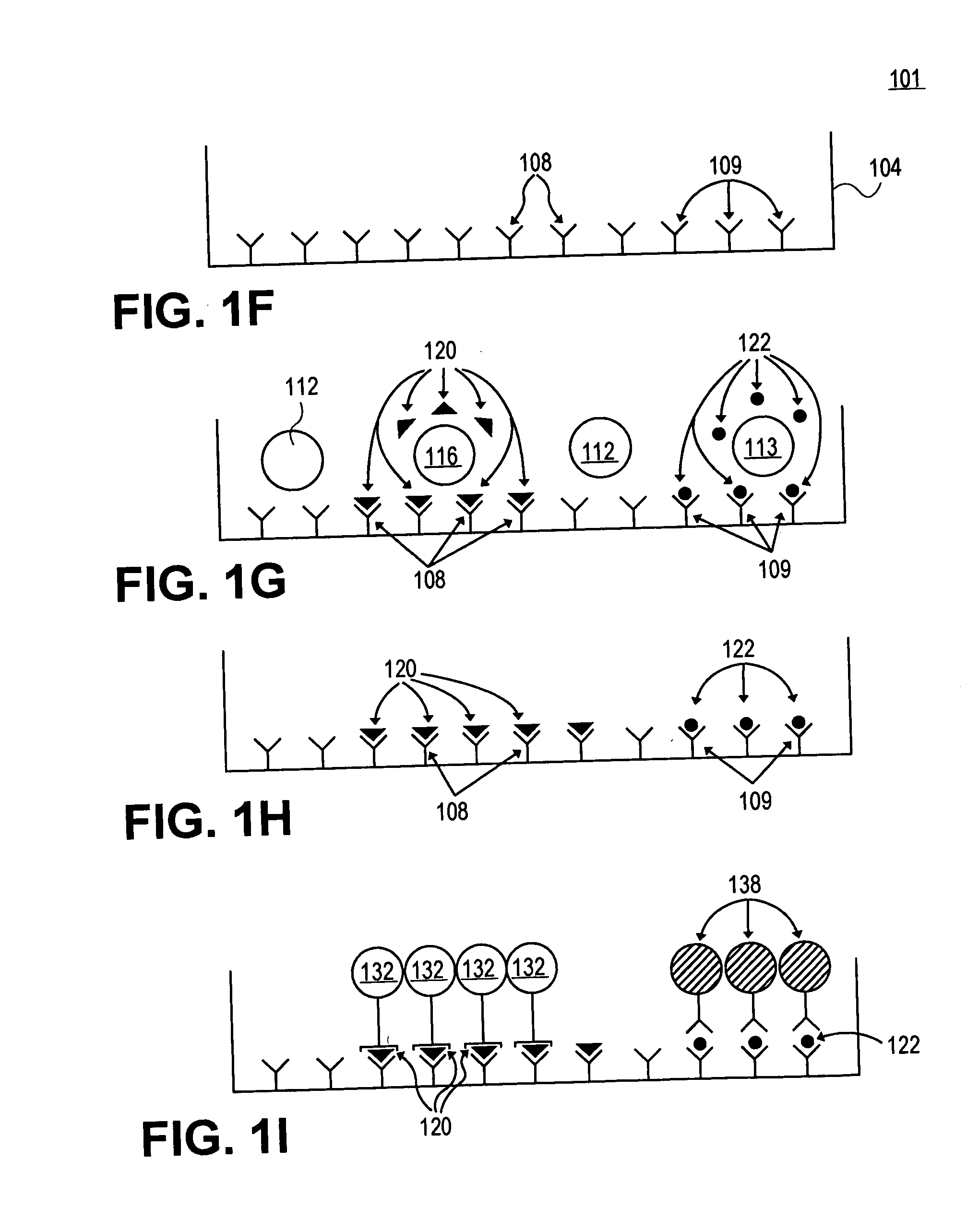
[0084] In this example, a capture surface is prepared as in Example 1, except that a second capture reagent and a second detection reagent / microsphere is used as well. To prepare the surface for a two-color assay, two (2) capture reagents (anti-IFNγ and anti-IL-5 antibody) are used simultaneously (for additional capabilities, still additional capture reagents can be employed for a “multicolor” assay).

[0085] Cells are again plated (about 5×10⁵ cells) and only the antigen specific T cells are stimulated by the antigen to release secretory products. However, in this case, the surface is capable of detecting both IFNγ and IL-5. After the culture period and washing described in Example 1, a second detection reagent attached to a second detection particle is employed together with the first detection reagent attached to a first detection particle. In this case, detection particles labeled with anti-IL-5 is added (alternatively, fluorochromes emitt...


PUM

PropertyMeasurementUnit
contact anglesaaaaaaaaaa
concentrationaaaaaaaaaa

Abstract

Embodiments of the present invention relate to devices and methods for detecting cellular products using detection particles having product-specific detection reagents and having a characteristic spectral feature. In particular, devices and methods are provided for measuring secreted cellular products including cytokines. Detection substrates, include microwells having product-specific capture reagents thereon or comprising hydrophobic membranes are described having greater capability to detect products from individual cells in a mixture of heterogeneous cells. With the use of multiple detection particles, multiple cellular products can be detected in a single well. Additionally, using the inherent spectral properties of detection particles, no enzymatic reactions are needed to visualize a secreted product, thereby increasing the sensitivity, reproducibility and ease of use.

Description

RELATED APPLICATION [0001] This application claims priority under 35 U.S.C. section 119(e) to U.S. Provisional Patent Application Ser. No. 60 / 489,451, filed Jul. 23, 2003, Paul Lehmann and Alexey Y. Karulin, titled “Microsphere Based Detection of Cellular Products.” This application is herein incorporated fully by reference.FIELD OF THE INVENTION [0002] The present invention relates to devices and methods for detecting secreted cellular products, and in particular for measuring secreted soluble cell products using nanoparticles and / or microparticles with associated capture reagents that recognize the cellular product. BACKGROUND [0003] Over the last thirty years, the immune system has been studied with better and better laboratory tools. Still, most knowledge of the immune response concerns antibody formation. This is understandable given that antibodies, including specific antibodies, are easily detectable in quantity in the serum of immunized individuals. Antibodies are products o...

Claims


Application Information

Patent Timeline
Patent Type & Authority Applications(United States)
IPC IPC(8): G01N33/50G01N33/533G01N33/58
CPCB82Y15/00G01N33/505G01N33/587Y10T436/25375G01N2333/5409G01N2333/57G01N33/588
Inventor LEHMANN, PAUL V.KARULIN, ALEXEY Y.KLEEN, THOMAS OLIVER
Owner CELLULAR TECH
Why Eureka
Try Eureka