

Controlled optoelectronic coupling in nanoparticle arrays
- Summary
- Abstract
- Description
- Claims
- Application Information
AI Technical Summary
Problems solved by technology
Method used
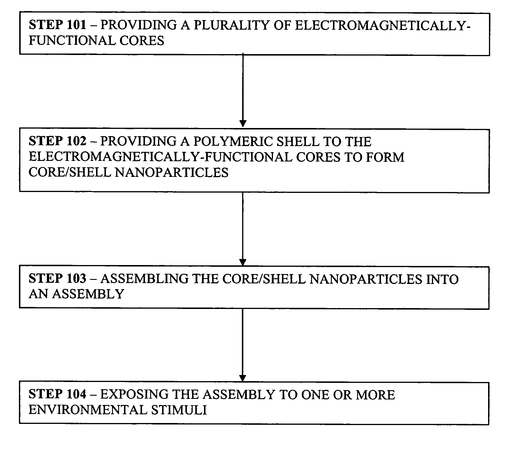
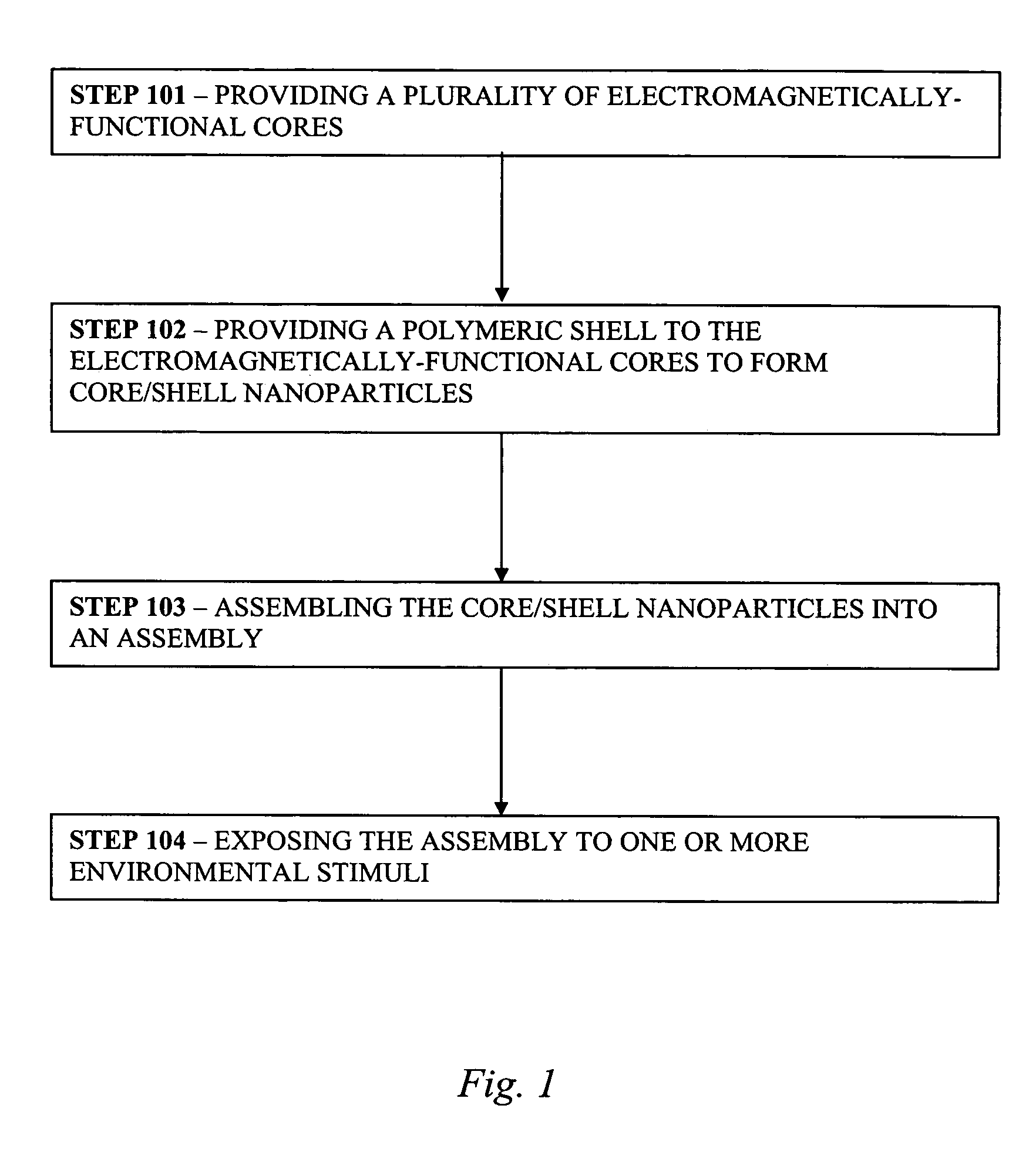
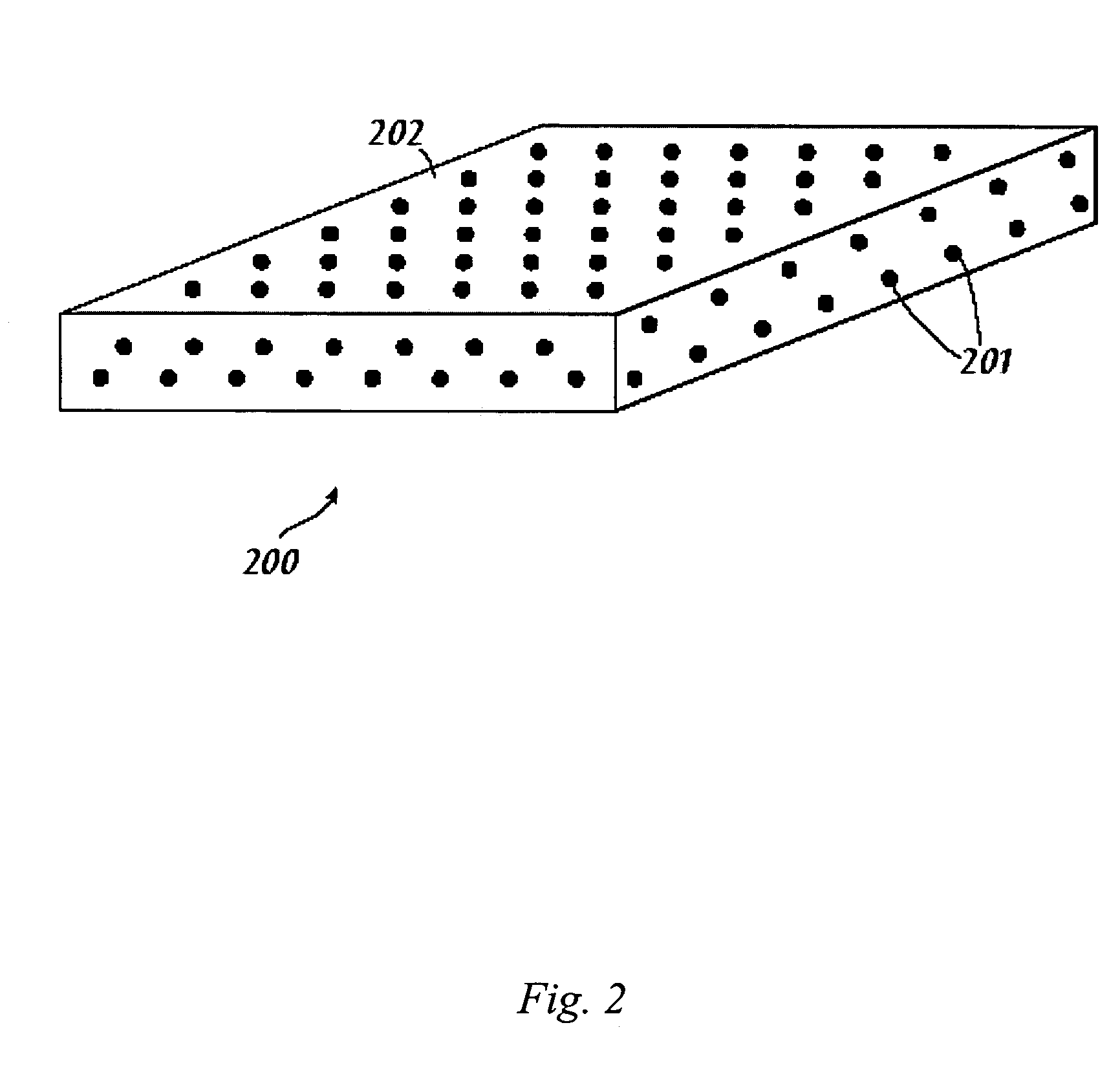
Image

Examples
example 1
[0051] This Example serves to illustrate both non-specific binding and grafting of polymer to core nanoparticles, and serves to illustrate the efficacy of disulfide linkages vs. thiol linkages in favoring one architecture over the other in binding polymer to gold nanoparticle cores, in accordance with some embodiments of the present invention.
[0052] The graft-to approach for particle functionalization is a more generally applicable technique, allowing each constituent of the composite core / shell particle to be prepared separately by well-established techniques, potentially allowing control of both particle size and polymer molecular weight (Zhu et al., J. Am. Chem. Soc., 2004, 126:2656; and Mangeney et al., J. Am. Chem. Soc., 2002, 124:5811). The pNIPA polymers used in this Example were prepared using the reversible addition fragmentation chain transfer (RAFT) method with the goal of providing polymer shells of defined thickness (Schilli et al., Macromolecules, 2002, 35:6819). Gold...
example 2
[0060] This Example serves to illustrate the synthesis of compound 4 used in the preceeding Example and as depicted in FIG. 12.
A) Materials
[0061] Reagents were purchased from Aldrich and used as received unless otherwise indicated. Anhydrous solvents were obtained from Aldrich. pNIPA-SH samples were purchased from Polymer Source, Inc.
B) Characterization
[0062] All nuclear magnetic resonance (NMR) spectra were obtained on a Bruker Avance 400 equipped with a 5 mm H / C dual probe. All spectra were obtained using standard parameters supplied with Bruker's XWINN software. These included a 30° flip angle, 1 second pulse delay, 10 kHz spectral width for proton and 30 kHz spectral width for 13C. Polystyrene standards in the range of 10 kD to 300 kD were used to establish a calibration. The molecular weight determination of pNIPA was carried out using ambient temperature gel-permeation chromatography (GPC) using a HP model 1050 LC system in-line to a HP model 1050 UV detector and a Varex m...
PUM
 Abstract
Description
Claims
Application Information