Intravascular imaging device and uses thereof

a technology of intravascular imaging and imaging device, which is applied in the field of probe-type imaging device, can solve the problems of difficult therapy deployment, complicated and expensive, and limited utility of these devices, and achieve the effect of enhancing diagnosis and treatment, and increasing sensitivity

Inactive Publication Date: 2006-02-23
ELMALEH DAVID R
View PDF31 Cites 39 Cited by
  • Summary
  • Abstract
  • Description
  • Claims
  • Application Information

AI Technical Summary

Benefits of technology

[0020] The probe-type imaging device of the invention and methods of use thereof overcome the limitations of other known imaging methods by providing for: 1) a light and/or scatter measurement of the intravascular system; 2) variable wave length for testing; 3) real-time read of wall thickness; 4) real-time imaging of the vascular lumen system; 5) a probe size that us...

Problems solved by technology

OCT visualization of vessel involves the use of complicated and expensive instrumentation as well as the repeated saline washes to view the vessel wall.
The utility of these devices is limited due to their complexity and use of mirrors, lens, and l...

Method used


Image

  • Intravascular imaging device and uses thereof
  • Intravascular imaging device and uses thereof
  • Intravascular imaging device and uses thereof

Examples


example 1

Imaging of Rabbit Aorta

[0128] A working scattered-light prototype of the probe-type imaging device was demonstrated using excised pieces of rabbit aorta and a modified confocal microscope. FIG. 17 illustrates an experimental probe setup. The drinking straw 170 (left) contained a double fiber optic probe 171 held by a mechanical positioner 172. Rabbit aorta 173 was pinned to a flexible base 174 placed on a microscope 175. A 10× objective was used to collect scattered light from delivery fiber 171 (wavelengths>540 nm). Although crude in construction (drinking straw, double fiber optic probe held by a mechanical positioner) the device records measurable differences in diffusely scattered light collected by a 10× objective at wavelengths greater than 510 nm. As shown in FIG. 18 light was delivered to the luminal surface of aorta 173. Scattered light was detected 10's of microns away from probe tip.

[0129] FIGS. 19A-B, illustrate visualization of vessel tissue obtained by placing the pr...

example 2

Imaging of Diseased and Normal Rabbit Aorta Tissue Regions

[0134] Light intensities detected from diseased and normal regions of rabbit aorta were examined using light from 488 nm fluorescent light, 532 nm and 1024 nm light from a solid state diode source as illustrated in FIG. 24. Shown in FIG. 24 illumination light was delivered to tissue via a single mode fiber 240 with a central core 241 of less than about 9 microns. A photodiode 242 placed near the fiber tip collected scattered and reflected light from the tissue.

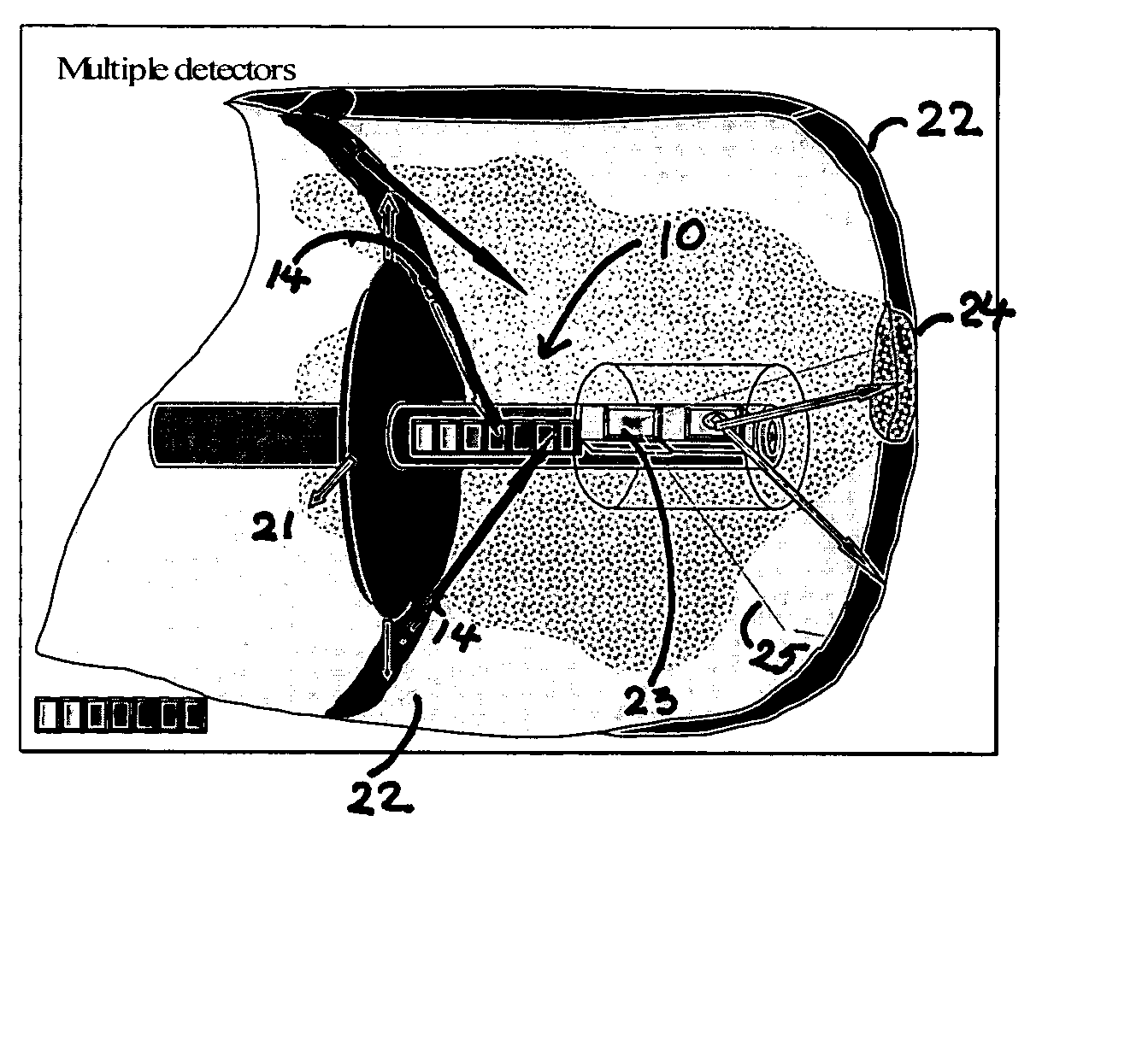
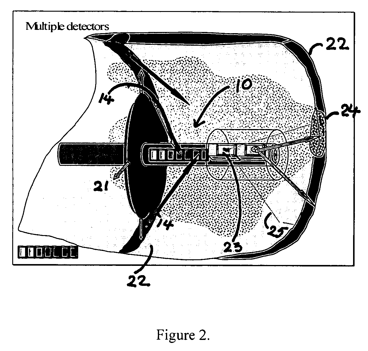
[0135]FIG. 25 is a schematic diagram of an experimental setup used to demonstrate scattered light imaging. A diseased experimental piece of aorta 250 was placed on microscope stage. A fiber optic imaging probe 251 was placed parallel, or at an oblique angle to the lumen surface. Light from a diode laser (not shown) passing through the fiber 251 illuminated a local region at the fiber end. Multiple photodetectors 252 placed above the fiber end detected the light (same ...


PUM


Abstract

The invention is directed to a probe-type imaging device useful to visualize interior surfaces, e.g., the lumen of blood vessels. Specifically, the probe-type device is particularly useful for direct tissue imaging in the presence or absence of molecular imaging agents.

Description

RELATED U.S. APPLICATION(S) [0001] The present application claims priority to U.S. Provisional Application Ser. No. 60 / 580,938, filed Jun. 18, 2004, which application is hereby incorporated herein by reference.TECHNICAL FIELD [0002] The present invention is directed to a probe-type imaging device useful to visualize interior surfaces, e.g., the lumen of blood vessels. Specifically, the probe-type imaging device is useful for direct tissue imaging in the presence or absence of molecular imaging agents. BACKGROUND [0003] Heart disease and stroke are the main cause of death in the United States. Most of the debilitating factors related to these diseases are rooted in vascular disorders. The main vascular insufficiencies are associated with vascular plaque formation and thrombus formation that block or decrease blood flow and, as a result, the oxygenated blood supply to these important organs. Medical device technologies and medication treatment are available to mechanically open the ar...

Claims


Application Information

Patent Timeline
IPC IPC(8): A61B6/00
CPCA61B5/0068A61B5/0071A61B5/02007A61B5/0086A61B5/0084A61K49/0034
Inventor ELMALEH, DAVID R.ROGERS, RICK A.BRISMAR, HJALMAR
Owner ELMALEH DAVID R
Why Eureka
Try Eureka