Combined intra-rectal optical-MR and intra-rectal optical-US device for prostate-, cevix-, rectum imaging diagnostics

a technology of intra-rectal optical and rectum, which is applied in the field of medical imaging, can solve the problems of not providing information about specific molecules (e.g., malignant vs. benign tissue) associated with the disease, and the optical imaging technology typically does not deliver sufficient anatomical delineation

Inactive Publication Date: 2006-05-11
SIEMENS MEDICAL SOLUTIONS USA INC
View PDF12 Cites 17 Cited by
  • Summary
  • Abstract
  • Description
  • Claims
  • Application Information

AI Technical Summary

Problems solved by technology

Anatomical imaging modalities, such as MR and CT, provide adequate anatomical delineation (e.g., bone and soft tissue), but typically do not provide information about specific molecules (e.g., malignant vs. benign tissue) associated with the disease.
However, optical imaging technologies typically do not deliver sufficient anatomical delineation.

Method used


Image

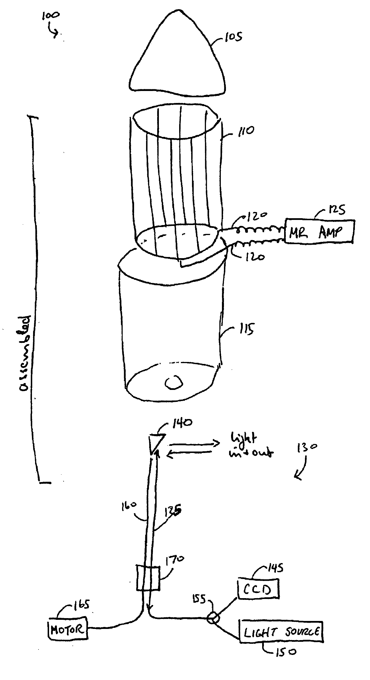
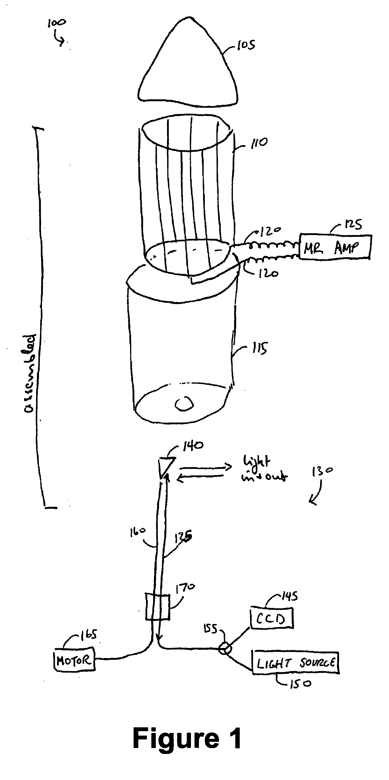
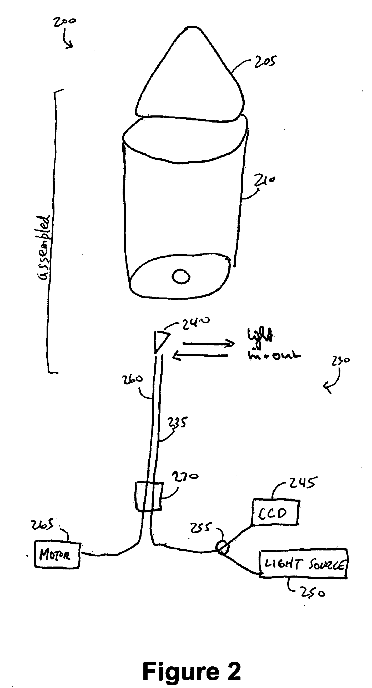
  • Combined intra-rectal optical-MR and intra-rectal optical-US device for prostate-, cevix-, rectum imaging diagnostics
  • Combined intra-rectal optical-MR and intra-rectal optical-US device for prostate-, cevix-, rectum imaging diagnostics
  • Combined intra-rectal optical-MR and intra-rectal optical-US device for prostate-, cevix-, rectum imaging diagnostics

Examples


Embodiment Construction

[0015] Illustrative embodiments of the invention are described below. In the interest of clarity, not all features of an actual implementation are described in this specification. It will of course be appreciated that in the development of any such actual embodiment, numerous implementation-specific decisions must be made to achieve the developers' specific goals, such as compliance with system-related and business-related constraints, which will vary from one implementation to another. Moreover, it will be appreciated that such a development effort might be complex and time-consuming, but would nevertheless be a routine undertaking for those of ordinary skill in the art having the benefit of this disclosure.

[0016] While the invention is susceptible to various modifications and alternative forms, specific embodiments thereof have been shown by way of example in the drawings and are herein described in detail. It should be understood, however, that the description herein of specific...


PUM


Abstract

We present, in exemplary embodiments of the present invention, a system combining optical imaging technologies with anatomical imaging technologies (e.g., MR, ultrasound). The system can be used for a variety of applications, including, but not limited to, (1) cancer diagnosis and staging; (2) image guidance; and (3) radiation therapy planning. Image guidance may include guiding a biopsy. For example, a prostatectomy potentially has severe side effects, such as impotence and incontinence. Thus, a histologically-confirmed diagnosis, such as one provided from a biopsy, may prevent unnecessary prostatectomy. Image guidance may also include guiding minimal invasive therapy, such as brachytherapy focused ultrasound. The present invention may be used to plan radiation therapy, for example, by detecting, and thus sparing, healthy tissue from radiation exposure.

Description

CROSS-REFERENCE TO RELATED APPLICATIONS [0001] This application claims priority to U.S. Provisional Application No. 60 / 541,020, which was filed on Feb. 2, 2004, and which is fully incorporated herein by reference.BACKGROUND OF THE INVENTION [0002] 1. Field of the Invention [0003] The present invention relates generally to the field of medical imaging, and, more particularly, to combined intra-rectal optical-magnetic resonance and combined intra-rectal optical-ultrasound device for prostate, cervix and rectum imaging. [0004] 2. Description of the Related Art [0005] Various diagnostic imaging methods are currently used for the diagnosis of prostate, cervix and rectum cancers. Modern diagnostic imaging techniques include magnetic resonance (“MR”), computer tomography (“CT”), ultrasound (“US”) and nuclear medicine (e.g., Positron Emission Tomography (“PET”), Single Photon Emission Computed Tomography (“SPECT”)). Anatomical imaging modalities, such as MR and CT, provide adequate anatomic...

Claims


Application Information

Patent Timeline
Patent Type & Authority Applications(United States)
IPC IPC(8): A61B6/00A61B1/04A61B5/00A61B5/055A61B8/12G01R33/28
CPCA61B1/0638A61B5/0077A61B5/415A61B5/418A61B8/12A61B8/4461A61B8/5238G01R33/285A61B1/0005A61B1/043A61B5/0035
Inventor RUECKMANN, BOGDAN VONGAREUS, RALPHHENGERER, ARNEWEISSLEDER, RALPHDURR, WILHELM
Owner SIEMENS MEDICAL SOLUTIONS USA INC
Why Eureka
Try Eureka