

Separation of denatured proteins in free solution
a free solution and protein technology, applied in the field of protein separation in free solution, can solve the problems of gel-based electrophoresis in microchannels being a challenge, and achieve the effect of greater compatibility
- Summary
- Abstract
- Description
- Claims
- Application Information
AI Technical Summary
Benefits of technology
Problems solved by technology
Method used
Image

Examples
example 1
Preparation of Proteins for Free Solution Electrophoresis
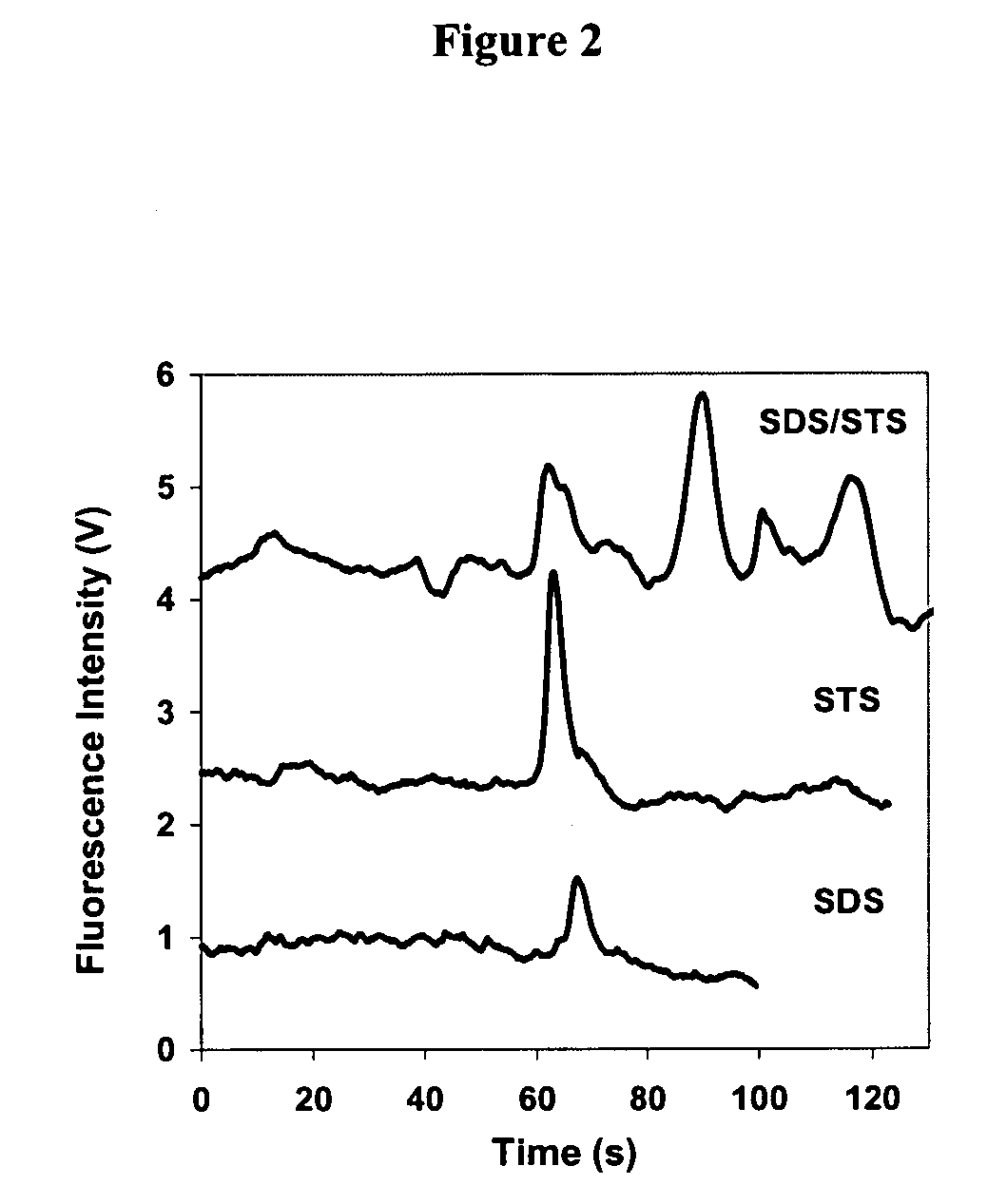
[0042] Surfactants with different compositions were used for the denaturation of proteins. The surfactants used in these experiments included: (1) Pierce SDS, a commercial alkyl sulfate mixture with multiple chain lengths available from Pierce (Rockford, Ill.; No. 28364, lot analysis: C12 73%, C14 24%, C16 3%, and C10+C18<1% w / w); (2) SDS (99%) from Sigma (St. Louis, Mo.); (3) STS (99%) from Lancaster Synthesis (Pelham, N.H.); and (4) SDS / STS, a binary mixture of SDS (99%) and STS (99%) (w / w=3:1).
[0043] The sample buffer for denaturation was 2.5 mM sodium borate (pH˜9.2) containing 1% 2-mercaptoethanol. In experiments involving Pierce SDS, pure SDS, and SDS / STS, the sample buffer contained 1.45% (w / v) surfactant. In experiments involving STS, the sample buffer contained 0.59% (w / v) STS due to its low solubility. Protein mixtures were denatured by heating in sample buffer at 95° C. for 5 minutes. Denaturation resulted in supp...
example 2
Free Solution Microchip Electrophoresis with Laser-Induced Fluorescence Detection
[0045] Microchips were fabricated on Pyrex 7740 wafers using photolithography, plasma etching, and thermal bonding techniques. FIG. 4 is an illustration of the design of the microchip. The microchannels had a depth of 3.8 μm and a half-depth width of 26 μm. The serpentine separation channel had a total length of 12 cm. The channel width at the turns was one half of the straight channel. The structure was previously reported for the minimization of geometric dispersion of analyte bands (see Molho et al., Anal. Chem. 73:1350 (2001); Ramsey et al., Anal. Chem. 75:3758 (2003)). The microfluidic chip was used with uncoated channel walls. Before the first use, the microfluidic chip was conditioned by flowing water and then 0.1 M NaOH for 20 minutes each. The running buffer was flowed for 20 minutes each time before the labeled-protein solution was loaded. After the experiment, the microchip was cleaned by ru...
example 3
Protein Separation Using Microchip Electrophoresis in Free Solution with Laser-Induced Fluorescence Detection
[0049] These examples illustrate the separation of denatured proteins using microfluidic chip electrophoresis in free solution. Electrophoresis was performed after proteins were denatured with a mixture of alkyl sulfates with different carbon chain lengths.
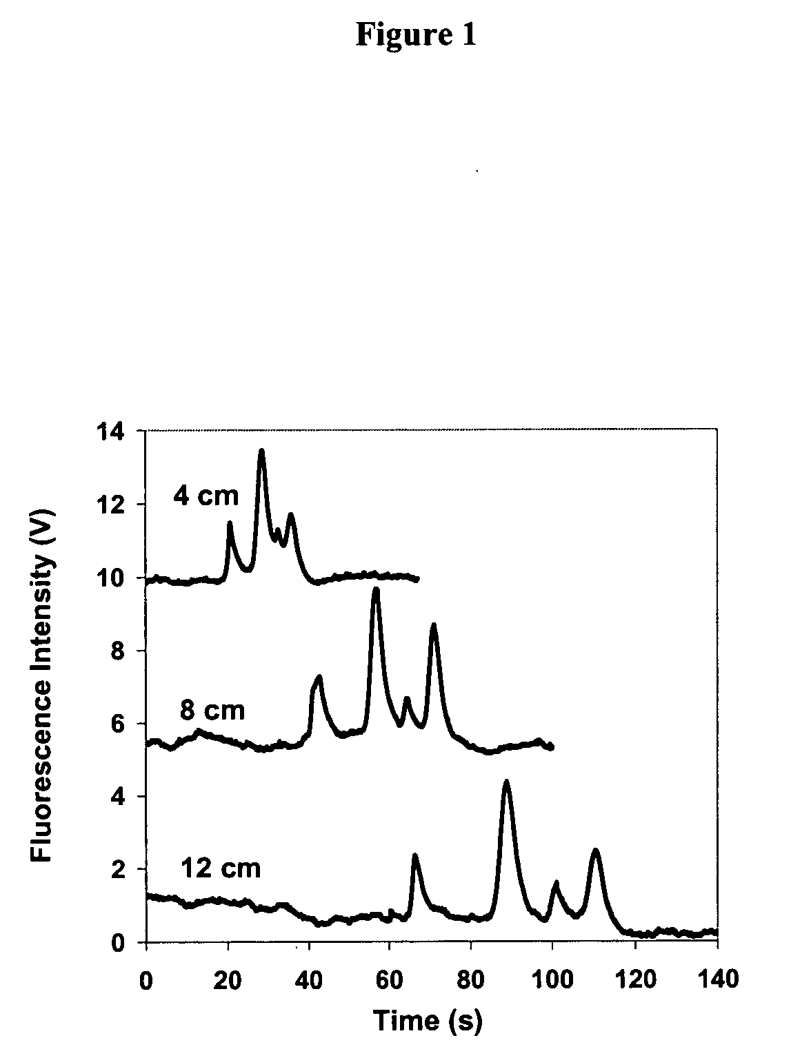
[0050] In one experiment, proteins were denatured using Pierce SDS and then separated using microchip electrophoresis in free solution. The running buffer consisted of 2.5 mM sodium borate+0.14% (w / v) Pierce SDS. The protein mixture consisted of about 1×10⁻⁶ M of each of four proteins: α-lactalbumin (14 kDa), ovalbumin (45 kDa), conalbumin (78 kDa), and β-galactosidase (116 kDa). Separation was conducted under an electric field of 436 V / cm in the separation channel, and fluorescence was detected at 4, 8, and 12 cm from the injection. Results are shown in FIG. 1. A single peak was observed for each denatured protein. The p...
PUM
| Property | Measurement | Unit |
|---|---|---|
| molecular weights | aaaaa | aaaaa |
| temperature | aaaaa | aaaaa |
| half-depth width | aaaaa | aaaaa |
Abstract
Description
Claims
Application Information