Methods for cancer imaging

a cancer and imaging technology, applied in the direction of diagnostic recording/measuring, ultrasonic/sonic/infrasonic diagnostics, sugar derivates, etc., can solve the problems of many biopsy methods invasive, cancer is dangerously fatal, and many cancer deaths are still

Inactive Publication Date: 2006-07-27
THRESHOLD PHARM INC
View PDF17 Cites 2 Cited by
  • Summary
  • Abstract
  • Description
  • Claims
  • Application Information

AI Technical Summary

Problems solved by technology

This sort of tumor progression makes cancer dangerously fatal.
Although there have been great improvements in diagnosis, general patient care, surgical techniques, and local and systemic adjuvant therapies, most deaths from cancer are still due to metastases that are either resistant to conventional therapies or are undetected by current diagnostic methods.
Many biopsy methods are invasive and require surgical removal of tissue for analysis.
While PET scanning is quite useful to image the entire patient for cancer, it requires expensive and cumbersome equipment for detection and construction of the positron used for the image.
Accordingly, PET scanning cannot be used for the detection of cancer in many clinical settings, such as in the physician's office during the time of physical examination or in the operating room at the time of surgery.
Drawbacks to this procedure reside in the experience of the examiner, and errors in diagnosis can be life threatening.
In instances where cancers are missed and then spread beyond the original site of disease, mortality can increase.
Conversely, some skin lesions are biopsied which are not cancerous, and the patients are thus subjected to unnecessary harm.
In some cases, biopsy samples are taken from the face, leading to cosmetic debility.
These tests provide guidance for surgery, but at the time of surgery, these images cannot be generated in real time to guide the surgeon to the tumor.
However, this procedure is often not conducted, as it requires a highly trained pathologist to be present at the surgery and to rapidly analyze the tissue sample while the patient remains on the operating table.
Unfortunately, despite such efforts, residual tumor will be left inside the patient about 15-25% of the time.
Studies have shown that these patients are at greater risk of dying of the cancer than those that have the tumor completely resected.
Because of this, these patients require further, often debilitating, costly therapy in an attempt to arrest and treat the cancer left in the patient at the time of surgery.
However, even with this enhanced method of screening, smaller tumors and cancer cells can go undetected.
These detection methods are limited in that they cannot be used in the general physician's practice for screening large numbers of patients nor can they be used at the time of surgery to locate the residual tumor.

Method used


Image

  • Methods for cancer imaging
  • Methods for cancer imaging
  • Methods for cancer imaging

Examples


example 1

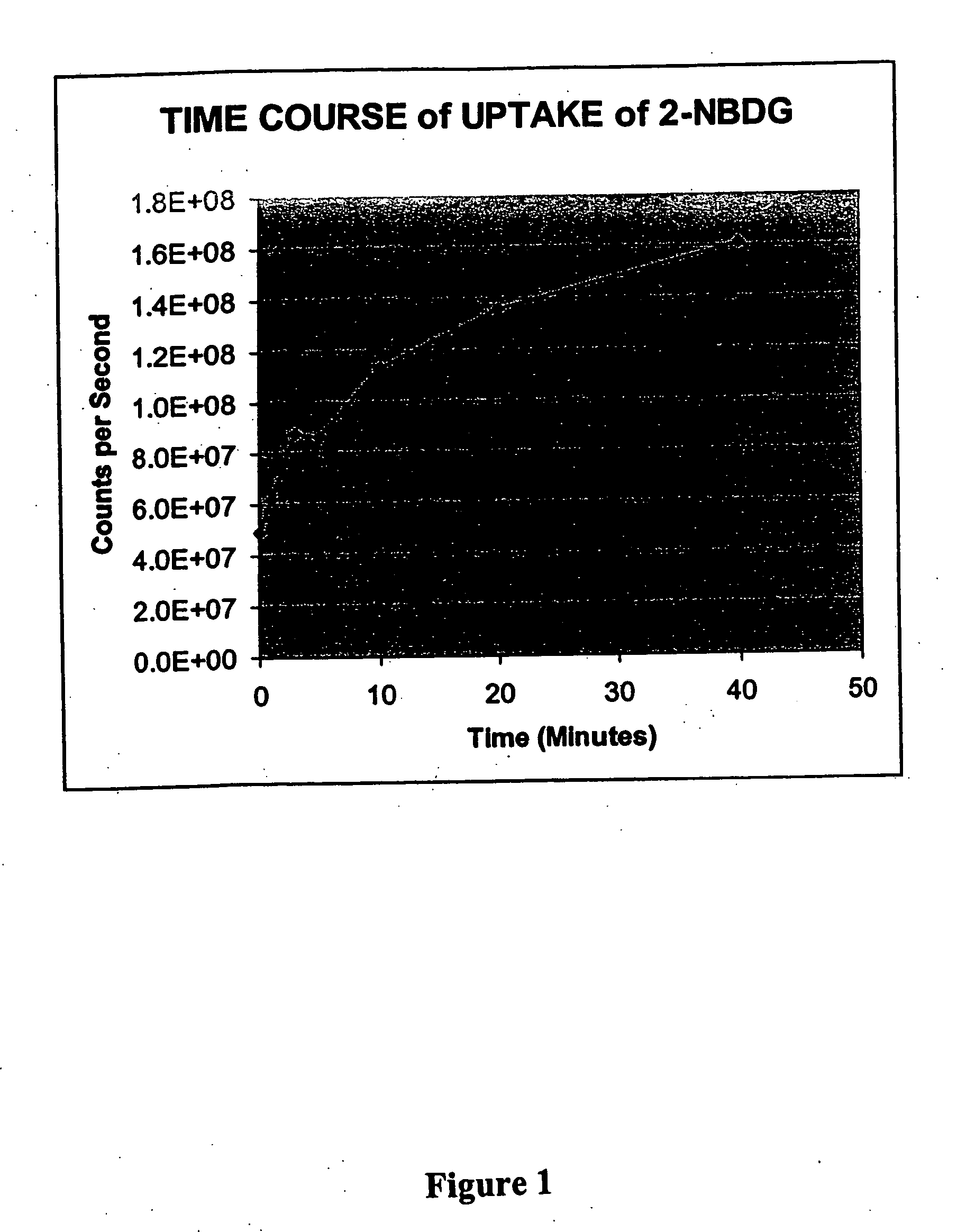
[0123] This example illustrates the time course of uptake of 2-NBDG in Raji lymphoma cells.

Materials:

[0124] Raji cells from culture of one t-75 flask. Phosphate-buffered saline plus 2.5 mM CaCl2, 1.2 mM MgSO4, 4 mM KCl and 5 mM glucose (PBS+). 2-NBDG can be prepared in accordance with the method reported in Yoshioka et al., 1996, Biochimica et Biophysica Acta 1289: 5-9, incorporated herein by reference.

Uptake Reaction:

[0125] Culture medium with cells (15 mL) was centrifuged for 10 min at 800×g. The pellet of cells was resuspended in PBS+ and centrifuged for 10 min at 800×g, and the resulting pellet was resuspended in 1.5 mL PBS+. Aliquots (100 uL) were placed into each of 8 tubes and incubated at 37° C. for 15 min. The fluorophore deoxyglucose conjugate 2-NBDG was added to each tube to provide a concentration of 200 uM. A tube was removed from the 37° C. bath at each of 0, 1, 3, 5, 10, 20, 30 and 40 min and immediately placed on ice. The cells were washed twice with PBS and re...

example 2

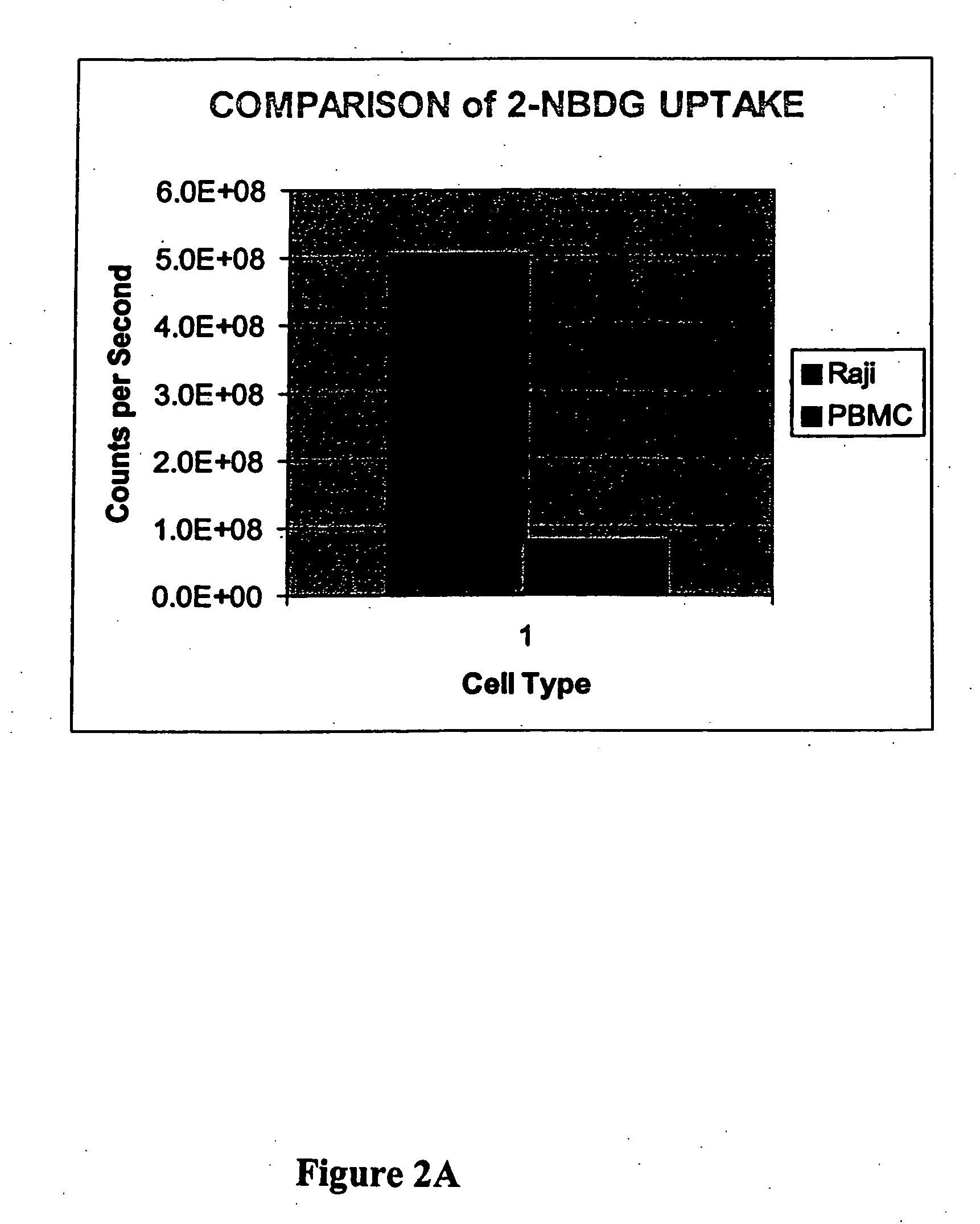
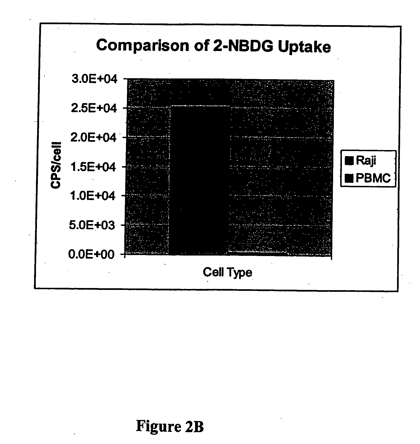
[0126] This example illustrates a comparison of 2-NBDG uptake in Raji lymphoma cells and peripheral blood white cells and illustrates the preferred uptake of a fluorophore deoxyglucose conjugate by a cancer cell as compared to a normal cell.

Materials:

[0127] Raji cells from culture one t-75 flask. Peripheral blood white cells from Stanford Blood Bank. Leukocytes were further separated by Ficoll separation (twice). These cells are referred to as PBMC peripheral blood mononuclear cells). Phosphate-buffered saline. 2-NBDG, 0.1 M in water.

Uptake Reaction:

[0128] Culture medium with cells (either Raji or PBMC, 15 mL) were centrifuged for 10 min at 800×g. The pellet of cells was resuspended in PBS and centrifuged for 10 min at 800×g, and the resulting pellet was resuspended in 1.5 mL PBS. Cells were counted and adjusted to 1×10⁶ per mL for both. Aliquots (100 uL) were placed into each of 2 tubes for each cell type, and the tubes were incubated at 37° C. for 15 min. The fluorescent deo...

example 3

[0129] This example illustrates the preparation of the fluorophore deoxyglucose conjugate of the invention FGC-002. In the conjugate, the fluorophore is BODIPY, and the glucose derivative is glucosamine.

[0130] Generally, the FGC-002 conjugate is prepared by treating 6-((4,4-difluoro-5,7-dimethyl-4-bora-3a,4a-diaza-s-indacene-3-propionyl)amino)hexanoic acid, succinimidyl ester (BODIPY from Molecular Probes, D-2184, MW 502) with an excess of D-glucosamine (Sigma) in an aprotic solvent with gentle heating. Isolation of the product (FGC-002) can be accomplished via chromatography.

[0131] More particularly, 2.2 mg (4 micromoles) of 6-((4,4-difluoro-5,7-diethyl-4-bora-3a,4a-diaza-s-indacene-3-propionyl)amino)hexanoic acid, succinimidyl ester was dissolved in 0.3 ml of DMF followed by the addition of 6 mg (28 micromoles) of glucosamine HCL dissolved in 0.3 ml of water and 3.9 microliters (28 micromoles) of triethyl amine. The reaction was stirred for 24 hrs at room temperature, evaporate...


PUM

PropertyMeasurementUnit
emission wavelengthaaaaaaaaaa
wavelengthsaaaaaaaaaa

Abstract

Methods are provided for cancer and pre-cancer detection by increased uptake of fluorophore glucose or deoxyglucose conjugates in cancerous and pre-cancerous cells relative to normal cells.

Description

CROSS-REFERENCES TO RELATED APPLICATIONS [0001] This application claims priority to provisional U.S. patent application Ser. No. 60 / 342,313, filed 21 Dec. 2001, which is incorporated herein by reference in its entirety.BACKGROUND OF THE INVENTION [0002] The term “cancer” generally refers to one of a group of more than 100 diseases that are caused by the uncontrolled growth and spread of abnormal cells and can take the form of solid tumors, lymphomas and non-solid cancers such as leukemia Unlike normal cells, which reproduce until maturation is attained and then only-reproduce as necessary to replace wounded cells, cancer cells grow and divide endlessly without differentiating to mature, functional cells, crowding out nearby cells and eventually spreading to other parts of the body. [0003] The most common sites in which cancer develops include the skin, lungs, female breasts, prostate, colon, rectum, bladder, uterus, blood-forming tissues, and lymphatic system. Cancer cells that have...

Claims


Application Information

Patent Timeline
Patent Type & Authority Applications(United States)
IPC IPC(8): A61K49/00C07H17/02A61B
CPCA61K49/0021A61K49/0041A61K49/0052
Inventor TIDMARSH, GEORGEMATTEUCCI, MARK
Owner THRESHOLD PHARM INC
Why Eureka
Try Eureka