Antibodies against lesion tissue

a technology of lesional tissue and antibodies, applied in the field of antibodies against lesional tissue, can solve problems such as difficulties in limitation, and achieve the effect of convenient use and higher probability

Inactive Publication Date: 2006-10-19
CHUGAI PHARMA CO LTD
View PDF6 Cites 51 Cited by
  • Summary
  • Abstract
  • Description
  • Claims
  • Application Information

AI Technical Summary

Benefits of technology

[0050] In this invention, any method may be used for isolating B cells which infiltrate cancer tissues. The preferable methods for isolating B cells include microdissection, a technique for excising specific cells from a tissue section. For example, target cells can be isolated from frozen tissue sections using the Laser Microdissection (LMD) system. A system for excising tissue sections with an ultraviolet laser is commercially available. The use of this system, which involves specifying the region to be excised on a computer image under microscopic observation, makes it possible to excise any region from a tissue section.
[0051] In this case, a large number of B cells can be isolated by observing a specimen under a microscope and selecting a portion enriched with B cells. Alternatively, a small number of B cells can be easily obtained by excising a region with low B cell density. As shown in the Examples, it is even possible to obtain a single cell.
[0052] In the present invention, B cells can be isolated from any specimen prepared for pathological analysis. For example, thin-section frozen specimens are preferable pathological specimens in this invention. As a pathological specimen, not only fresh tissues but also specimens fixed with paraformaldehyde (PFA) or such can be used. Accordingly, it is also possible to obtain antibody genes from, for example, preserved pathological specimens using the...

Problems solved by technology

This limitation poses difficulties for, for example, obtaining antibodies produced by B cells which have infiltrated cancer tissues.
However, since B cell populations in peripheral blood...

Method used


Image

  • Antibodies against lesion tissue
  • Antibodies against lesion tissue
  • Antibodies against lesion tissue

Examples


example 1

LMD Isolation of a Single Cancer Tissue-Infiltrating B Cell

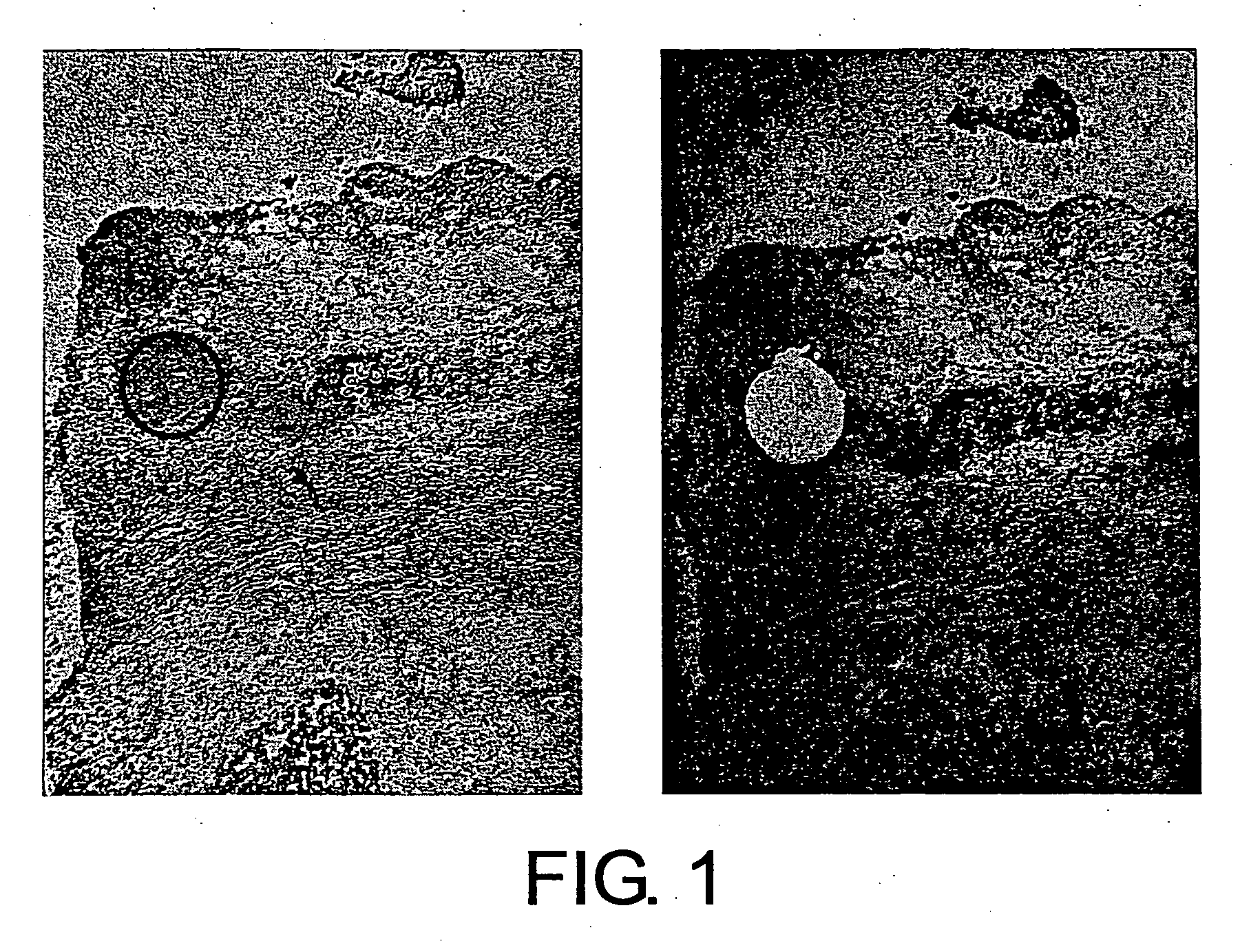
[0140] A fresh human tissue (breast cancer tissue) was sliced into a suitable size to prepare frozen blocks using an OCT compound (Tissue-tek), and fixed prior to freezing with fixatives such as paraformaldehyde-lysine-periodate, as necessary. Next, thin specimen sections were prepared from each frozen block and attached onto LMD slides (Matsunami Glass Co.). Resulting thin-section frozen specimens were dried in air, fixed with fixatives such as acetone, and stained with toluidine blue (Muto Pure Chemicals Co., Ltd) or such. After staining, plasma cells were excised with a laser microdissection system (Leica AS-LMD), and recovered using a recovery buffer (RLT solution attached to QIAGEN RNeasy Mini Kit). Plasma cells before and after the excision are shown in FIGS. 1, 5, 7, 9, 11, 13, and 15. In all figures, the left photograph shows the state of the cells before excision, and the right photograph shows the state of the cel...

example 2

RNA Preparation and cDNA Synthesis

[0141] A suspension of approximately one to five B cells excised from a thin-section LMD specimen was mixed with a suspension of about 300 carrier cells which do not express any antibody genes. Total RNAs were prepared from the resultant mixture, using the RNeasy Mini Kit (QIAGEN) according to the manufacturer's instructions. When 50 cells or more were excised from the thin-section specimen, total RNAs were prepared without the addition-of carrier cells. cDNA was synthesized using all 35 μl of the RNA eluate fraction as a template, and Sensiscript Reverse Transcriptase (QIAGEN) according to the manufacturer's instructions. cDNA synthesis reaction was performed on an 80-μl scale using 40 ng of Oligo dT primers (Promega) and 0.8 μg of random hexamers (Invitrogen) as the reverse transcription primers at 37° C. for 1 h. When cDNAs thus synthesized were not immediately subjected to PCR, they were stored at −80° C.

EXAMPLE 3

Cloning of Human Antibody Var...

example 4

Preparation of Single-Stranded Antibody Molecules

[0145] The linker sequences to be used for preparing single-stranded antibody genes were produced according to the method of Marks et al. (J. Mol. Biol. (1991) 222, 581-597). The template DNA sequences and the nucleotide sequences of primers used in the preparation are shown below. Linker fragments synthesized using PCR amplification were confirmed by agarose gel electrophoresis, and bands containing the respective fragments were excised and purified.

Template DNA sequence (Template linker) / SEQ ID NO: 1515′-GGACAATGGTCACCGTCTCTTCAGGTGGTGGTGGTTCGGGTGGTGGTGGTTCGGGTGGTGGCGGATCGGACATCCAGATGACCCAGTCTCC-3′Nucleotide sequences of primers:Reverse JH for linker / SEQ ID NO: 152 to 155 1 LJH1_25′-GCACCCTGGTCACCGTCTCCTCAGGTGG-3′ 2 LJH35′-GGACAATGGTCACCGTCTCTTCAGGTGG-3′ 3 LJH4_55′-GAACCCTGGTCACCGTCTCCTCAGGTGG-3′ 4 LJH65′-GGACCACGGTCACCGTCTCCTCAGGTGG-3′Reverse VK for linker / SEQ ID NO: 156 to 161 5 LVK15′-GGAGACTGGGTCATCTGGATGTCCGATCCGCC-3′ 6 LVK25...


PUM

PropertyMeasurementUnit
Volumeaaaaaaaaaa

Abstract

Methods for isolating polynucleotides encoding antibodies against lesional tissues are provided, wherein the methods comprise the steps of: (a) isolating a B cell(s) that infiltrates into a lesional tissue of interest; and (b) obtaining an antibody-encoding polynucleotide from the isolated B cell(s). The lesions may be a cancer tissue or such. Antibody genes can be obtained without depending on B cell cloning. Accordingly, it is also possible to obtain genes encoding human-derived antibodies which are difficult to clone. Genes that encode antibodies against cancer can be obtained using cancer tissues as the lesion.

Description

TECHNICAL FIELD [0001] The present invention relates to antibodies against lesional tissues and methods for producing the same. BACKGROUND ART [0002] Lymphocytes are widely known to infiltrate cancer tissues (Document 1 / Hurliamnn et al. (1985) Int J Cancer 35:753; Document 2 / Whiteside et al. (1986) Cancer Immunol Immunother 23:169; Document 3 / Wolf et al. (1986) Otolaryngol Head Neck Surg 95:142; Document 4 / Husby et al. (1976) J Clin Invest 57:1471; Document 5 / Vose et al. (1979) Int J Cancer 24:579). Experimental and clinical data suggest that lymphocyte infiltration of cancer tissues is associated with host immunoreactions against cancer (Document 5 / Rosenberg et al. (1988) New Engl J Med 319:1676; Document 6 / Van Pel et al. (1995) Immunol Reviews 145:229; Document 7 / Kreider et al. (1984) Cancer Metastasis Rev 3:53). [0003] In the host immune defense system against cancer, cytotoxic T cells (CTLs) are the effector cells that directly kill cancer cells (Document 8 / Nobholz and ...

Claims


Application Information

Patent Timeline
IPC IPC(8): C07K16/30G01N33/574C07H21/04C12P21/06C12N5/06C07K16/00C07K16/32C12N15/12
CPCC07K16/00C07K16/30C07K2317/622C07K2317/21C07K16/3015A61P35/00
Inventor TSUCHIYA, MASAYUKISUZUKI, MASAMIYOSHIDAFUJI, ETSUKOMATSUBARA, KOUICHITSUNODA, HIROYUKI
Owner CHUGAI PHARMA CO LTD
Why Eureka
Try Eureka