
RT-PCR detection for differential diagnosis of field isolates or lapinized vaccine strain of classical swine fever virus (CSFV) in samples

a diagnostic kit and classical swine fever virus technology, applied in the field of rt-pcr detection for differential diagnosis of field isolates or lapinized vaccine strains of classical swine fever virus (csfv) samples, can solve the problems of inability to differentiate vaccine virus from field isolates, inability to detect vaccine virus, interference with laboratory diagnostic methods for detection of csfv, etc., to achieve rapid differentiation

Inactive Publication Date: 2006-12-21
NAT INST FOR ANIMAL HEALTH COUNCIL AGRI EXECUTIVE YUAN
View PDF2 Cites 7 Cited by
  • Summary
  • Abstract
  • Description
  • Claims
  • Application Information

AI Technical Summary

Benefits of technology

[0008] For solving the problem of the interference from the lapinized CSF vaccine viruses with the laboratory diagnosis of CSF. This invention relates to a method and a diagnostic kit for detection of CSFV and differentiation of field isolates and lapinized vaccine strains, by RT-PCR, alone or in combination with nest-PCR. By a pair (or pairs) of CSFV specific primers designed from the conserved region of the 3′-untranslated region of CSFV which there are an insertion of 12˜13 nucleotides in the 3′-untranslated region of the lapinized CSF vaccine viruses, in comparison with the corresponding region of field isolates of CSFV, to perform the nucleotide expansion methods, such as RT-PCR or n-PCR, and to separate the PCR products by electrophoresis (the better embodiment is by using the 3-4% agarose gels) for direct analysis of the results. The size of the RT-PCR amplicons can be compared directly by electrophoresis without further processing the complicated enzymatic digestion and nucleotide sequencing to determine the existence of the field isolates and lapinized CSF vaccine virus and to differentiate between them. Therefore, the field isolates of CSFV can be rapidly differentiating from lapinized CSF vaccine viruses.
[0015] The term “nest polymerase chain reaction (nest-PCR or n-PCR)” refers to the improved design which can increase the sensitive of PCR, i.e. the secondary PCR which concerns the design of a pair of primers on the PCR product to perform the secondary PCR, the sensitivity can thus increase by 100 to 1000 times.
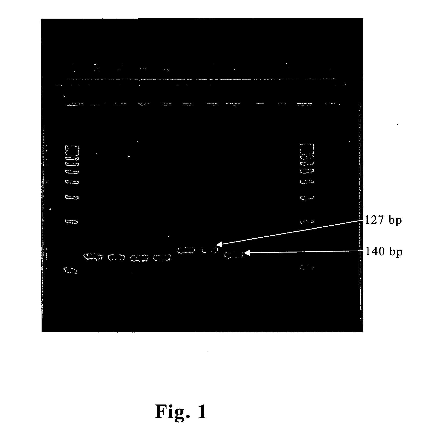
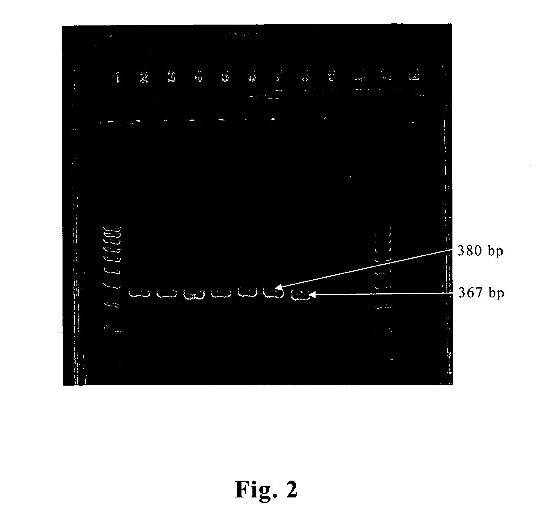
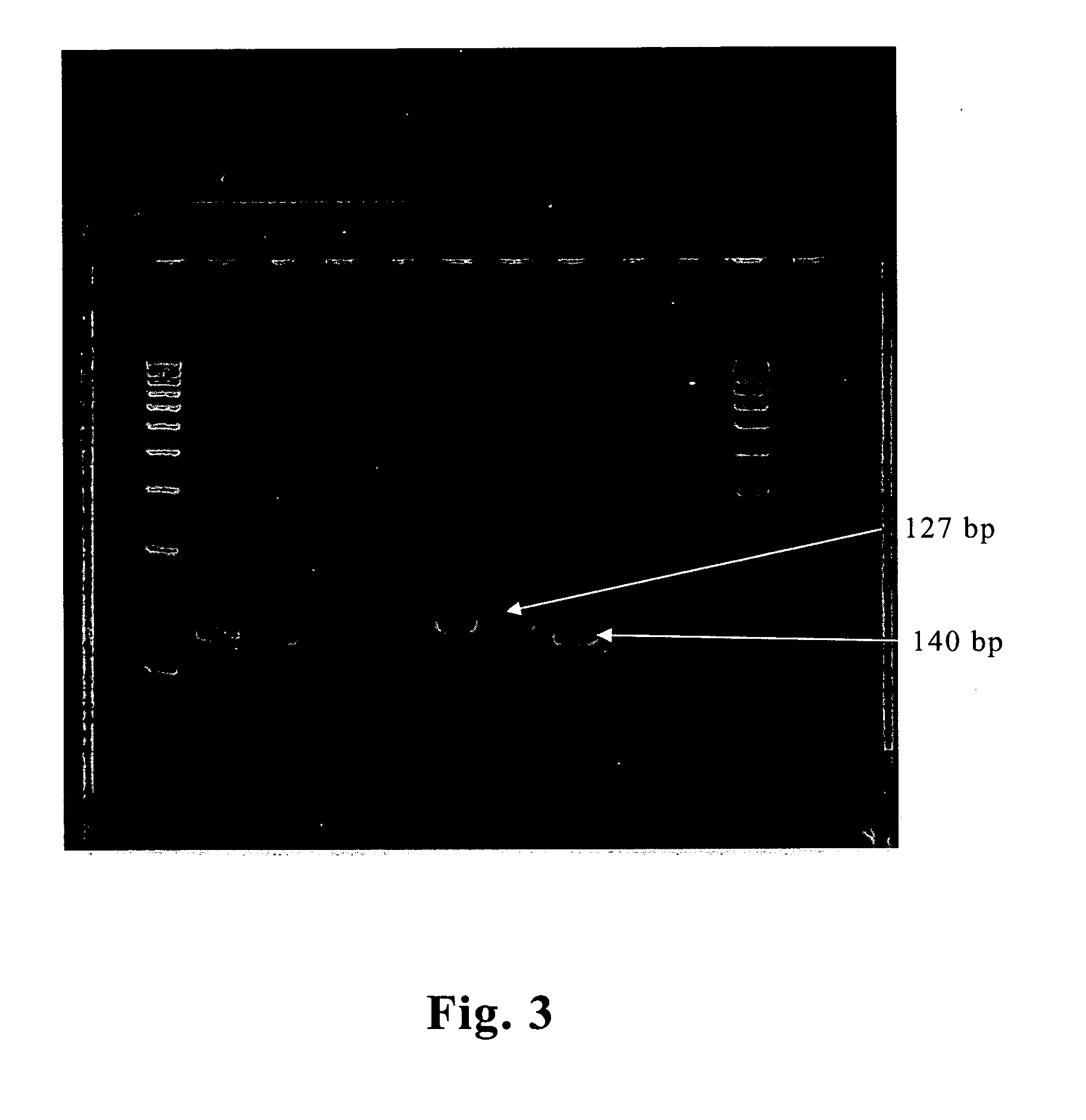
[0020] In the preferred embodiment of this invention, the specific primers of CP-3F (5′-ACCCTRTTGTARATAACACTA-3′) and CP-3R (5′-GTTAAAAATGAGTGTAGTGTGGTA-3′) designed from the conserved region are used to amplify the different genotypes of CSFV to perform RT-PCR amplification. The length of the RT-PCR product after amplification is 127 bp for field isolates of CSFV and 140 bp for LPC vaccine virus. Therefore, the field isolates of CSFV and the lapinized vaccine strain can directly be distinguished from the result of the electrophoresis analysis without performing the DNA sequencing. In another embodiment of this invention, for increasing the sensitivity of the test, a nest-PCR (n-PCR) CSFV testing method is designed as to firstly amplify the CSFV specific primers CP-5 (5′-GTAGCAAGACTGGAAATAGGTA-3′) and CP-6 (5′-AAAGTGCTGTTAAAAATGAGTG-3′) of different genotypes of CSFV for the first RT-PCR amplification, then the product of the first amplification (the length for field isolates of CSFV is 367 bp and LPC vaccine virus is 380 bp respectively); is taken as template to perform nest-PCR using CP-3F and CP-3R as primers. The length of the obtained PCR product is 127 bp for field isolates of CSFV and 140 bp for LPC vaccine virus. In one other embodiment, using CP-3F (5′-ACCCTRTTGTARATAACACTA-3′) and CP-9R (5′-GTACCAGTTCTTRCTCATTCAATA-3′) as primers, the amplified product of nest-PCR is 89 bp for field isolates of CSFV and 102 bp for LPC vaccine virus; only that the PCR product using this pair of primers is less. The nest-PCR is of high sensitivity and can conduct a high-quality testing result to chronic classical swine fever with low virus tilter or CSFV carrier pigs with normal appearance.

Problems solved by technology

Domestic pigs and wild boar are susceptible to CSFV.
Unfortunately, the vaccine virus can be detected and can't be differentiated from field isolates of CSFV by ELISA and RT-PCR in samples of pigs.
Unfortunately, the use of laboratory diagnostic methods to detect CSFV could be interfered by vaccine virus when attenuated vaccines are in use.
Wild boars distributed in the woods and they are difficult to be caught for injecting CSF vaccine.
Therefore, diagnosis with the commonly used diagnostic methods such as immuno-fluorescence stain, ELISA and RT-PCR can be interfered by the vaccine virus.
However, no real-time RT-PCR for distinguishing the field isolates of CSFV from the lapinized CSF vaccine viruses has been established.
The disadvantage of these methods includes the laborious process of the methods and the incapability of screening large field samples.

Method used


Image

  • RT-PCR detection for differential diagnosis of field isolates or lapinized vaccine strain of classical swine fever virus (CSFV) in samples
  • RT-PCR detection for differential diagnosis of field isolates or lapinized vaccine strain of classical swine fever virus (CSFV) in samples
  • RT-PCR detection for differential diagnosis of field isolates or lapinized vaccine strain of classical swine fever virus (CSFV) in samples

Examples


example 1

[0027] A one-step RT-PCR for distinguishing the field isolates of CSFV from the lapinized CSF vaccine viruses.

[0028] (1) Design of the Primers

[0029] A pair of amplification primers is designed from the conserved sequence within 3′-untranslated region of CSFV. This primer is a CSFV specific primer, which can only amplify the CFS virus but not its two close relatives bovine viral diarrhea virus (BVDV) and border disease virus (BDV). This primer is also one of the universal primers, which can be used to amplify different genotypes of CSFV. The nucleotide sequence of this primer pair is as follows:

CP-3F(5′-ACCCTRTTGTARATAACACTA-3′)CP-3R(5′-GTTAAAAATGAGTGTAGTGTGGTA-3′)

[0030] (2) Virus Sources

[0031] The lymphatic tissues of internal organs of swine such as tonsil, lymph node and spleen are ground with a mortar, then MEM medium (minimal essential medium) is added to form a 10% (w / v) suspension. After 20 minutes of 3000×g centrifugation, the top layer is retrieved for performing the RT...

example 2

[0036] For increasing the sensitivity of the test, another method using nest-PCR is designed for distinguishing the field isolates of CSFV from the lapinized CSF vaccine virus in addition to the one- step RT-PCR to detect the CSFV. The method is as follows:

[0037] (1) Design of the Primers

[0038] A pair of amplification primers is designed from the outer side of the amplification region of the CP-3F and CP-3R primers. This pair of primers is CSFV specific primers, which only amplifies the CSFV but not the two close relatives, BVDV and BDV. This pair of primers is one of the universal primers designed from the conserved sequences of CSFV, i.e. it can be used for CSF virus of all genotypes. The nucleotide sequence of this pair of primers is as follows:

CP-55′-GTAGCAAGACTGGAAATAGGTA-3′CP-65′-AAAGTGCTGTTAAAAATGAGTG-3′

[0039] (2) Single-tube RT-PCR

[0040] Take 5 μL of the prepared nucleic acid sample, add the pair of CSFV specific primers CP-5 and CP-6 (20 pm), 5 μL of 10× Super Thermal ...


PUM

PropertyMeasurementUnit
Fractionaaaaaaaaaa
Sizeaaaaaaaaaa

Abstract

The present invention provides a rapid RT-PCR detection method and a diagnostic kit for differentiating field isolates of classical swine fever virus (CSFV) from lapinized CSF vaccine viruses in samples. In order to detecting different genotypes of CSF virus, this invention uses a pair (or pairs) of CSF virus specific primers designed from the conserved sequences within the 3′-untranslated region of CSFV which contains an insertion of 12˜13 nucleotides (poly T) in the region of the lapinized CSF vaccine virus, in comparison with the corresponding region of field isolates of CSFV. By the RT-PCR or the RT-PCR followed by a nest-PCR, field isolates of CSFV and lapinized CSF vaccine viruses in samples could be detected directly and quickly and / or differentiated in electrophoresis without further enzymatic digestion or DNA sequencing.

Description

FIELD OF THE INVENTION [0001] This invention relates to a method and a diagnostic kit for detection of the classical swine fever virus (CSFV), in particular the differentiation field isolates and lapinized vaccine strains of CSFV thereof, by reverse transcription-polymerase chain reaction (RT-PCR), alone or in combination with nest-PCR (n-PCR), in which at least a pair of CSFV-specific primers, each of which was designed based on the conserved sequences within the 3′-untranslated region of CSFV's genome, are used. BACKGROUND OF THE INVENTION [0002] Classical swine fever (CSF), previously referred to hog cholera (HC), is an important infectious disease of swine caused by CSFV. Domestic pigs and wild boar are susceptible to CSFV. The virus belongs to the genus Pestivirus within the family Flaviviridae, which also includes bovine viral diarrhea virus (BVDV) and border disease virus (BDV) (Matthaeus, ZbL Vet. Med. 328: 126-132, 1981). They are antigenically and structurally closely rela...

Claims


Application Information

Patent Timeline
IPC IPC(8): C12Q1/70C12Q1/68C12P19/34
CPCC12Q1/701
Inventor PAN, CHU-HSIANGJONG, MING-HWA
Owner NAT INST FOR ANIMAL HEALTH COUNCIL AGRI EXECUTIVE YUAN
Learn More