Combined visual-optic and passive infra-red technologies and the corresponding systems for detection and identification of skin cancer precursors, nevi and tumors for early diagnosis

a technology of infra-red and visual-optic technology, applied in the field of non-invasive methods and devices for identifying pathological skin lesions, can solve the problems of limited number of sights that can be biopsied, requiring intrusive tissue removal, and a large number of such expensive painful tests

Inactive Publication Date: 2007-03-29
SKIN CANCER SCANNING
View PDF4 Cites 49 Cited by
  • Summary
  • Abstract
  • Description
  • Claims
  • Application Information

AI Technical Summary

Benefits of technology

[0033] According to still further features in the described preferred embodiments, the step step of adapting the spectral analysis includes choosing

Problems solved by technology

Biopsies have many obvious disadvantages: firstly biopsies require intrusive removal of tissue that can be painful and expensive.
Only a very limited number of sights can be biopsied in one session and patients are not likely to put up with a large number of such expensive painful tests.
Storage and transportation increase the cost, increases the possibility that samples will be mishandled, destroyed or lost, and also causes a significant time delay in receiving results.
This time delay means that examination follow up requires bringing the patient back to the doctor for a separate session.
This increases the inconvenience to the patient, the cost and the risk that contact will be lost or the disease will precede to a point of being untreatable.
Furthermore, the waiting period causes significant anxiety to the patient.
Finally, interpretation of biopsies is usually by microscopic analysis, which results in qualitative subjective results, which are not well suited to consistent interpretation.
There is, nevertheless, a limit to the amount of time, money or inconvenience that a basically healthy patient is willing to dedicate to routine screening procedures.
X-ray technology, which has been used successfully for detection of anomalies inside the human-body since the early 60's, is not suited for earlier detection of skin cancer because, due to it's the dangerous effects of X-ray radiation on human health, it cannot be used often enough (weekly or monthly), for diagnostics of patients with skin anomalies which need intensive reexamination over short-time periods.
Acoustic active methodologies, which are useful for detection of structures inside the human body, are also non-effective for early diagnosis cancerous skin anomalies.
Precancerous skin lesions are often of microscopic dimensions (on the order of millimeters or micrometers), which cannot be detected and identified by use acoustic methods (which are limited to detecting structures larger than the wavelength of sound on the order of centimeters).
Microwave technologies are very complicated and radiate the human body with microwave radiation, which may have dangerous effects.
Nevertheless, all of the widely known techniques such as optical imaging, optical spectral analysis, and thermal imaging have disadvantages making them not fully appropriate for detection and identification of skin cancer and cancer precursors.
Using reflectance spectra alone, it would be difficult to differentiate between various skin conditions since different skin diseases have similar reflectance spectra.
Nevertheless, being a purely spectral method limited to the visible frequency band, this method does not give important information about the geometry of a lesion.
Also some lesions can be difficul

Method used


Image

  • Combined visual-optic and passive infra-red technologies and the corresponding systems for detection and identification of skin cancer precursors, nevi and tumors for early diagnosis
  • Combined visual-optic and passive infra-red technologies and the corresponding systems for detection and identification of skin cancer precursors, nevi and tumors for early diagnosis
  • Combined visual-optic and passive infra-red technologies and the corresponding systems for detection and identification of skin cancer precursors, nevi and tumors for early diagnosis

Examples


second embodiment

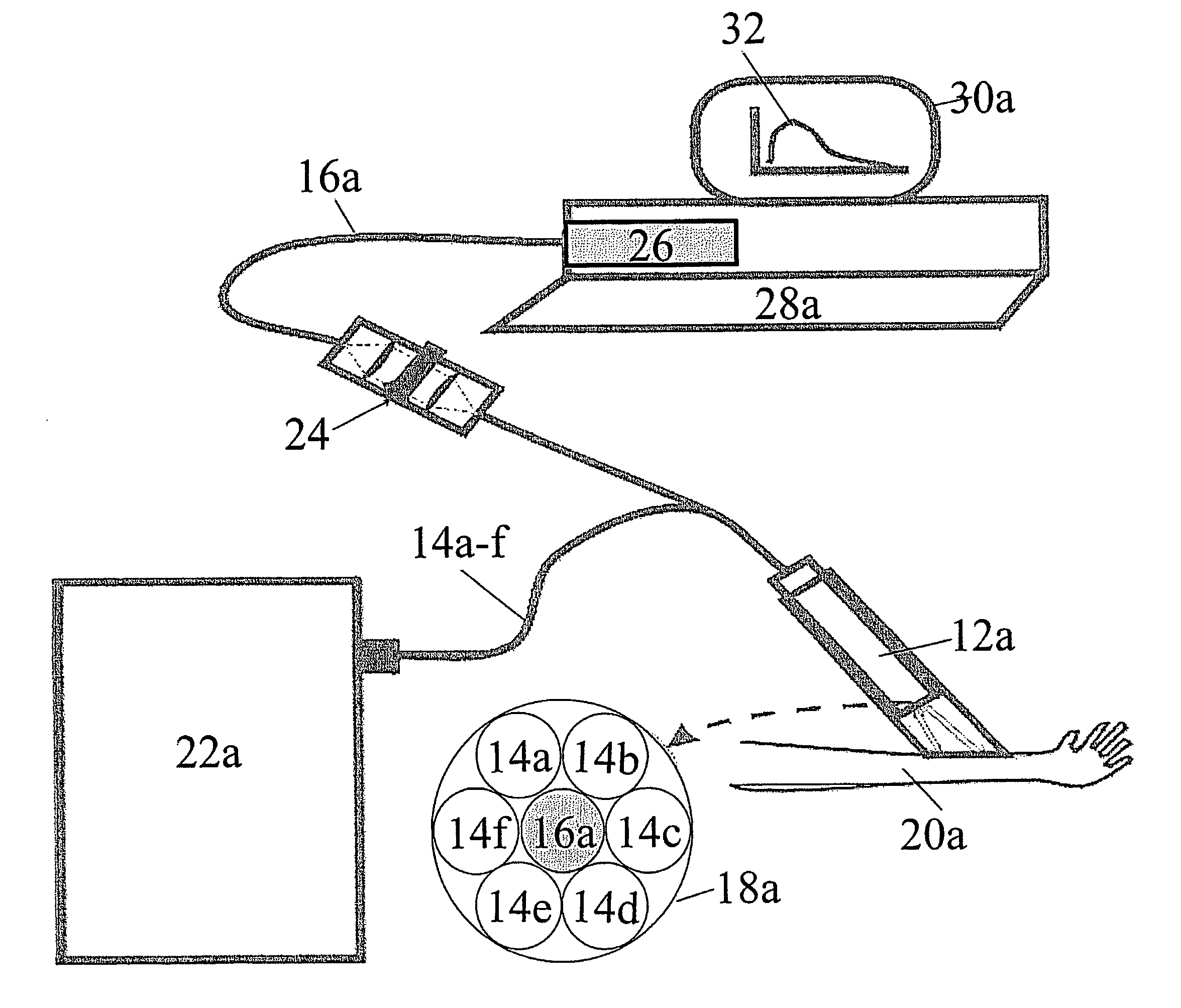
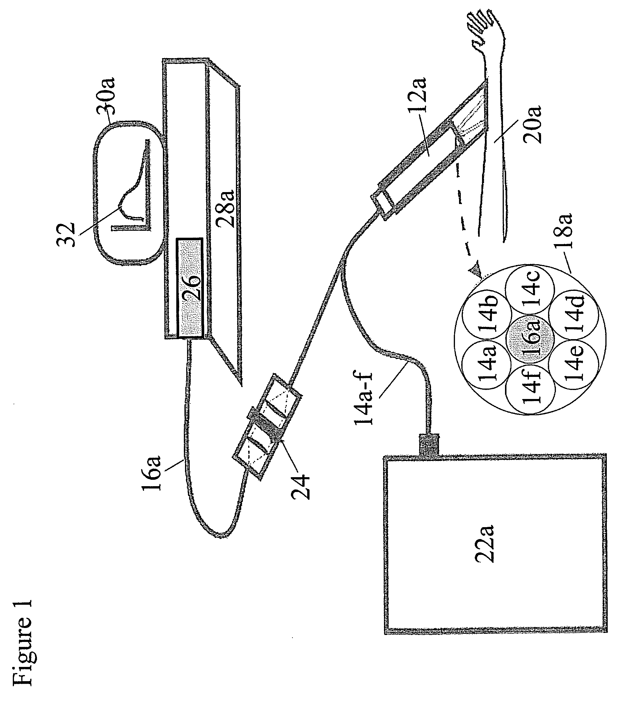
[0073]FIG. 6 illustrates the current invention. In the embodiment of FIG. 6, the skin 20b of a patient is investigated using a probe 12b having an illumination fiber 14g connected to a light source 22b. Probe 12b also contains a pick-up fiber 16b connected to a spectrometer 502. Spectrometer 502 measures simultaneously measures radiation in multiple bands in the visible, NIR and MIR bands using a detector system 504 which may be an array of multiple detectors, each detector measuring a different frequency band. Alternatively, detector system 504 can be a interferometer producing an interference spectrum which is interpreted by a processor, which is a PC 28b by means of Fourier transform analysis. Under any conditions the measurements of detector system 504 are sent to PC 28b via interface electronics and PC 28b displays the results as a spectrogram on a monitor 30b. PC 28b also is connected to a first control cable 506a to control light source 22b to provide illumination either in t...

third embodiment

[0074]FIG. 7 shows a scanner assembly 600 according to the current invention Particularly scanner assembly 600 includes an active visible sensor assembly 602, which is a bundle of five optical fibers, four illumination fibers 14h-14k and a pick up fiber 16c shown in cross section 18b. Visible light does not appreciably penetrate skin, therefore the visible sensor assembly 602 is focused by lens 610c onto a point 612 on the surface of skin 20c. Scanner assembly 600 also includes two passive MIR sensor assemblies 602604a and 604b, which are focused by lenses 610a and 610b respectively from opposite angles at a point 7 mm below point 612. Thus as scanner assembly moves along in scanning direction 606, visible sensor assembly 602 detects discoloration (or fluorescence) of the skin surface along a line, while simultaneously MIR sensor assemblies 604a and 604b measure black body MIR radiation from two directions along the same line in order to gauge the depth of a lesion 614. Thus the loc...


PUM


Abstract

A device and method to non-invasively identify pathological skin lesions. The method and device detect and identify of different kinds of skin nevi, tumors, lesions and cancers (namely, melanoma) by combined analyses of visible and infra-red optical signals based on integral and spectral regimes for detection and imaging leading earlier warning and treatment of potentially dangerous conditions.

Description

[0001] This is a continuation-in-part of U.S. Provisional Patent Application No. 60\708389, filed Aug. 16, 2005.FIELD AND BACKGROUND OF THE INVENTION [0002] The present invention relates to a non-invasive method and device to identify pathological skin lesions. More specifically the present invention relates to a method and device for non-intrusive detection and identification of different kinds of skin nevi, tumors, lesions and cancers (namely, melanoma) by combined analyses of visible and infra-red optical signals based on integral and spectral regimes for detection and imaging leading earlier warning and treatment of potentially dangerous conditions. [0003] Commonly suspicious lesions are biopsied to determine their status. Biopsies have many obvious disadvantages: firstly biopsies require intrusive removal of tissue that can be painful and expensive. Only a very limited number of sights can be biopsied in one session and patients are not likely to put up with a large number of s...

Claims


Application Information

Patent Timeline
IPC IPC(8): A61B6/00
CPCA61B5/0059A61B5/0064A61B5/7257A61B5/444A61B5/445A61B5/0071A61B6/00
Inventor ZILBERMAN, ARKADIISMOLYAK, YAFIMBLAUNSHTEIN, NATHANDEKEL, BEN ZIONYARKONY, AVRAHAM
Owner SKIN CANCER SCANNING
Why Eureka
Try Eureka