
Raman instrument for measuring weak signals in the presence of strong background fluorescence

a background fluorescence and weak signal technology, applied in the direction of instruments, spectrometry/spectrophotometry/monochromators, optical radiation measurement, etc., can solve the problem of low levels of carotenoids and related substances such as retinoids as high risk factors for malignant lesions, and the signal intensity of raman spectroscopy is very low, and the effect of raman scattered light intensity

Active Publication Date: 2007-04-05
NU SKIN INT
View PDF21 Cites 21 Cited by
  • Summary
  • Abstract
  • Description
  • Claims
  • Application Information

AI Technical Summary

Benefits of technology

[0016] While multiple embodiments are disclosed, still other embodiments of the invention will become apparent to those skilled in the art from the following detailed description, which shows and describes illustrative embodiments of the invention. As will be realized, the invention is capable of modifications in various obvious aspects, all without departing from the spirit and scope of the invention. Accordingly, the drawings and detailed description are to be regarded as illustrative in nature and not restrictive.

Problems solved by technology

In addition, low levels of carotenoids and related substances such as retinoids have been assessed as high risk factors for malignant lesions.
Such techniques require removal of large amounts of tissue sample from the patient for subsequent analysis and processing, which typically takes at least twenty-four hours to complete.
Thus, this technique is both invasive and slow and also expensive.
One difficulty associated with Raman spectroscopy is the very low signal intensity which is inherent to Raman scattered light.
The Raman signal can be separated from Rayleigh scattered light through the use of filters, gratings, or other wavelength separation devices; however, this can have the effect of further weakening the measured Raman signal through the additional attenuation that can occur when the light passes through a wavelength separation device.
In practice, the Raman scattered light is extremely difficult to detect.
Lasers have been used as a light source to increase light intensity, but this can cause burning or degradation of the sample.
Yet another difficulty associated with Raman measurements is that the substances of interest in the skin not only scatter incident light, but can absorb and subsequently fluoresce with substantial intensity.
This fluorescence often comprises a very strong, spectrally broad signal that tends to “drown out” or overwhelm the Raman spectral peaks, increasing the difficulty of identification and quantification of the substances.
The disadvantage of fluorescence measurements is that since many different molecules fluoresce in broad bands of wavelengths, such measurements cannot be used to identify conclusively the presence or concentration of a particular substance.
The spectral resolution necessary for this analysis severely limits the amount of light that can be collected from the tissue with a standard spectrometer, which the system relies on.
Lasers are a good source of monochromatic light that can be focused to a small spot for the Raman excitation, but lasers with wavelengths in the needed blue-green spectral region are expensive and somewhat difficult to keep stable in environments with varying physical conditions.
Moreover, estimating the level of background light beneath the Raman peak based on measurements of light to either side of the peak is susceptible to error that can vary with specific hardware variability.

Method used


Image

  • Raman instrument for measuring weak signals in the presence of strong background fluorescence
  • Raman instrument for measuring weak signals in the presence of strong background fluorescence
  • Raman instrument for measuring weak signals in the presence of strong background fluorescence

Examples


Embodiment Construction

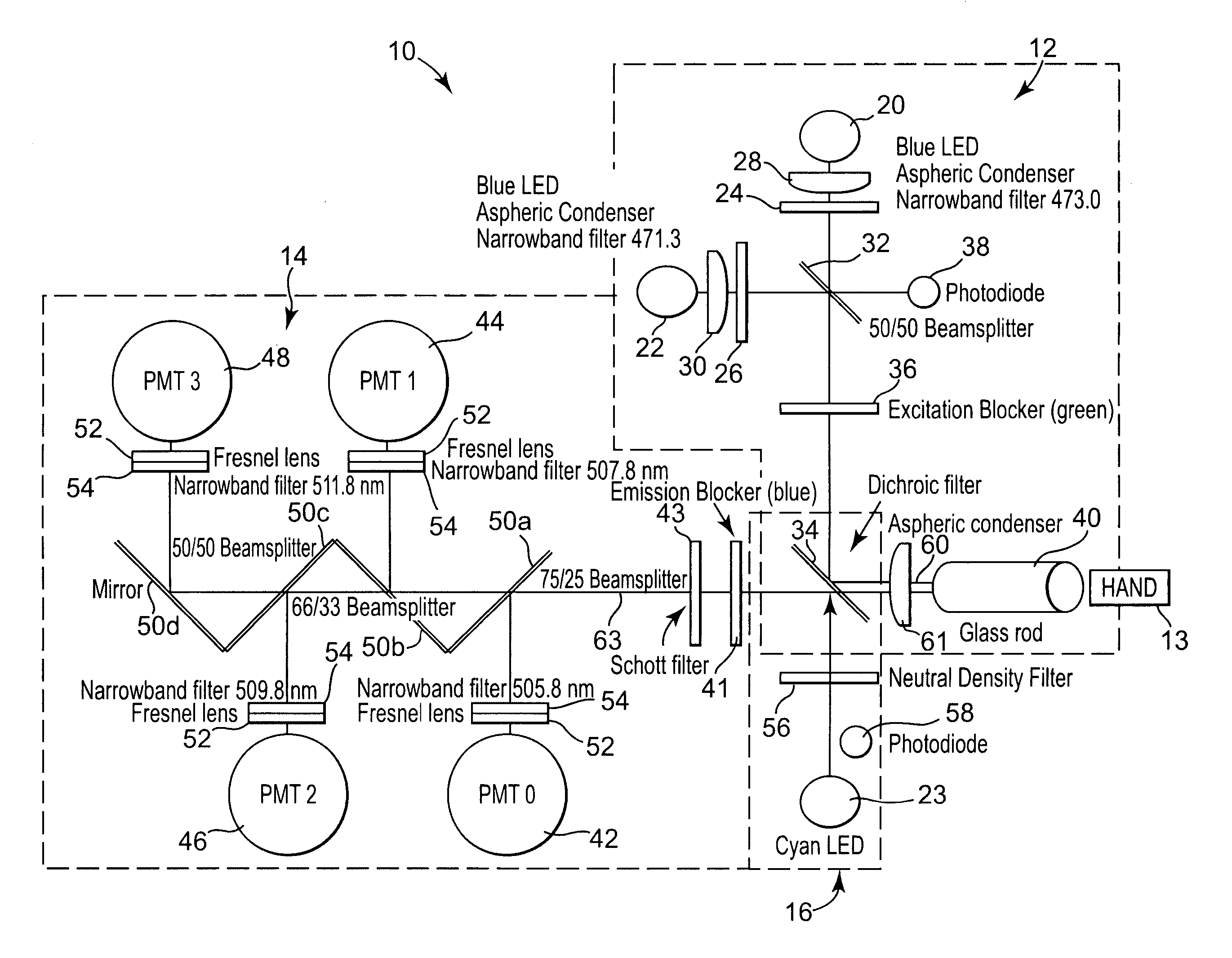
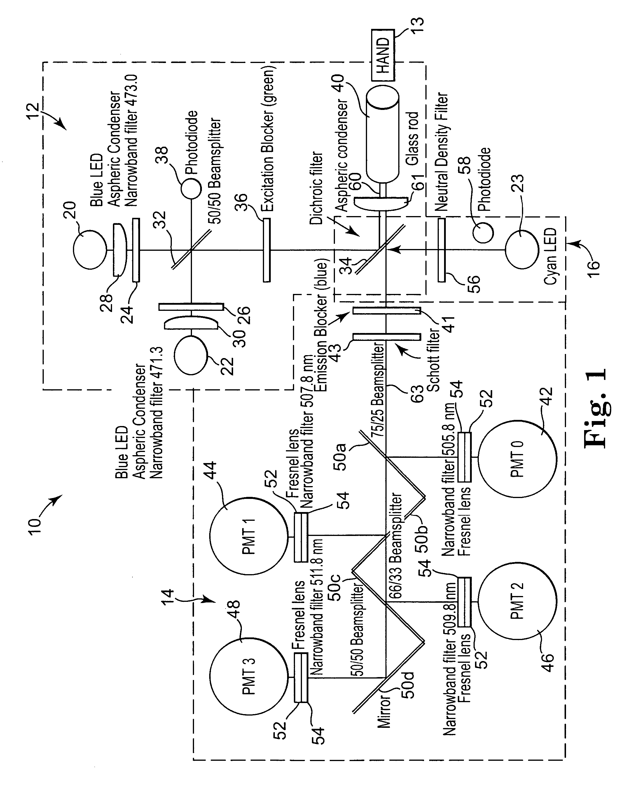
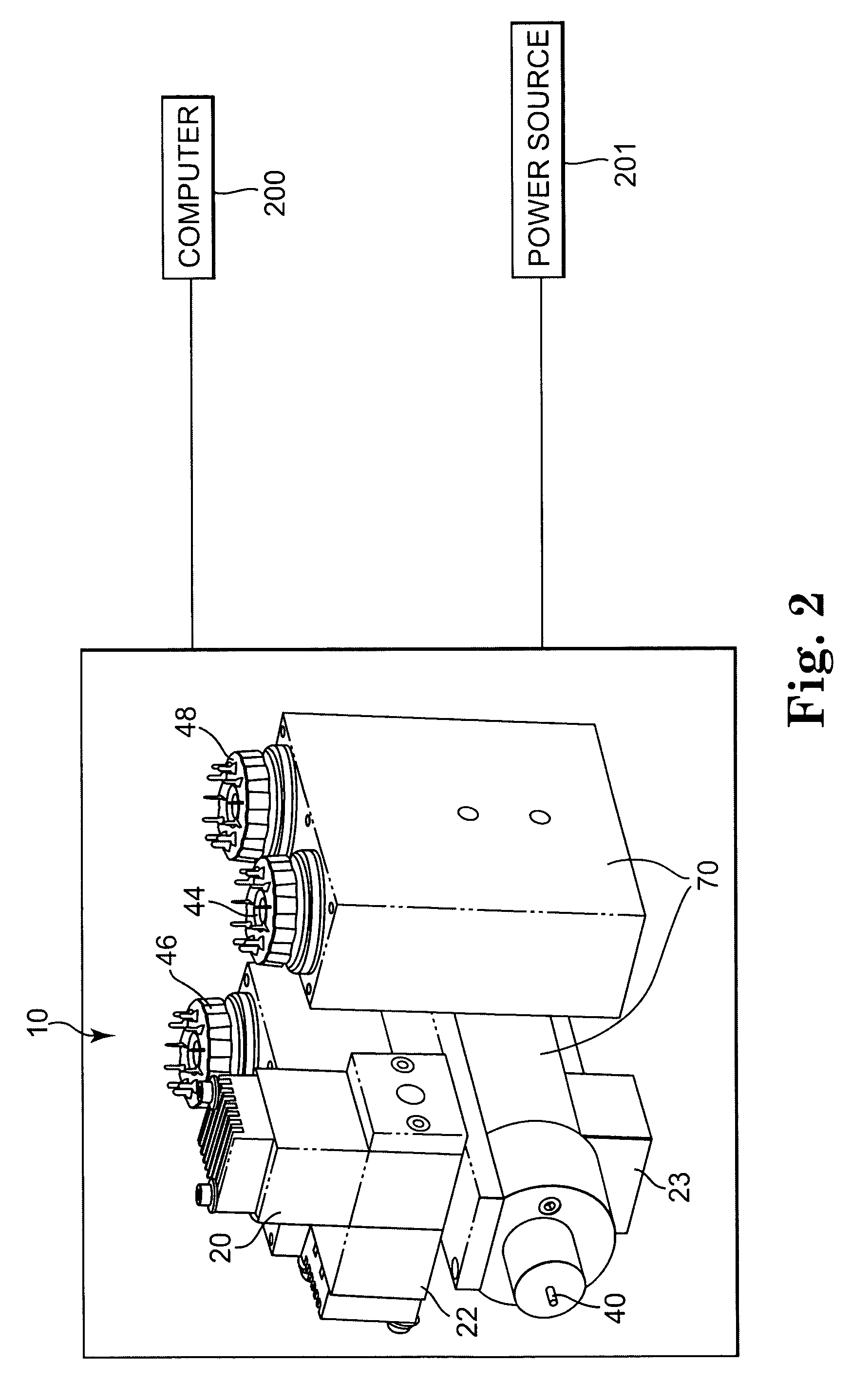
[0037] A method and apparatus for the measurement of the levels of carotenoids and other related substances or other selected molecules in biological tissue or a target sample, such as living skin is provided. The apparatus comprises an excitation portion, an analyzer portion, and a calibration portion. The apparatus enables noninvasive, rapid, safe, and accurate determination of the levels of carotenoids (including their isomers and metabolites) and similar substances in tissue. This information can be a marker for conditions where carotenoids or other antioxidant compounds may provide useful information.

[0038] Examples of biological tissues which can be measured non-invasively with the technique of the invention include human skin on a hand. It is anticipated that measurements may also be made for cervix, colon, and lungs. With the addition of appropriate sample presentation accessories, it is expected that bodily fluids that can be measured will include saliva, whole blood, and ...


PUM


Abstract

A method for measuring a chemical concentration in tissue has two measurement steps. First, generating a first light and illuminating a portion of the tissue with the first light; capturing a first reflected light from the tissue; directing the first reflected light to a plurality of light sensors, each light sensor measuring light at a different wavelength, that wavelength being proximate to a wavelength of an expected Raman shift wavelength for the chemical in the tissue; and obtaining a measurement from each of the light sensors, each measurement being specific to the first reflected light through that light sensor. Second, generating a second light and illuminating a portion of the tissue with the second light; capturing a second reflected light from the tissue; directing the second reflected light to the plurality of light sensors, each light sensor measuring light at a different wavelength that wavelength being proximate to a wavelength of an expected Raman shift wavelength for the chemical in the tissue; and obtaining a measurement from each of the light sensors, each measurement being specific to the second reflected light through that light sensor. The measurements of the first reflected light and the measurements of the second reflected light are used to calculate a concentration of the chemical in the tissue.

Description

FIELD OF THE INVENTION [0001] The present invention relates a method and apparatus for measuring levels of chemical compounds found in biological tissue. More specifically, the invention relates to a method and apparatus for the noninvasive detection and measurement of levels of carotenoids or other selected molecules in biological tissue. BACKGROUND OF THE INVENTION [0002] Carotenoids are plant pigments available from the diet that may have important effects in the human body. For example, it is thought that many carotenoids seem to have positive effects on immune function, which can be critical in stopping cancer. Carotenoids also have an antioxidant effect which may protect against heart disease. Further, some studies have shown that carotenoids may help reduce the risk of degenerative diseases, such as age-related macular degeneration. [0003] Recently, researchers have focused on the carotenoids alpha-carotene (in carrots), lycopene (in red fruits and vegetables, such as tomatoe...

Claims


Application Information

Patent Timeline
IPC IPC(8): A61B5/05A61B6/00
CPCA61B5/0059A61B2560/0223G01J3/02G01J3/0291G01J3/44G01N21/274G01N21/65G01N2021/6419G01N2021/6421A61B5/00
Inventor FERGUSON, SCOTTFRALICK, JOHN F.BERGESON, SCOTT DOUGLASPEATROSS, JUSTIN
Owner NU SKIN INT
Learn More