Method and apparatus for optical imaging via spectral encoding

a technology of optical imaging and spectral encoding, applied in the direction of spectroscopy diagnostics, instruments, catheters, etc., can solve the problems of limiting the options for screening for pre-neoplastic conditions, significant challenges in identification and diagnosis, and removal of tissue from patients

Active Publication Date: 2007-04-12
THE GENERAL HOSPITAL CORP
View PDF99 Cites 65 Cited by
  • Summary
  • Abstract
  • Description
  • Claims
  • Application Information

AI Technical Summary

Benefits of technology

[0015] In yet further exemplary embodiments of the present invention, a method for positioning or directing an electromagnetic radiation within an anatomical structure is provided, which can include scanning at least a portion of the anatomical structure using the electromagnetic beam, and using a signal which may b...

Problems solved by technology

Because precancerous growth and early stage cancers often arise on a microscopic scale, they can present significant challenges for identification and diagnosis.
Although this approach may be regarded as a current standard for microscopic diagnosis, it requires the removal of tissue from the patient and significant processing time to generate slides.
However, the lack of an appropriate imaging technology can greatly limits options for screening for pre-neoplastic conditions (e.g. metaplasia) and dysplasia.
In addition, an inability to identify areas of dysplasia and carcinoma in situ has led to screening procedures such as, e.g., random biopsy of the prostate, colon, esophagus, and bladder, etc., which can be highly undesirable and indiscriminate.
Resolutions approaching 20 μm have been achieved by these technologies, but fundamental physical limitations can still prevent their application in patients.
High NA imaging, however, can be particularly sensitive to aberrat...

Method used


Image

  • Method and apparatus for optical imaging via spectral encoding
  • Method and apparatus for optical imaging via spectral encoding
  • Method and apparatus for optical imaging via spectral encoding

Examples


Embodiment Construction

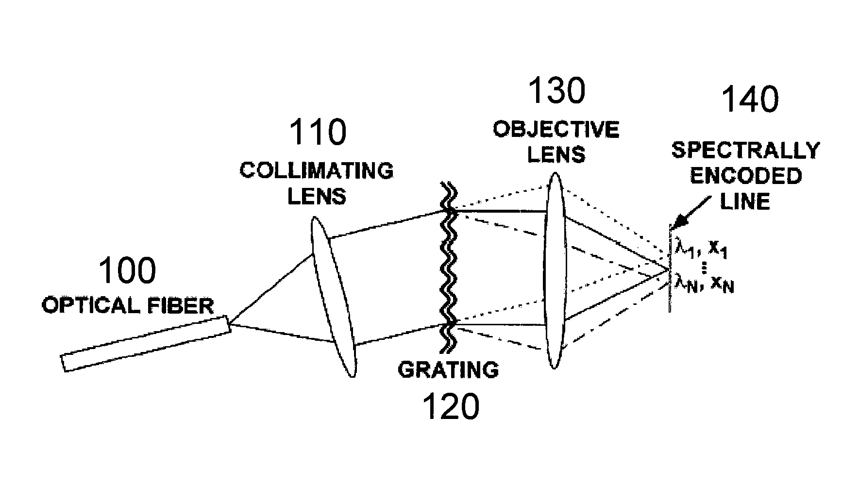
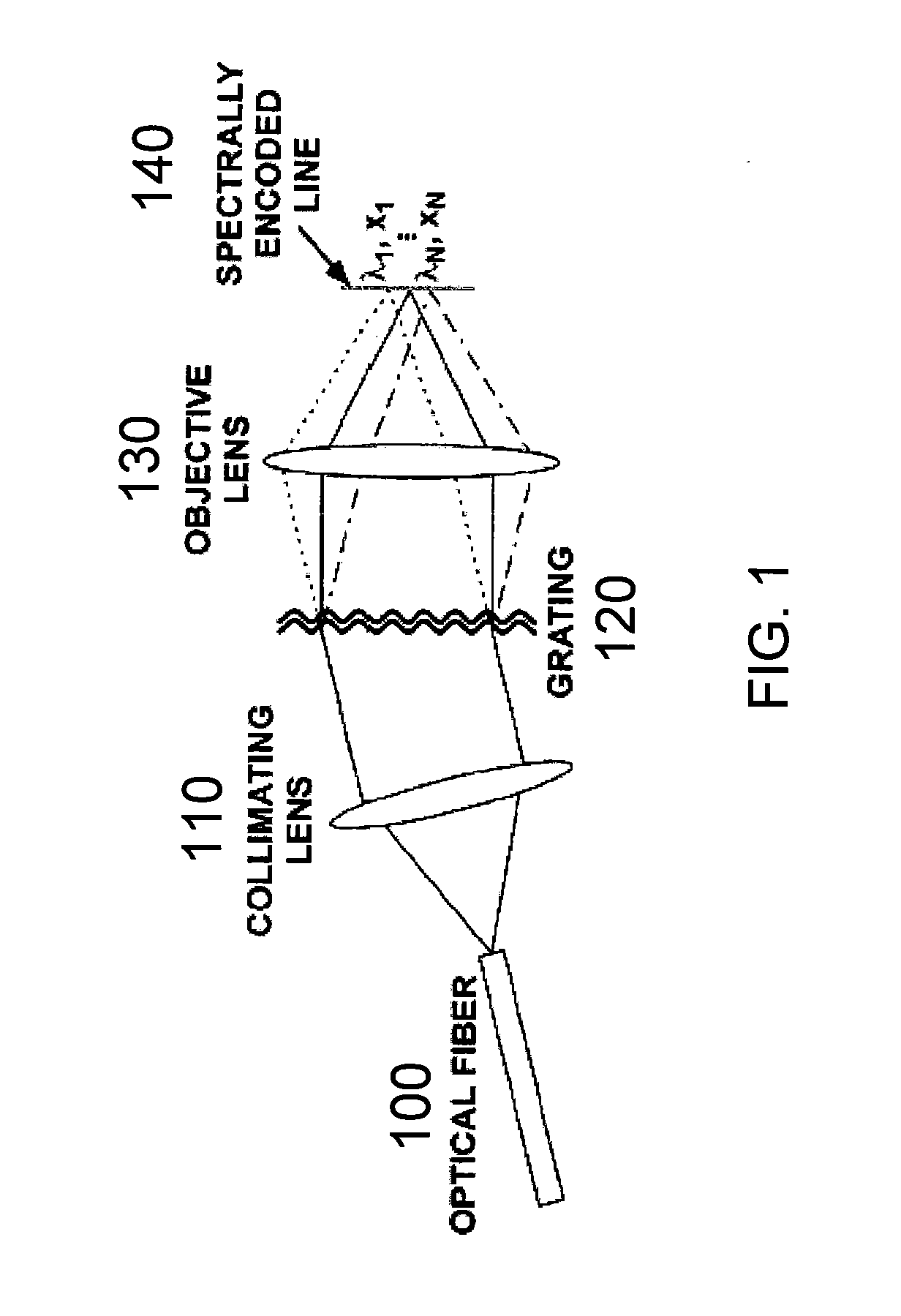
[0090] In accordance with exemplary embodiments of the present invention, a method and apparatus for endoscopic confocal microscopy is provided which circumvents the need for miniature, high-speed scanning mechanisms within a probe. Spectrally encoded confocal microscopy (“SECM”) is a wavelength-division multiplexed confocal approach that may be used. SECM utilizes a broad bandwidth light source and can encode one dimension of spatial information in the optical spectrum.

[0091] An exemplary SECM technique is shown in FIG. 1. The output from a single-mode optical fiber 100, which may be located at a distal end of a probe, can be collimated by a collimating lens 110, and then illuminate a dispersive optical element (such as, e.g., a transmission diffraction grating 120). An objective lens 130 can then focus each diffracted wavelength to a distinct spatial location within the specimen, resulting in a transverse line focus 140 where each point on the line may be characterized by a disti...


PUM


Abstract

Method, apparatus and arrangement according an exemplary embodiment of the present invention can be provided for generating an image of at least one portion of an anatomical structure. For example, the portion can have an area greater than about 1 mm2, and the image can have a resolution a transverse resolution that is below about 10 μm. For example, light can be scanned over such portion so as to generate first information which is related to the portion, where the light may be provided through a diffraction arrangement to generate a spectrally dispersed line. Method, apparatus and arrangement according to a further exemplary embodiment of the present invention can be provided for positioning a radiation or optical beam within an anatomical structure based on signals generated by scanning a portion of the structure using the same or a different beam.

Description

CROSS-REFERENCE TO RELATED APPLICATION(S) [0001] This application is based upon and claims the benefit of priority from U.S. patent application Ser. No. 60 / 721,802, filed Sep. 29, 2005, the entire disclosure of which is incorporated herein by reference.FIELD OF THE INVENTION [0002] The present invention relates to devices and methods for comprehensive optical imaging of epithelial organs and other biological structures via spectral encoding. BACKGROUND OF THE INVENTION [0003] Radiological techniques such as X-ray computed tomography (“CT”), magnetic resonance imaging (“MRI”), and ultrasound can enable noninvasive visualization of human pathology at the organ level. Although these modalities may be capable of identifying large-scale pathology, the diagnosis of cancer can require the evaluation of microscopic structures that is beyond the resolution of conventional imaging techniques. Consequently, biopsy and histopathologic examination may be required for diagnosis. Because precancer...

Claims


Application Information

Patent Timeline
IPC IPC(8): G02B21/06
CPCA61B5/0062G01N2021/1765A61B5/0068A61B5/0073A61B5/0075A61B5/0084A61B5/6852G01N21/4795G01N21/6458G01N23/046G02B21/0028G02B23/2423G02B23/243G02B23/2461G02B23/2476G01B9/02G01B9/02064G01B9/02027G01B9/02091G01B9/02049G01B9/02087G01B9/04G01N21/25G01N21/6486G01N2223/419G01N21/27G01N33/4833A61B5/0066G01N33/48
Inventor TEARNEY, GUILLERMO J.YELIN, DVIRBOUMA, BRETT EUGENE
Owner THE GENERAL HOSPITAL CORP
Why Eureka
Try Eureka