Dynamic imaging of biological cells and other subjects

a biological cell and dynamic imaging technology, applied in the field oframan spectroscopy and microscopic imaging of biological cells, can solve the problems of limited in its utility to the particular conditions, difficult to understand the effects of a drug on an individual cell or small group of cells on a biochemically relevant scale, and the inability to apply univariate analysis broadly

Active Publication Date: 2007-08-09
CHEMIMAGE CORP
View PDF34 Cites 47 Cited by
  • Summary
  • Abstract
  • Description
  • Claims
  • Application Information

AI Technical Summary

Benefits of technology

[0019] The present invention permits rapid observation of the sample with full spatial information, and allows the monitoring of the evolution and changes in the sample that are naturally proceeding or occurring (i.e., under equilibrium conditions), as well as those that are additionally forced or imposed by creating a non-equilibrium condition via an external means (e.g., one or more external fields or forces applied to the sample). In certain embodiments, the external means may be applied to a specific location within the sample (rather than the whole sample).

Problems solved by technology

However, it has been difficult to understand the effects of a drug on an individual cell or small group of cells on a biochemically-relevant scale.
(i.e., a single strong Raman peak purportedly corresponding to paclitaxel and the particular deconvolution scheme), be limited in its utility to the particular conditions present in their experimental system.
Such univariate analysis is unlikely to be broadly applicable.
For instance, it is an inappropriate methodology to use for analysis of systems that exhibit more complex Raman spectra than that purportedly exhibited by the system described by Ling et al.
Furthermore, rotation of the system displaces the image, requiring realignment of the Raman and optical images.
A significant drawback of imaging systems that rely on detection of fluorescence is that such systems exhibit relatively low spectral resolution, owing to the broad spectral width of most, if not all, fluorescent emissions.
Another drawback is that many molecules and cellular components of interest do not fluoresce.
Furthermore, live and dead cells often exhibit intense fluorescence background emissions that can interfere with fluorescent observation of a target of interest.
Because most fluorescent emissions are not specific for the molecular species emitting the radiation, such background can complicate or prevent efforts to meaningfully interpret fluorescence.
Spectral imaging is low resolution and cannot spectrally resolve individual molecular species.
The technology described in this patent is appropriate for analysis of large signals, such as fluorescent emissions, but is inappropriate for analysis of weaker signals, such as Raman scattered radiation.
The patent does not disclose whether the methods can be used to detect scattering bodies smaller than tissue lesions.
Furthermore, the patent does not disclose combining spatial resolution data with optical imaging data, since the patent is directed to planar tomographic analysis of macroscopic tissues.

Method used


Image

  • Dynamic imaging of biological cells and other subjects
  • Dynamic imaging of biological cells and other subjects
  • Dynamic imaging of biological cells and other subjects

Examples


example

[0082] The invention is now described with reference to the following Example. This Example is provided for the purpose of illustration only, and the invention is not limited to this Example, but rather encompasses all variations which are evident as a result of the teaching provided herein.

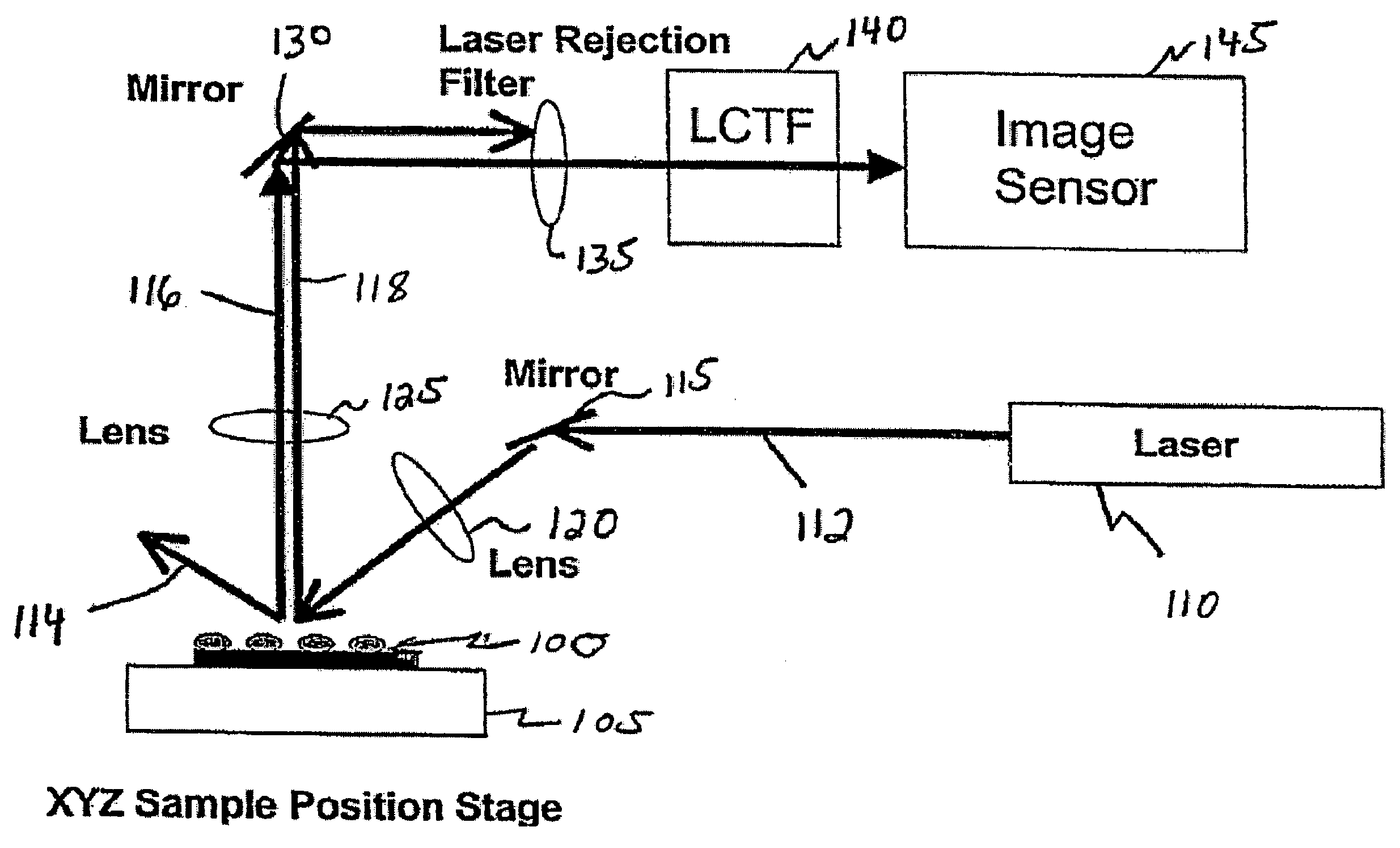
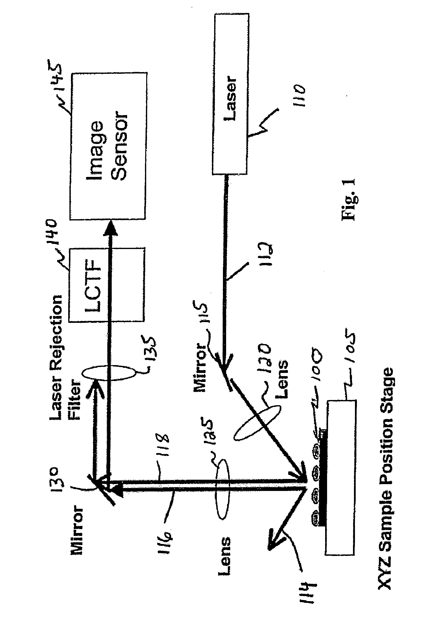
[0083] Raman chemical imaging of a Raman-active compound in a breast cancer cell.

[0084] A Raman-active compound was contacted with a breast cancer cell. Subsequent Raman chemical imaging of the cell was performed. Raman spectra obtained from the cell prior to and after contacting it with the compound indicated significant differences in Raman spectral features at about 900 cm−1 and at about 2250 cm−1. Raman chemical imaging of the cell after contacting it with the compound demonstrated the location of the compound exhibiting the 2250 cm−1 feature in the cell cytoplasm.


PUM

PropertyMeasurementUnit
Raman shiftaaaaaaaaaa
timeaaaaaaaaaa

Abstract

The invention relates to methods of dynamic chemical imaging, including methods of cellular imaging. The method comprises illuminating at least a portion of a cell with substantially monochromatic light and assessing Raman-shifted light scattered from the illuminated portion at a plurality of discrete times. The Raman-shifted light can be assessed at a plurality of Raman shift (RS) values at each of the discrete times, and the RS values can be selected to be characteristic of a pre-selected component at each of the discrete times. Multivariate analysis of Raman spectral features of the images thus obtained can yield the location and chemical identity of components in the field of view. This information can be combined or overlaid with other spectral data (e.g., a visible microscopic image) obtained from the field of view.

Description

BACKGROUND OF THE INVENTION [0001] The invention relates generally to the fields of Raman spectroscopy and microscopic imaging of biological cells. The invention also relates to dynamic chemical imaging in general. [0002] Understanding what is occurring, particularly on a molecular scale, in and among biological cells permits one to understand the behavior of the cells and influence their behavior in desired ways. For instance, most drugs exert their pharmacological effects based on their effects on cells. However, it has been difficult to understand the effects of a drug on an individual cell or small group of cells on a biochemically-relevant scale. Instead, macroscopic effects of a drug on a tissue or an entire organism have been observed and extensive experimentation and educated guessing has been required to understand the biochemical basis of a drug's effect. A significant need exists for methods of better observing the interactions of cells with each other and with the chemic...

Claims


Application Information

Patent Timeline
Patent Type & Authority Applications(United States)
IPC IPC(8): G01J3/44G01N21/65
CPCG01J3/2803G01J3/32G01J3/36G01J3/44G01N21/359G01N21/6486G01N21/65G01N21/658G01N2021/656G01N21/6456
Inventor MAIER, JOHN S.TREADO, PATRICK J.TUSCHEL, DAVIDVOIGT, THOMAS C.
Owner CHEMIMAGE CORP
Why Eureka
Try Eureka