Methods And Apparatus For Preserving The Endothelium In Isolated Hollow Organs And Biological Vessels

a technology for endothelium and biological vessels, applied in the field of methods and apparatus for treating and preserving endothelium in isolated hollow organs, can solve the problems of remarkable damage to endothelium function, autologous vascular substitutes often not providing satisfying transplant results, and significant loss of functions, so as to prevent ablation or destruction of endothelium and infiltrate into very small vessels and vascular segments.

Inactive Publication Date: 2007-08-30
BIOTEST SERUM INST GMBH
View PDF4 Cites 21 Cited by
  • Summary
  • Abstract
  • Description
  • Claims
  • Application Information

AI Technical Summary

Benefits of technology

[0090] With the expression “endothelium-protective perfusion solution” is meant a solution which is useful for treatment of endothelial tissue in isolated hollow organs or biological vessels, wherein the consistency of the endothelial tissue is maintained. The application of the endothelium-protective perfusion solution prevents an ablati

Problems solved by technology

These numbers make clear that even an autologous vascular substitute, although it has several advantages in comparison to other kind of vascular implants, is associated with considerable loss of functions.
Therefore, an autologous vascular substitute can often not provide a satisfying result for transplantation.
It was found by the inventors of the present invention that such a treatment procedure which has been commonly utilized in practice, may result in a remarkable damage of the endothelial function, resulting in a total destruction of the endothelial tissue.
However, the damage or destruction of endothelial tissue is one reason why biological vascular and organ implants may become obstructed subsequent to their transplantation, just as mentioned above.
A destruction of the endothelial layer—such as one that is caused, for example, by the above-mentioned treatment of autologous vascular implants—often causes

Method used


Image

  • Methods And Apparatus For Preserving The Endothelium In Isolated Hollow Organs And Biological Vessels
  • Methods And Apparatus For Preserving The Endothelium In Isolated Hollow Organs And Biological Vessels
  • Methods And Apparatus For Preserving The Endothelium In Isolated Hollow Organs And Biological Vessels

Examples


example 1

Preferred Method for Endothelium-Preserving Treatment of Isolated Hollow Organs or Biological Vessels

[0205] In one embodiment of the method of the invention, a perfusion solution with the following composition was used when blood vessels were to be treated:

[0206] Physiological electrolyte solution with 127 mM NaCl; 4.6 mM KCl; 1.1 mM MgSO4; 1.2 mM KH2PO4; 24 mM histidin-Cl; 2 mM CaCl2. Prior to addition of CaCl2 the pH value of the solution was adjusted under atmospheric conditions at pH=7.40. Further, the perfusion solution contained a 10 vol-% lipoprotein-free, hemolysin-free, homologous serum preparation from a pool of blood preparations (obtained from healthy donors), 2.5 mM L-glutamine, 2 mM Na-pyruvate, 8 mM glucose, 200 U / ml penicillin and 0.2 mg / ml streptomycin, low molecular heparin (fraxiparin purchased from Pharmacia Ltd.: 100 μl / 100 ml complete solution) and, in addition, 50 μM of each of uric acid and ascorbate.

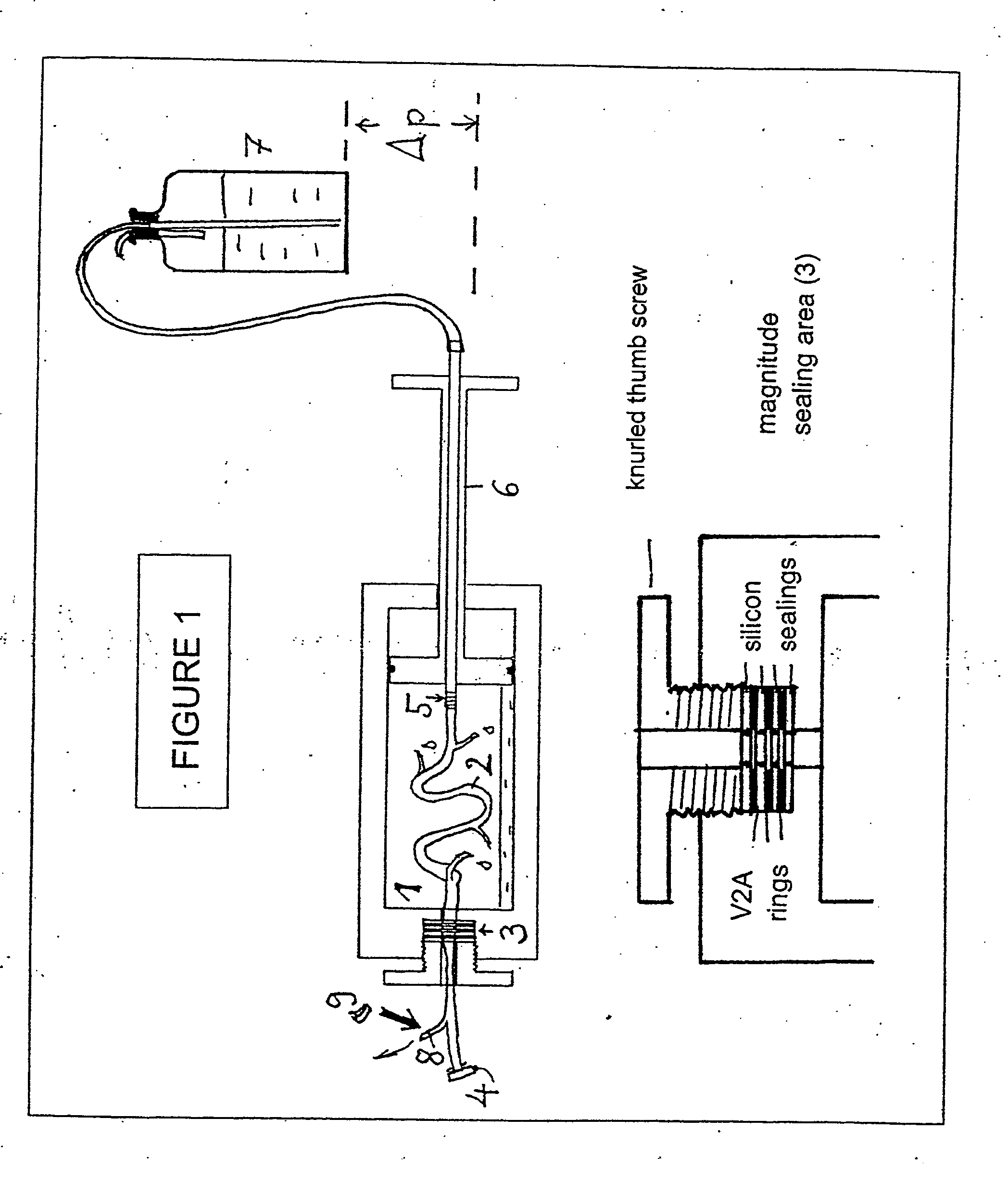
[0207] In the method for endothelium-preserving treatmen...

example 2

Regeneration of Endothelial Tissue from Vena Saphena by Treatment a with One Embodiment of the Endothelium-Preserving Perfusion Solution

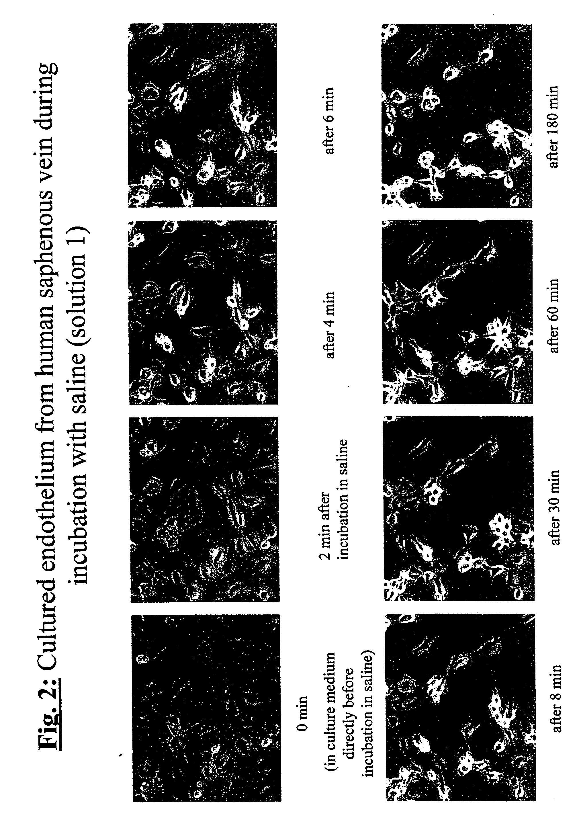
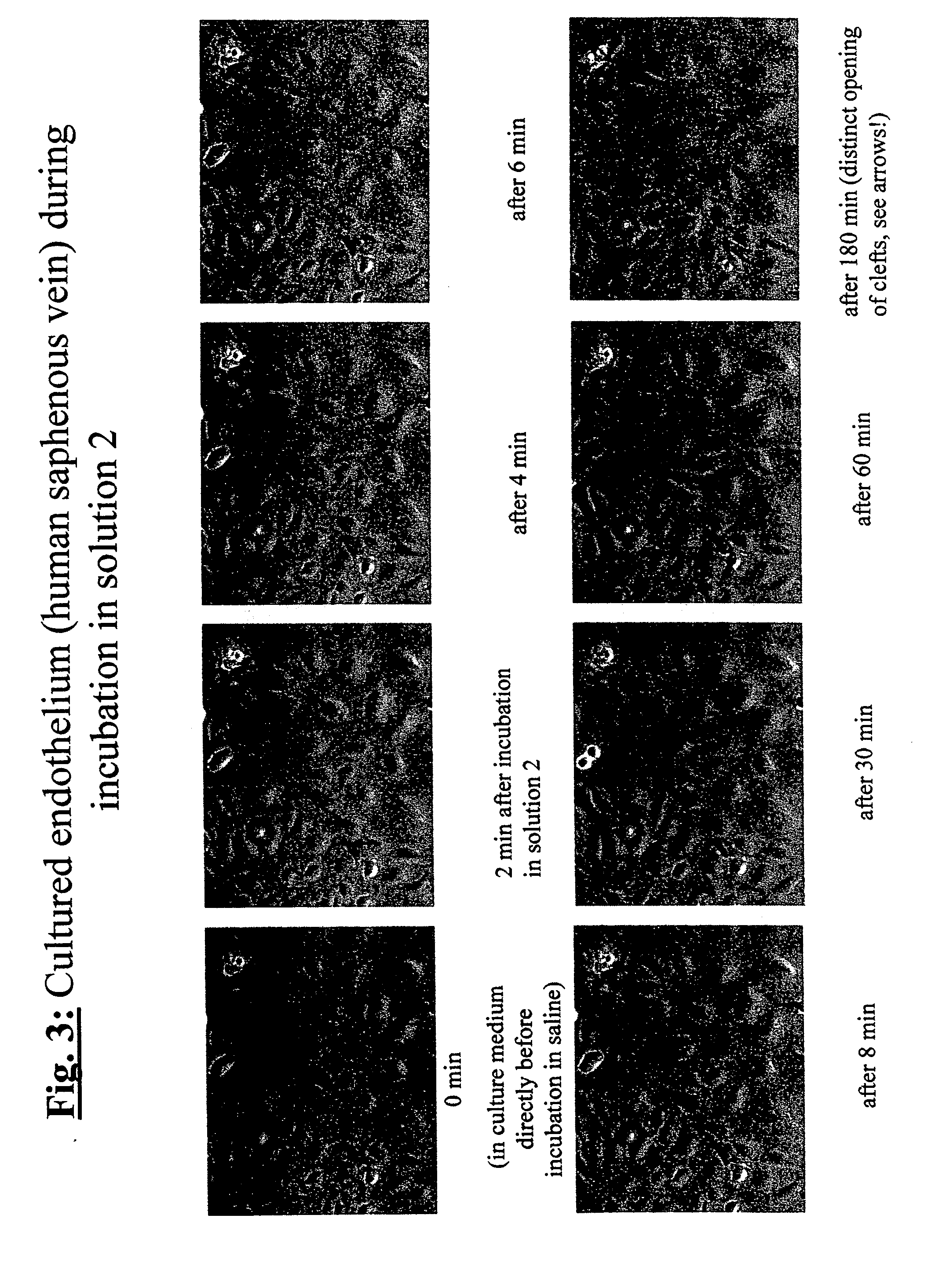
[0208] In order to demonstrate the efficiency of the endothelium-protective perfusion solution and the method of the invention, isolated endothelial cells obtained from human Vena saphena were microscopically observed during incubation with different embodiments of the perfusion solution of the invention following treatment of the cells and the efficiency was documented by serial or video time-lapse microphotography.

[0209] Endothelial cells from non-required rest pieces of human Vena saphena were selectively removed by collagenase incubation and grown in minimal essential medium (e.g. “Dulbecco minimal essential medium”, DMEM) containing 10% v / v fetal calf serum under water vapor-saturated atmospheric environment under addition of 5% v / v carbon dioxide at a temperature of 37° C. (“incubator conditions”) until confluence. Then, the selected plates ...

example 3

Preferred Perfusion Solution or Incubation Solution which Includes Quercetin

[0220] In the following example, the effect of quercetin, a naturally occurring anti-inflammatory-acting flavonoid on the endothelium of the smallest body vein (venules) was investigated in detail (see FIG. 5). To do this, different isolated human hearts of donors were rinsed thoroughly in the presence and absence of quercetin. This solution had the following composition: Isotonic electrolyte solution (127 mM NaCl; 4.6 mM KCl; 1.1 mM MgSO4; 1.2 mM KH2PO4; 24 mM histidin-Cl; 2 mM CaCl2 (pH to 7.40 prior to addition of CaCl2); 0.1% albumin and 2.5 mM L-glutamine. The perfusion solution also contained: 2 mM Na-pyruvate, 8 mM glucose, 200 U / ml penicillin and 0.2 mg / ml streptomycin, low molecular heparin (fraxiparin from Pharmacia Ltd.: 100 μl / 100 ml complete solution) and, in addition, 100 μM freshly added quercetin.

[0221] For the investigation of myocardial arteries and venules (which are particularly effecte...


PUM


Abstract

The invention relates to a method and an apparatus for the endothelium-preserving treatment of isolated hollow organs, especially biological vessels such as blood vessels and lymphatic vessels by means of endothelium-protective perfusates or incubation solutions containing at least 0.1 percent by weight of native albumin and a nutrient substrate, preferably L-glutamine. Also disclosed are the use of the endothelium-protective perfusates for preparing hollow organs or biological vessels as a transplant for the treatment of organ diseases or vascular diseases, the use thereof for repairing endothelial lesions in isolated hollow organs and/or biological vessels, and the use thereof for preserving organs and/or vessels.

Description

BACKGROUND OF THE INVENTION [0001] The present invention relates to a method and an apparatus for the treatment and preservation of endothelial tissue in isolated hollow organs, in particular in isolated biological vessels such as blood vessels and lymphatic vessels, by using an albumin-containing endothelium-protective perfusion solution or incubation solution, the use of such an endothelium-protective perfusion solution for the preparation of hollow organs or biological vessels as grafts for the treatment of organ or vessel diseases, its use for repair of endothelial lesions in isolated hollow organs and / or biological vessels and its use for preservation of organs and / or vessels. [0002] An occlusion of blood vessels that is caused by arteriosclerosis is one reason for a plurality of organ diseases in Western countries. A transplantation of vessels, i.e. the replacement of morbid, narrowed or closed biological vessels by healthy vascular substitutes has therefore a high clinical si...

Claims


Application Information

Patent Timeline
IPC IPC(8): A01N1/02A61B17/00
CPCA01N1/02A61B2017/00969A01N1/0247A01N1/0226
Inventor NEES, STEPHANNEES, FRIDERIKE
Owner BIOTEST SERUM INST GMBH
Why Eureka
Try Eureka