Structurally modified acellular tissue engineering scaffolds and methods of production

Active Publication Date: 2007-10-25
WAKE FOREST UNIVERSITY
View PDF14 Cites 69 Cited by
  • Summary
  • Abstract
  • Description
  • Claims
  • Application Information

AI Technical Summary

Benefits of technology

[0013]Preferably, the advential layer or capsular layer on the tissue is removed (completely, at least in part, or at least a major portion thereof), prior to the decellularizing step, or between the decellularizing and oxidizing step, or after the oxidizing step, so that cell seeding efficiency is facilitated when cells are seeded (in vitro before implantation or in vivo after implantation) on the bioscaffold.

Problems solved by technology

Many synthetic and naturally-derived scaffolds also lack the initial mechanical strength to permit immediate motion and rehabilitation after implantation, leading to subsequent adhesion formation, decreased range of motion, and poor functional outcomes (Cao et al.
As an example, tissue engineered tendons produced in vitro have been shown to be weaker than native adult tendons and would not be expected to withstand rehabilitation after implantation (Garvin et al.
However, these grafts have a micro-architecture that is quite dense and in which appreciable amounts of cellular debris remain, even in freeze-dried grafts.
Experience in the preparation and use of allogeneic and xenogeneic (i.e., tissue taken from an animal of a different species) grafts for tissue regeneration has shown that dense grafts with residual cells are difficult to seed in vitro, instigate prolonged inflammatory responses in vivo, and require longer times to incorporate into native tissue and remodel.
In clinical practice, this phenomenon has been observed using human allograft tissue and has been shown to result in a delay of remodeling and integration of implanted allografts in comparison to autograft tissue (Jackson et al.
However, the products of recent in vivo and in vitro studies have not produced a construct with sufficient biomechanical strength to withstand immediate rehabilitation after implantation.
However, because the cells are essentially broken into small fragments and washed away, the porosity of these bioscaffolds is not ideal for repopulating with new cells, whether they are provided via in vitro seeding or by host cell infiltration.
Consequently, seeding cells uniformly throughout such bioscaffolds is extremely difficult, and in some cases impossible.
In addition, ingrowth of seeded or host cells proceeds via a cell-mediated remodeling process that requires considerable time.

Method used


Image

  • Structurally modified acellular tissue engineering scaffolds and methods of production
  • Structurally modified acellular tissue engineering scaffolds and methods of production
  • Structurally modified acellular tissue engineering scaffolds and methods of production

Examples


example 1

Nerve Scaffolds

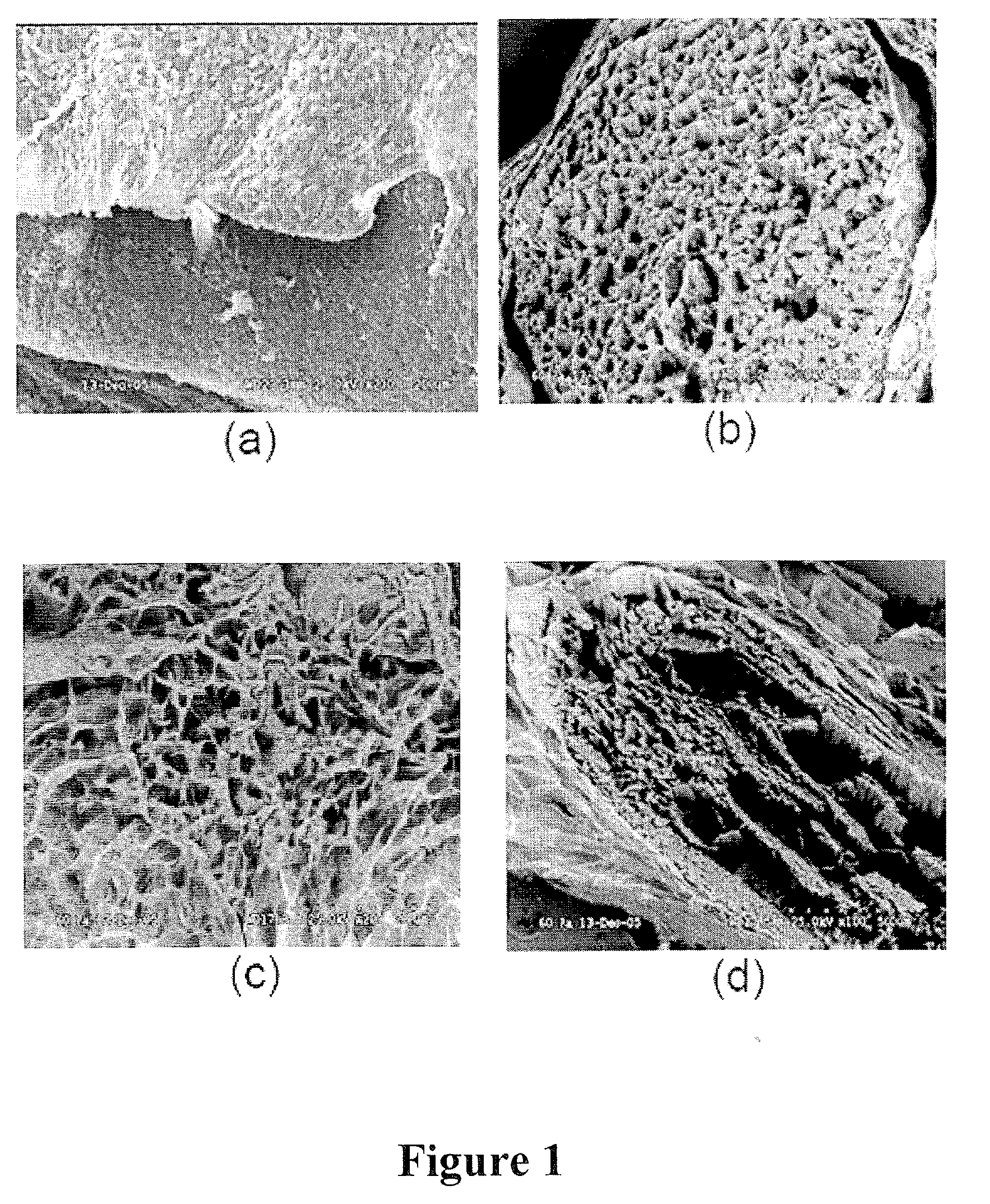
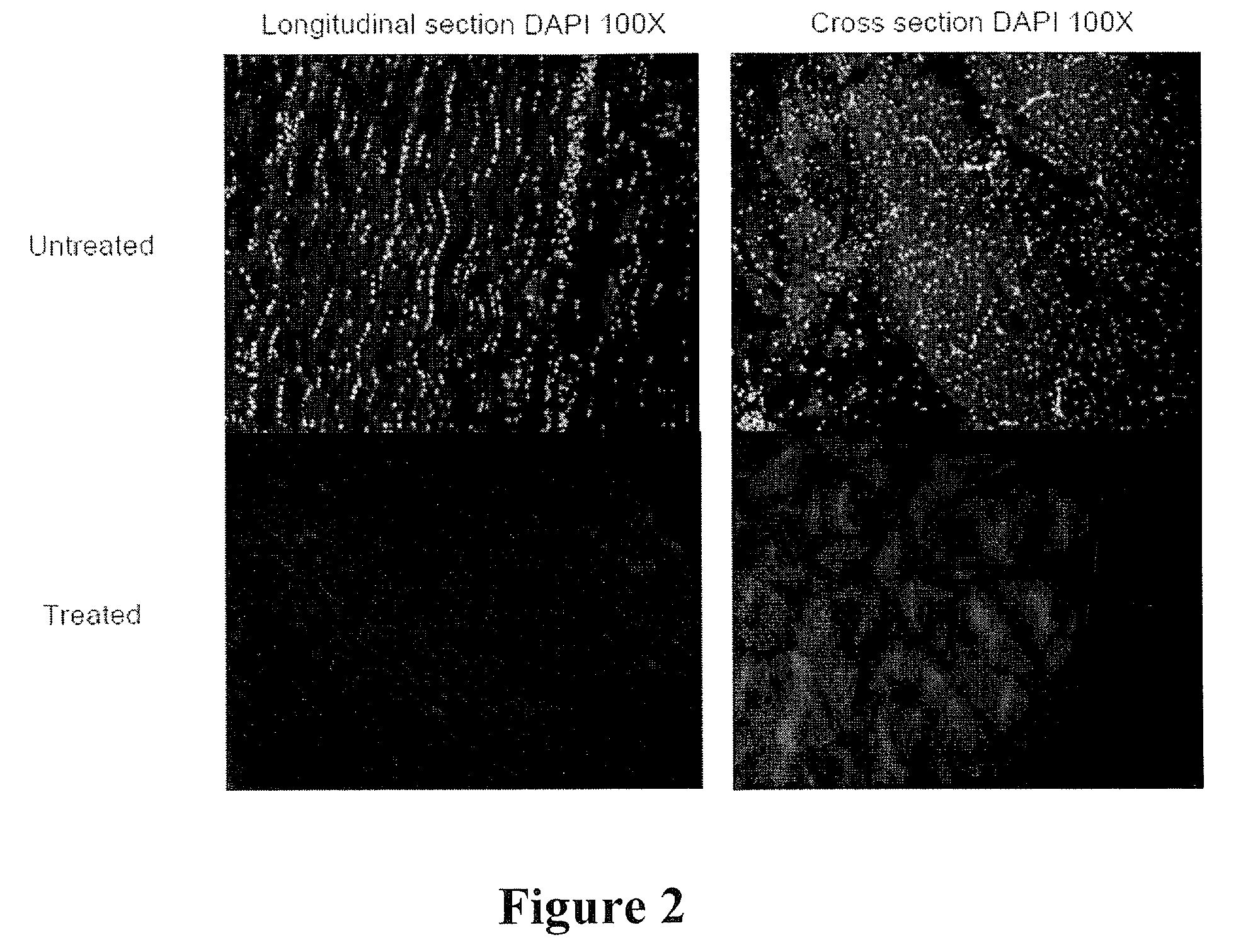
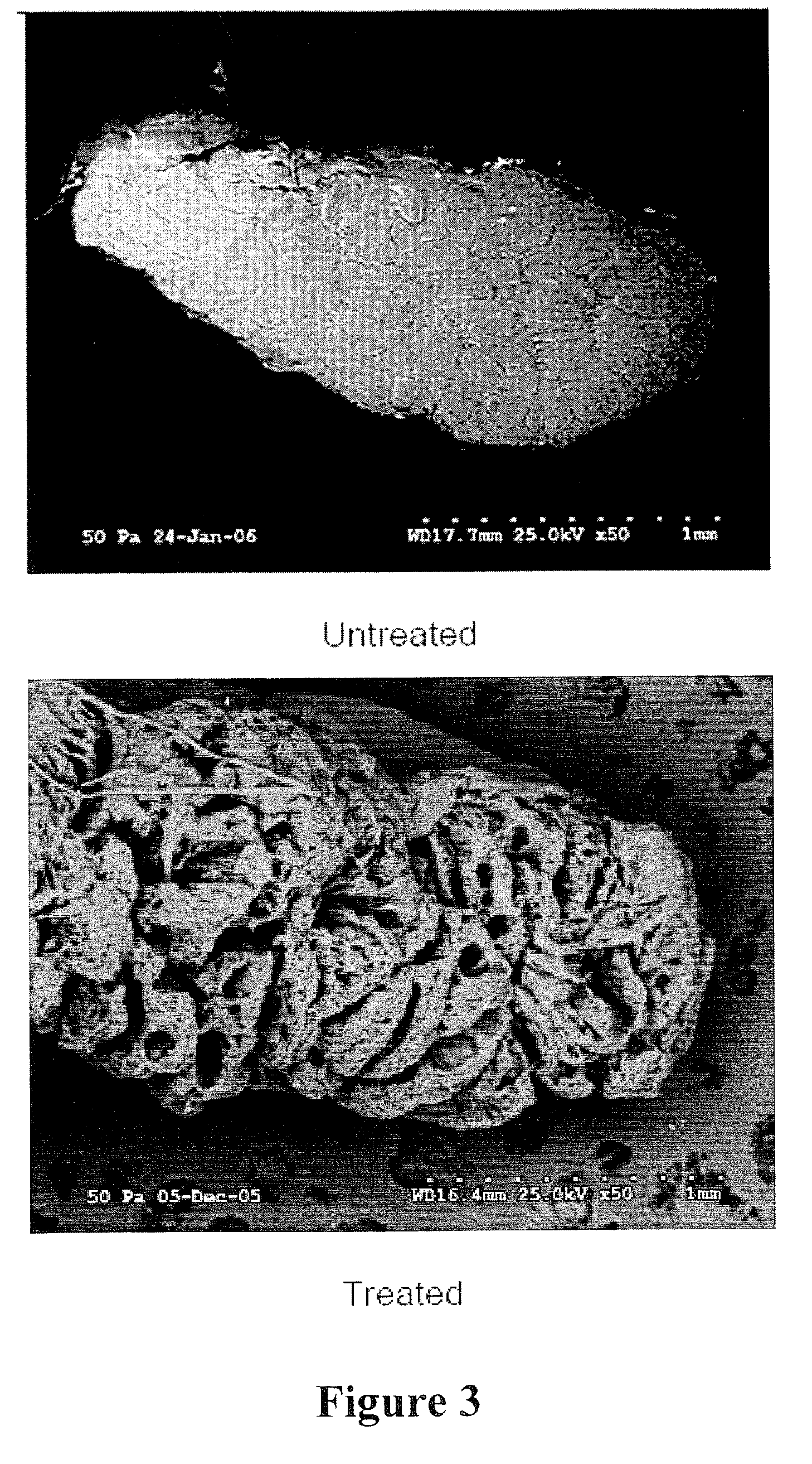
[0084]Sciatic nerves were harvested from white New Zealand rabbits. The tissue was placed in deionized (DI) water and shaken at 4° C. for 48 hours to break open the cells. Cell fragments were removed by extraction at 4° C. with 0.5% Triton X-100 in 0.05% sodium hydroxide solution for 48 hours. Residual reagents were removed by shaking in DI water at 37° C. for 96 hours. Efficient removal of the cellular material was confirmed by histological, microscopic, and residual DNA analyses. To increase the porosity and interconnectivity, the acellular nerves were treated at 37° C. for 24 hours with peracetic acid at concentrations of 1, 5, and 10 w / v %. Residual oxidant was removed by repeated extractions with DI water. Finally, the bioscaffolds were freeze dried prior to analysis by scanning electron microscopy (SEM).

[0085]The SEM results are summarized in FIG. 1. These data demonstrate the efficacy of oxidant treatment on peripheral nerve bioscaffolds. In cross section, the ...

example 2

Tendon Scaffolds

[0086]The feet of 56 day old Leghorn chickens were provided by Wayne Farms (Dobson, N.C.), placed on ice immediately after receipt, and stored at 4° C. until harvest. The feet were cleaned and disinfected using aseptic techniques. The flexor digitorum profundus (FDP) tendon from the long digit was removed and placed into a sterile, 15 ml conical tube (Becton Dickinson, Bedford, Mass.).

[0087]A. Preparation of Decellularized / Oxidized FDP Tendon Scaffolds. Immediately after harvest, FDP tendons were transferred under aseptic conditions from 15 ml conical tubes to individual clean, autoclaved, 100 ml glass, screw-top bottles (Gibco, Grand Island, N.Y.). 100 ml of DNase-free / RNase-free, distilled water (Gibco) was added to each sample. The bottle was placed onto a rotating shaker (Barnstead MaxQ400, Dubuque, Iowa) at 200 rpm, 37° C., for 24 hours. After 24 hours, the water was discarded and the cycle was repeated. At the conclusion of the second cycle, the water was disca...

example 3

Tendon Scaffolds

[0103]Experiments with the human Achilles tendon have produced similar results as those reported in Example 2.

[0104]A. Preparation of Decellularized / Oxidized Achilles-derived Tendon Scaffolds. Freeze-dried human Achilles tendon allografts from multiple donors were provided and stored at 25° C. until use. Freeze-dried human Achilles tendon allografts were transferred under aseptic conditions to individual clean, autoclaved, 1000 ml glass flasks. 1000 ml of DNase-free / RNase-free, distilled water (Gibco) was added to each sample. The flask was placed onto a rotating shaker (Barnstead MaxQ400, Dubuque, Iowa) at 200 rpm, 37° C., for 24 hours. After 24 hours, the water was discarded and the cycle was repeated. At the conclusion of the second cycle, the water was discarded and 500 ml of 0.05% trypsin-EDTA (Gibco) was added. The sample was placed onto the rotating shaker at 200 rpm, 37° C. for 1 hour. At the end of the cycle, the trypsin solution was discarded and 500 ml of ...


PUM


Abstract

Methods are provided for producing a bioscaffold from natural tissues by oxidizing a decellularized tissue to produce a bioscaffold having pores therein. The pore size and porosity is increased to better accommodate intact cells so that live cells can better infiltrate and inhabit the bioscaffold. The bioscaffold may be freeze-dried or lyophilized, sterilized and (optionally) aseptically packaged for subsequent use. A further aspect of the present invention is a bioscaffold produced by the processes described herein. Methods of treatment using the bioscaffold as a graft or as a biomedical implant for implantation are also provided. Also provided are methods of seeding a bioscaffold with mammalian cells, wherein the seeding carried out either in vitro or in vivo, and wherein a bioscaffold produced as described herein is utilized for said seeding.

Description

RELATED APPLICATIONS[0001]This application claims the benefit under 35 U.S.C. § 119(e) of U.S. Provisional Patent Application Ser. No. 60 / 794,037, filed Apr. 21, 2006, the disclosure of which is incorporated herein by reference in its entirety.FIELD OF THE INVENTION[0002]The present invention concerns tissue engineering scaffolds and methods of making the same.BACKGROUND OF THE INVENTION[0003]Bioscaffold engineering has the potential to vastly improve the treatment of tissue injuries, especially those associated with tumor, trauma, and congenital deficiencies where autograft tissue (i.e., tissue harvested from the recipient patient) or allograft tissue (i.e., tissue harvested from a different subject of the same species) might not be available in sufficient quantity for reconstruction. Current tissue-engineering strategies have relied on scaffolds derived from both synthetic (e.g., polyglycolic acid) and naturally-derived (e.g., collagen) materials to form the cell-scaffold construc...

Claims


Application Information

Patent Timeline
IPC IPC(8): A61K35/30A61F13/00A61K35/22A61K35/32A61K35/34A61K35/39A61K35/407A61K35/44
CPCA61K35/22A61K35/30A61K35/32A61K35/34A61K35/39A61K35/407C12N2533/90A61L27/3604A61L27/3687A61L27/38A61L27/56A61L2430/40C12N5/0068A61K35/44
Inventor VAN DYKE, MARK E.CHRIST, GEORGE J.WHITLOCK, PATRICK W.
Owner WAKE FOREST UNIVERSITY
Why Eureka
Try Eureka