Method And System For Vascular Elastography

a vascular elastography and vascular technology, applied in the field of vascular tissue characterization, can solve the problems of plaque rupture and thrombosis, potential limitation of non-invasive characterization of vessel walls with extracorporal ultrasound probes, and difficulty in interpretation of motion parameters

Inactive Publication Date: 2007-12-06
UNIV JOSEPH FOURIER +1
View PDF1 Cites 47 Cited by
  • Summary
  • Abstract
  • Description
  • Claims
  • Application Information

AI Technical Summary

Benefits of technology

[0029] Other objects, advantages and features of the present invention will become more apparent upon reading the following non restrictive description of preferred embodiments thereof, given by way of example only with reference to the accompanying drawings. It is to be noted that the examples presented hereafter were based on the analysis of the ultrasound RF signals. The present invention is not restricted to RF or B-mode signals and may be applied to any new ultrasound modalities providing tissue movements. The RF signals may be seen as the raw data from which all current imaging modalities available on the market were developed.

Problems solved by technology

It is known, for example, that the presence of plaque stiffens the vascular wall, and that the heterogeneity of its composition may lead to plaque rupture and thrombosis.
However, most of the conventional methods in elastography only provide the map of the strain distribution in the direction of the ultrasound beam propagation (axial strain, or radial strain in endovascular elastography).
This can set a potential limitation in non-invasive characterization of vessel walls with an extracorporal ultrasound probe, since the ultrasound beam propagates axially whereas the tissue motion runs radially.
Indeed, it has been shown that motion parameters might be difficult to interpret when tissue motion and the ultrasound beam do not occur in the same orientation.
However, the proposed technology is not restricted to this application and concerns imaging of the mechanical structures of small vessels in humans and small animals such as rats and mice.
However, those experiments were conducted ex vivo and required the sacrifice of the animals.
Atherosclerosis, which is a disease of the intima layer of arteries, remains a major cause of mortality in western countries.
Unfortunately no imaging modality, currently in clinical use, allows the access to these properties.
This makes IVUS, alone, insufficient to predict the plaque mechanical behavior.
However, no further validation of the spectral approach was so far conducted in EVE.
In vivo applications of EVE are subjected to many difficulties.
In such conditions, tissue displacements may be misaligned with the ultrasound beam, introducing substantial decorrelation between the pre- and the post-tissue-compression signals.
Regarding that, 1D estimators may not be optimal if such decorrelation is not appropriately compensated for.
Another potential difficulty, that is associated with EVE in vivo applications, stems from the eventual cyclic catheter movement in the vessel lumen.
Owing to the pulsatile blood flow motion, catheter instability may constitute another source of signal decorrelation between pre- and post-compression signals.

Method used


Image

  • Method And System For Vascular Elastography
  • Method And System For Vascular Elastography
  • Method And System For Vascular Elastography

Examples


first embodiment

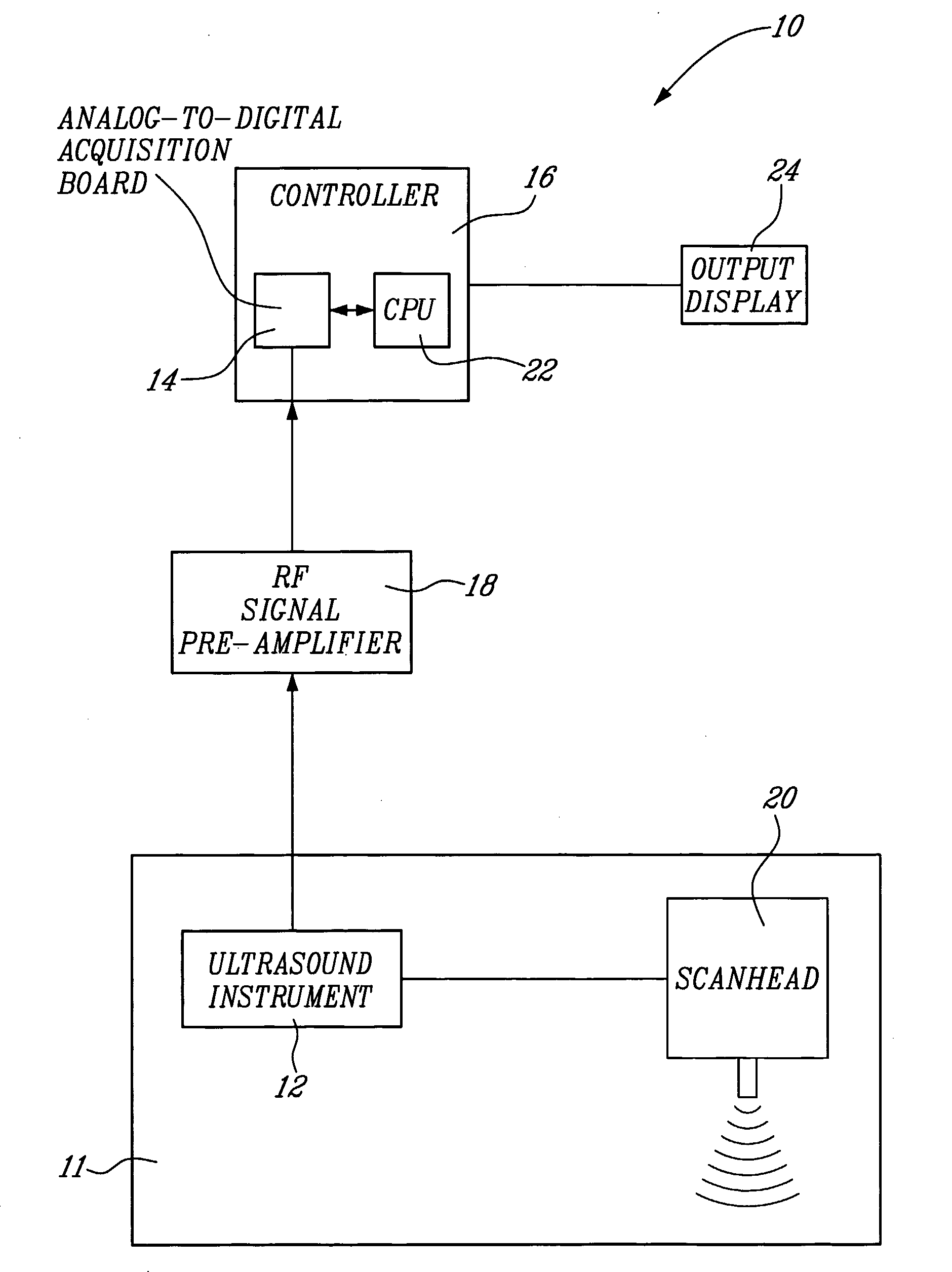
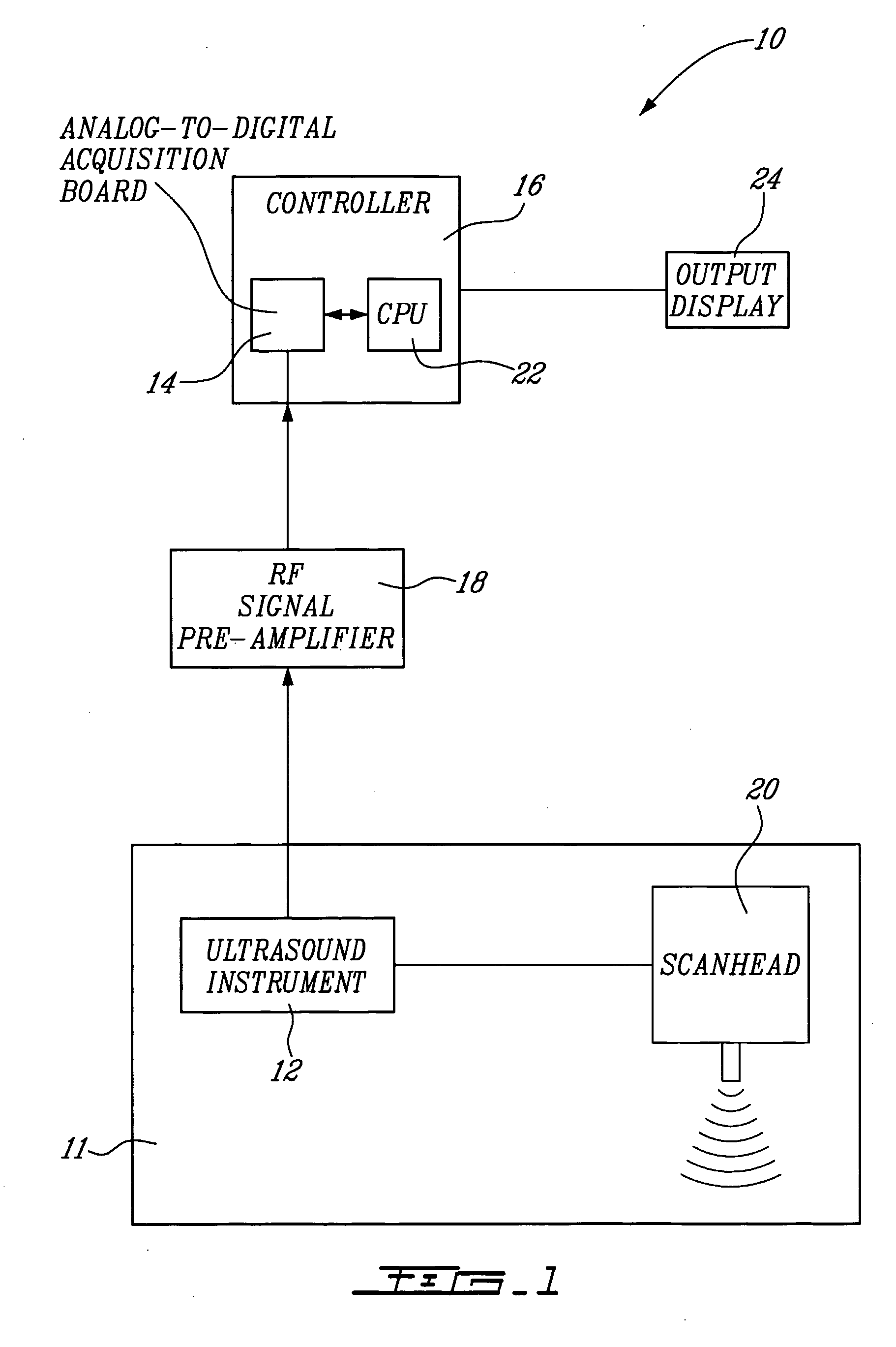
[0057] A system 10 for vascular elastography according to a first aspect of the present invention will now be described with reference to FIG. 1. More specifically, the system 10 allows for non-invasively characterizing arteries. Whereas not restricted to, this system allows predicting risks of vascular tissue rupture due to the presence of atherosclerotic plaques and potentially vascular aneurysms. Since vascular tissue rupture due to atherosclerotic plaques and aneurysms is believed to be well known in the art, it will not be described herein in more detail.

[0058] The system 10 comprises an ultrasound system 11 including an ultrasound instrument 12 provided with a scanhead 20 including an ultrasound transducer. The instrument 12 is coupled to an analog-to-digital acquisition board 14 of a controller 16 via a radio-frequency (RF) pre-amplifier 18.

[0059] For NIVE, the ultrasound instrument 12 is configured for extracorporal measurement, while for MicroNive, it is in the form of an ...

third embodiment

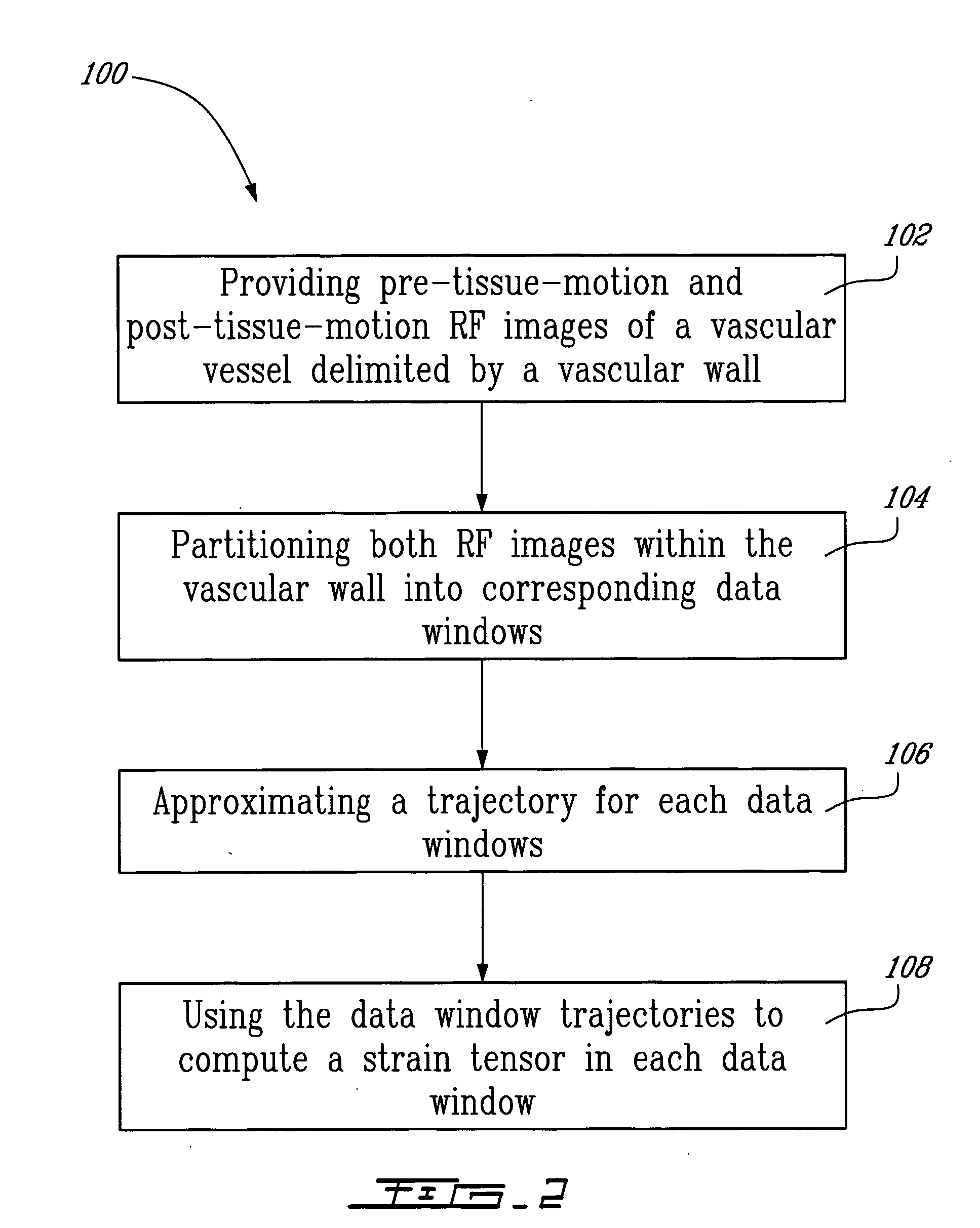
[0131] A method for endovascular elastography (EVE) according to a third illustrative embodiment of the second aspect of the present invention will now be described. Since the method according to this third embodiment is similar to the method illustrated in FIG. 2, and for concision purposes, only the differences between these two methods will be described herein.

[0132] The first step of the method is to acquire intravascular RF images using a catheter. Following the example of IVUS, and as schematically illustrated in FIG. 15, a transducer is placed at the tip of the catheter and cross-sectional imaging of a vessel is generated by sequentially sweeping the ultrasound beam over a 360° angle. It is to be noted that, in the ideal situation illustrated in FIG. 15, the ultrasound beam runs parallel with the vascular tissue motion, i.e. in the (r,φ) coordinate system.

[0133] Mechanical parameters (radial strain, in this case) are then estimated from analyzing the kinematics of the vascul...

second embodiment

[0149] The method for endovascular elastography according to the present invention has also been validated in vitro using a fresh excised human carotid artery. The experimental set-up 50 used in the validation is illustrated in FIG. 23. The set-up 50 includes a system 52 for endovascular elastography according to the first aspect of the present invention.

[0150] The system 52 comprises an ultrasound scanner 54 in the form of a CVIS (ClearView, CardioVascular Imaging System Inc.) ultrasound scanner, working with a 30 MHz mechanical rotating single-element transducer (not shown), a digital oscilloscope 56, more specifically the model 9374L from LECROY, and a pressuring system 58.

[0151] The extremities 60-62 of an artery 64 are fixed to two rigid sheaths by watertight connectors 66, separated according to the original longitudinal dimension of the vessel 64 before excision. The intravascular catheter 68, part of the system 52 was introduced through the proximal sheath into the lumen of...


PUM


Abstract

The method for vascular elastography comprises: i) obtaining a sequence of radio-frequency (RF) images including pre-tissue-motion and post-tissue-motion images in digital form of a vessel delimited by a vascular wall; the pre-tissue-motion and post-tissue-motion images being representative of first and second time-delayed configuration, of the whole vessel; ii) partitioning both the pre-tissue-motion and post-tissue-motion images within the vascular wall into corresponding data windows; approximating a trajectory between the pre- and post-tissue-motion for corresponding data windows; and using the trajectory for each data window to compute the full strain tensor in each data window, which allow determining the Von Mises coefficient. The method can be adapted for non-invasive vascular elastography (NIVE), for non-invasive vascular micro-elastography (MicroNIVE) on small vessels, and for endovascular elastography (EVE).

Description

FIELD OF THE INVENTION [0001] The present invention relates to vascular tissue characterization. More specifically, the present invention is concerned with a method and system for vascular elastography imaging. BACKGROUND OF THE INVENTION [0002] In the early nineties, Ophir et al., (1991) introduced elastography, which is defined as biological tissue elasticity imaging. Primary objectives of elastography were to complement B-mode ultrasound as a screening method to detect hard areas in the breast [Garra et al., 1997]. [0003] Within the last few years, elastography has also found application in vessel wall characterization using endovascular catheters [Brusseau et al., (2001); de Korte et al., (1997-2000b)]. Indeed, changes in vessel wall elasticity may be indicative of vessel pathologies. It is known, for example, that the presence of plaque stiffens the vascular wall, and that the heterogeneity of its composition may lead to plaque rupture and thrombosis. As indicated below, the me...

Claims


Application Information

Patent Timeline
Patent Type & Authority Applications(United States)
IPC IPC(8): A61B8/00A61B8/08G06T7/00G06T7/20
CPCA61B5/0066A61B5/02007A61B8/08A61B8/0891A61B8/485G06T2207/30241G06T7/0012G06T7/20G06T2207/10132G06T2207/30101A61B8/587
Inventor MAURICE, ROCHCLOUTIER, GUYOHAYON, JACQUESSOULEZ, GILLES
Owner UNIV JOSEPH FOURIER
Why Eureka
Try Eureka