Serum-free media and their uses for chondrocyte expansion

a chondrocyte and serum-free technology, applied in the field of cell and tissue culture, can solve the problems of difficult control, limited amount of starting cell material available for autologous chondrocyte implantation, and physical debilitation, and achieve the effects of promoting cell proliferation, enhancing cell attachment and proliferation, and maintaining the capacity of chondrocytes

Inactive Publication Date: 2007-12-20
GENZYME CORP
View PDF10 Cites 19 Cited by
  • Summary
  • Abstract
  • Description
  • Claims
  • Application Information

AI Technical Summary

Benefits of technology

[0012] Among other advantages, the invention allows one to avoid the use of serum in chondrocyte cultures, enhance cell attachment and proliferation under serum-free conditions, and / or to maintain the capacity of chondrocytes to re-express cartilage-specific phenotype. In one aspect, the invention provides DM that is sufficient for the initial attachment of cells to a culture substratum, thereby eliminating a need for a serum-containing medium in the initial stage of cell culture. Another aspect of the invention provides defined serum-free cell culture media that promote proliferation of cells such as chondrocytes without use of serum at any stage during cell culture. Yet another aspect of the invention provides cell culture media that may be used to prime chondrocytes prior to implantation into a subject or included as a redifferentiation-sustaining medium to chondrocytes embedded in a matrix intended for implantation into cartilage defects. Another aspect of the invention provides a method of culturing a chondrocyte to a state that is suitable for treating a patient suffering from a cartilage defect. Additional advantages of the invention will be set forth in part in the description which follows, and in part will be obvious from the description, or may be learned by practice of the invention.

Problems solved by technology

Damage to cartilage produced by trauma or disease, e.g., rheumatoid and osteoarthritis, can lead to serious physical debilitation.
However, even though serum is widely used for mammalian cell culture, there are several problems associated with its use: (1) serum contains many unidentified or non-quantified components and therefore is not “defined;” (2) the composition of serum varies from lot to lot, making standardization difficult for experimentation or other uses of cell culture; (3) many of the serum components affect cell attachment, proliferation, and differentiation making it difficult to control these parameters; (4) some components of serum are inhibitory to the proliferation of specific cell types and to some degree may counteract its proliferative effect, resulting in sub-optimal growth; and (5) serum may contain viruses and other pathogens which may affect the outcome of experiments or provide a potential health hazard if the cultured cells are intended for implantation in humans.
The amounts of starting cell material available for autologous chondrocyte implantation are generally limited.
Attempts to culture articular chondrocytes at subconfluent densities in DM have been only partially successful.

Method used


Image

  • Serum-free media and their uses for chondrocyte expansion
  • Serum-free media and their uses for chondrocyte expansion
  • Serum-free media and their uses for chondrocyte expansion

Examples


example 1

IL-6 Increases Cell Yield and Proliferation of Primary Human Chondrocytes

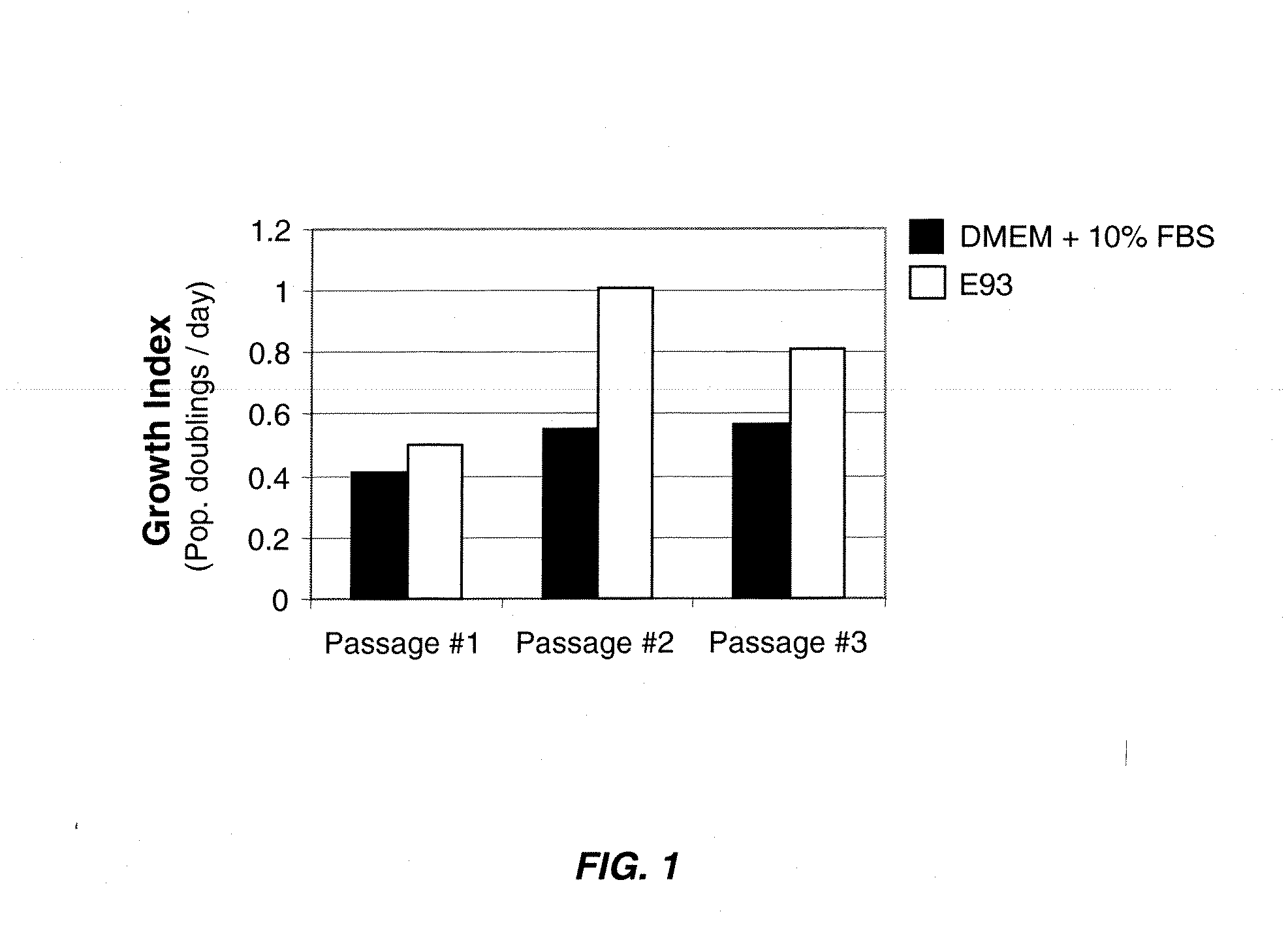
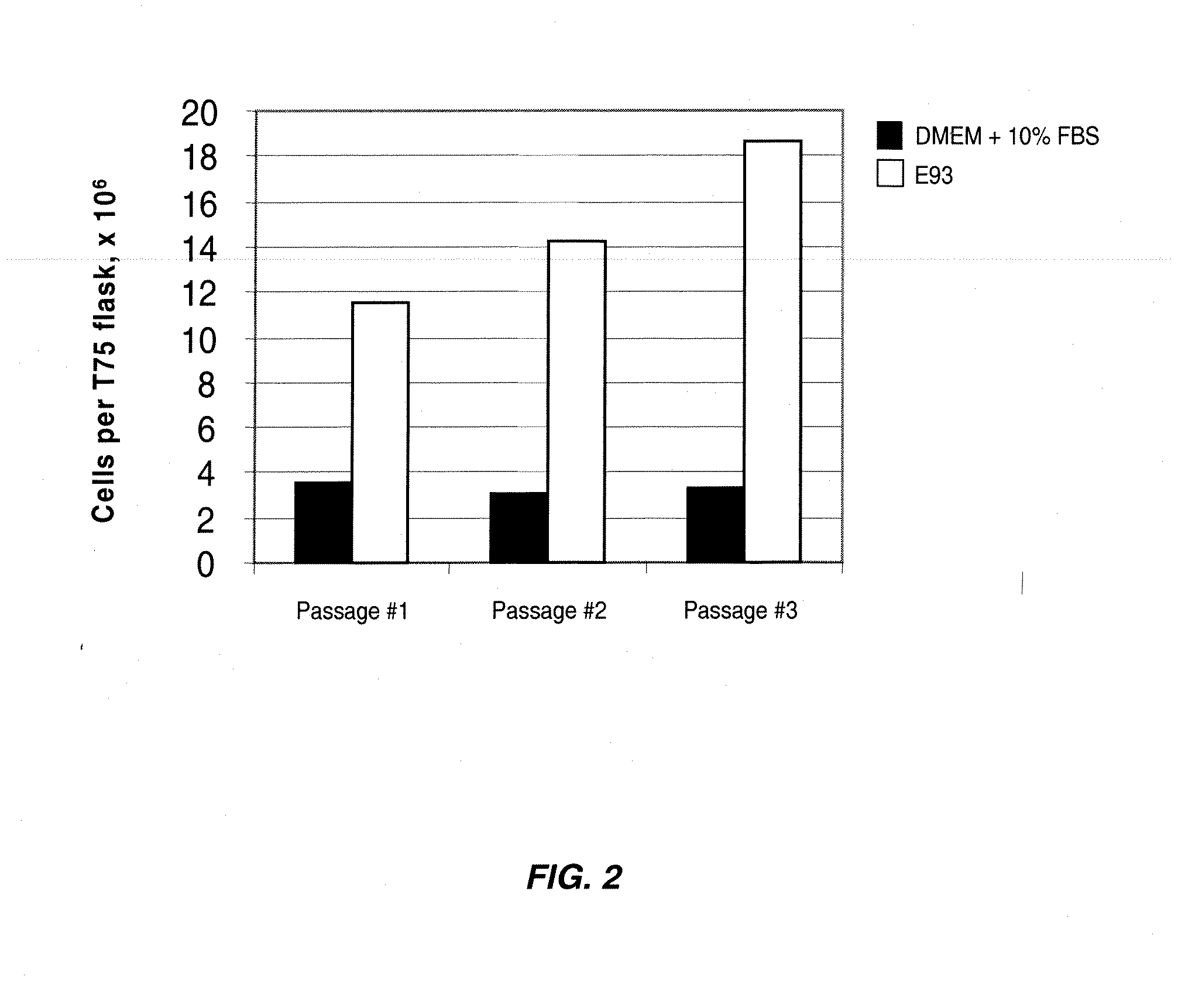
[0084] Primary human chondrocytes were isolated from biopsies of articular cartilage by mincing of the sample followed by enzymatic digestion with 0.25% protease type XIV (Streptomyces griseus) for one hour and then 0.1% collagenase overnight at 37° C. Cells were recovered by centrifugation for five minutes at 1,000×g and resuspended in the appropriate test medium. Cells grown in DMEM+10% FBS were plated at a density of 3,000 cells per cm2 and cells grown in serum-free medium were plated at a density of 5,000 cells per cm2. T75 flasks were used for all experiments. The following media were tested: [0085] 1) DMEM+10% FBS [0086] 2) cDRF / P / L as defined in Table 8 [0087] 3) cDRF / P / L as defined in Table 8, supplemented with 0.2 ng / ml IL-6 [0088] 4) cDRF / P / L as defined in Table 8, supplemented with 1.0 ng / ml IL-6

[0089] Cells were passaged upon reaching 50% to 80% confluence. Cells grown in DMEM+10% FBS were rinsed ...

example 2

OSM Increases Cell Yield and Proliferation of Primary Human Chondrocytes

[0092] Primary human chondrocytes were isolated from biopsies of articular cartilage by mincing of the sample followed by enzymatic digestion with 0.25% protease type XIV (Streptomyces griseus) for one hour and then 0.1% collagenase overnight at 37° C. Cells were recovered by centrifugation for five minutes at 1,000×g and resuspended in the appropriate test medium. Cells grown in DMEM+10% FBS were plated at a density of 3,000 cells per cm2 and cells grown in serum-free medium were plated at a density of 5,000 cells per cm2. T75 flasks were used for all experiments. The following media were tested: [0093] 1) DMEM+10% FBS [0094] 2) cDRF / P / L as defined in Table 8 [0095] 3) cDRF / P / L as defined in Table 8, supplemented with 0.1 ng / ml OSM [0096] 4) cDRF / P / L as defined in Table 8, supplemented with 0.5 ng / ml OSM [0097] 5) cDRF / P / L as defined in Table 8, supplemented with 1.0 ng / ml OSM

[0098] Cells were passaged upon r...

example 3

LIF Increases Cell Yield and Proliferation of Primary Human Chondrocytes

[0100] Primary human chondrocytes were isolated from biopsies of articular cartilage by mincing of the sample followed by enzymatic digestion with 0.25% protease type XIV (Streptomyces griseus) for one hour and then 0.1% collagenase overnight at 37° C. Cells were recovered by centrifugation for five minutes at 1,000×g and resuspended in the appropriate test medium. Cells grown in DMEM+10% FBS were plated at a density of 3,000 cells per cm2 and cells grown in serum-free medium were plated at a density of 5,000 cells per cm2. T75 flasks were used for all experiments. The following media were tested: [0101] 1) DMEM+10% FBS [0102] 2) cDRF / P / L as defined in Table 8 [0103] 3) cDRF / P / L as defined in Table 8, supplemented with 0.1 ng / ml LIF [0104] 4) cDRF / P / L as defined in Table 8, supplemented with 0.5 ng / ml LIF [0105] 5) cDRF / P / L as defined in Table 8, supplemented with 2.0 ng / ml LIF

[0106] Cells were passaged upon r...


PUM

PropertyMeasurementUnit
concentrationaaaaaaaaaa
molecular weightaaaaaaaaaa
concentrationaaaaaaaaaa

Abstract

The present invention provides defined serum-free cell culture media useful in culturing fibroblasts, especially articular chondrocytes, that avoid problems inherent in the use of serum-containing media. The defined media comprise platelet-derived growth factor (PDGF), chemically defined lipids, oncostatin M (OSM), interleukin-6 (IL-6), leukemia inhibitory factor (LIF), or combinations of these compounds. In another aspect, the present invention also provides tissue culture methods that comprise incubating chondrocytes in the defined serum-free media. The methods enhance attachment and proliferative expansion of chondrocytes seeded at low density while maintaining their redifferentiation potential.

Description

[0001] This application claims priority to U.S. application No. 60 / 805,307, filed on Jun. 20, 2006, which is incorporated herein by reference in its entirety.FIELD OF THE INVENTION [0002] The present invention relates to the field of cell and tissue culture. More specifically, the invention relates to methods and compositions for ex vivo propagation of cells capable of forming cartilaginous tissue intended for treatment or repair of cartilage defects. BACKGROUND OF THE INVENTION [0003] Articular cartilage is composed of chondrocytes encased within the complex extracellular matrix produced by those chondrocytes. The unique biochemical composition of this matrix provides for the smooth, nearly frictionless motion of articulating surfaces of the joints. With age, tensile properties of human articular cartilage change as a result of biochemical changes. After the third decade of life, the tensile strength of articular cartilage decreases markedly. Damage to cartilage produced by trauma ...

Claims


Application Information

Patent Timeline
Patent Type & Authority Applications(United States)
IPC IPC(8): C12N5/08C12N5/02C12N5/077
CPCC12N2501/135C12N2501/2306C12N2501/39C12N2501/237C12N2501/235C12N2500/36C12N2501/58C12N2501/115C12N5/0655A61K35/32C12N2533/74A61P19/00A61P19/02
Inventor DUGUAY, STEPHENSEYMOUR, BARBARA
Owner GENZYME CORP
Why Eureka
Try Eureka