

Gene Therapy for Skin Disorders Using Needleless Syringes
a technology of skin disorders and needleless syringes, which is applied in the field of skin disorders treating methods, can solve the problems of not responding, in shape or appearance, signs of healing, and none of these agents are satisfactory in terms of effectiveness, safety, prevention, etc., and achieve the effect of avoiding pain and the risk of infection
- Summary
- Abstract
- Description
- Claims
- Application Information
AI Technical Summary
Benefits of technology
Problems solved by technology
Method used
Image

Examples
example 1
Verification of Introduction Sites
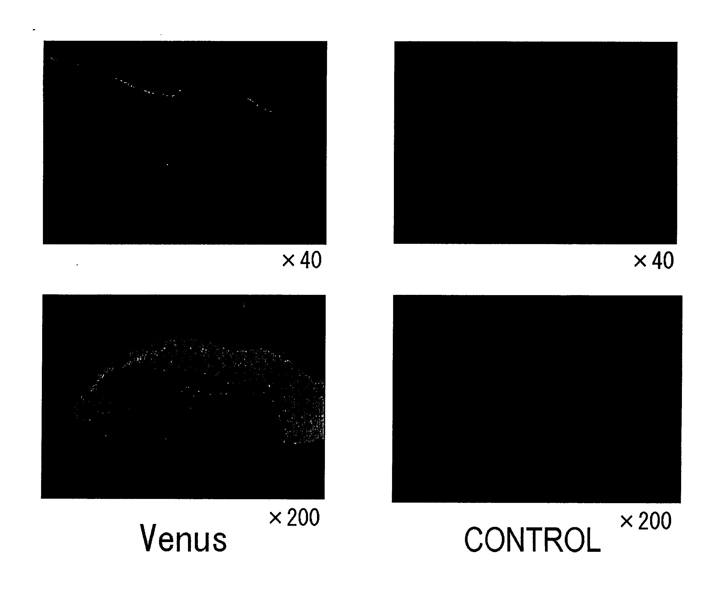
Verification of sites introduced with Yellow Fluorescence Protein (Venus) plasmid (Venus / pCS2), by non-needle injection into rat skin (see FIG. 1).
Methods:
[0035] (1) The dorsal skin of rats was shaved and then depilated using Kanebo epilat hair remover cream. Then 100 μg / 100 μl of Yellow Fluorescence Protein (Venus) / pCS2 was injected using ShimaJET. For comparison, plasmid (pCS2) without Venus was used as a control.
(2) The rats were sacrificed after 24 hours, and the skin at the injection site was collected.
(3) The tissues were placed into OCT compound and frozen rapidly in liquid nitrogen, then sectioned and observed under a fluorescence microscope.
Results:
Expression of Yellow Fluorescence Protein (Venus) was detected only in epidermal tissues.
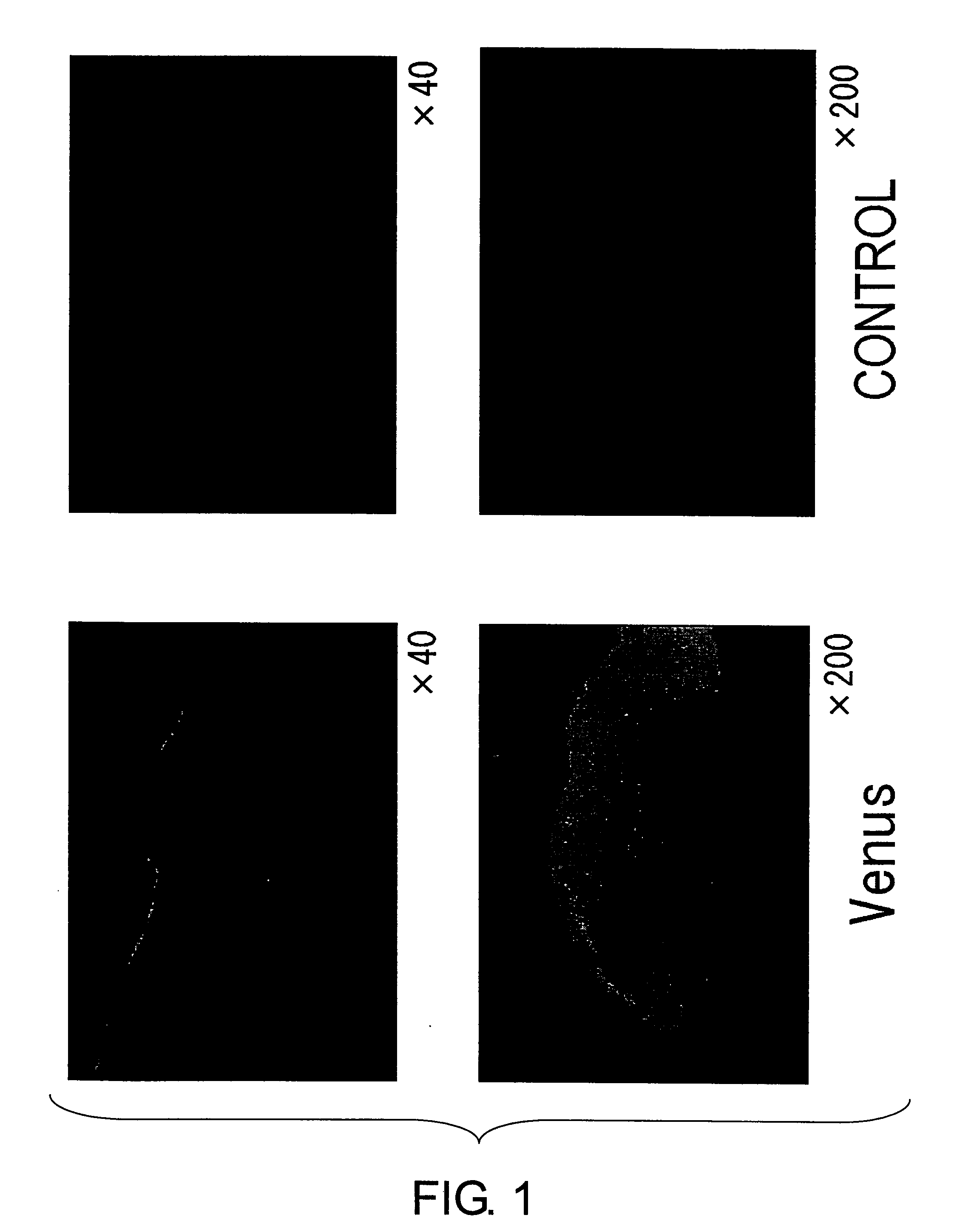
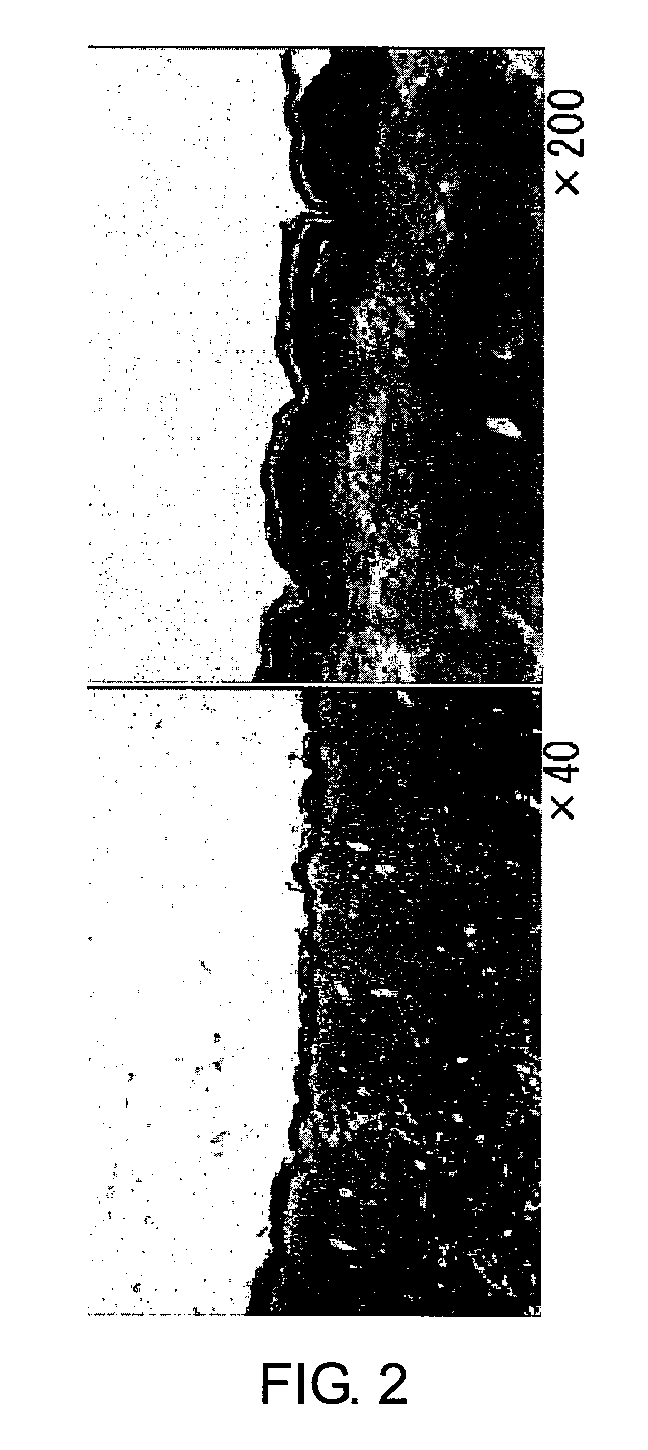
Verification of sites introduced with lacZ plasmid (pcLacZ) by non-needle injection into rat skin (see FIG. 2).
Methods:
(1) The dorsal skin of rats was shaved and depilated. Then, 200 μg / 200...
example 2
Evaluation of Introduction Efficiency
Luciferase activity assay (see FIG. 3)
Method:
(1) The dorsal skin of rats was shaved and depilated. Then, 50 μg / 50 μl or 100 μg / 100 μl of luciferase plasmid (pGL3 luc) was injected into the rat's skin using ShimaJET.
For comparison, rats were intradermally needle-injected with an equal amount of the plasmid using a 26G syringe.
(2) The rats were sacrificed after 24 hours, and about two square centimeters of the skin centered on the injection site was collected. As a control, skin was collected from untreated rats.
(3) 1 ml of Luciferase Lysis Buffer (Promega) was added, and the skin was cut with scissors into the smallest pieces as possible.
(4) The skin was frozen rapidly at −80° C. for ten minutes, and then thawed at room temperature. This treatment was repeated twice.
(5) After centrifugation at 5000 rpm for ten minutes, the supernatant was collected.
(6) Luciferase activity was assayed (Berthold LB9507).
[0037]FIG. 3 shows total lu...
example 3
Assessment of Wound Healing Effect
Preparation of a Rat Model of Impaired Wound Healing
[0038] A rat model of impaired wound healing was prepared by administering water-soluble prednisolone (prednisolone sodium succinate: Pz, Shionogi & CO., LTD) as a steroid to rats. Steroid administration to the steroid-administration rat model causes disorders in wound tensile strength, epithelialization, angiogenesis, wound contraction, and so on, resulting in retardation of wound healing.
[0039] (1) 7-week-old male Wister rats were needle-injected intramuscularly with water-soluble prednisolone (Prednisolone Sodium Succinate, Shionogi & CO., LTD) at a dose of 30 mg / kg. After three days (on the day of wound creation), water-soluble prednisolone was again needle-injected intramuscularly at a dose of 30 mg / kg. The control group was needle-injected intramuscularly with PBS.
[0040] (2) After shaving and depilating the dorsal skin of rats, wounds were created by removing entire layers of skin in a c...
PUM
| Property | Measurement | Unit |
|---|---|---|
| temperature | aaaaa | aaaaa |
| area | aaaaa | aaaaa |
| volume | aaaaa | aaaaa |
Abstract
Description
Claims
Application Information