

Nerve regeneration employing keratin biomaterials
a biomaterial and nerve regeneration technology, applied in the direction of prosthesis, peptide/protein ingredients, peptide sources, etc., can solve the problems of limited graft availability, poor regeneration of motor nerves through sensory nerve grafts, and grafting remains a technically demanding procedur
- Summary
- Abstract
- Description
- Claims
- Application Information
AI Technical Summary
Benefits of technology
Problems solved by technology
Method used
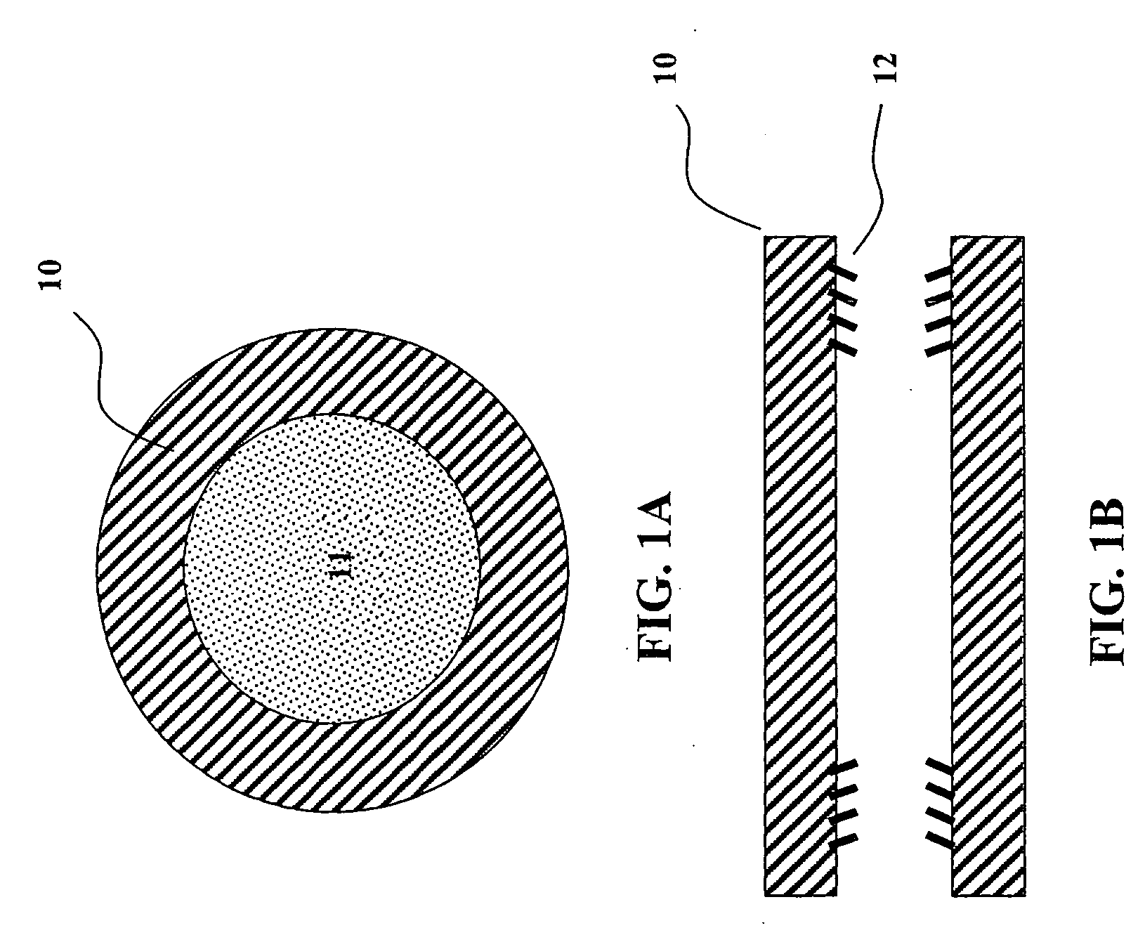
Image

Examples
example 1
Keratin Preparation
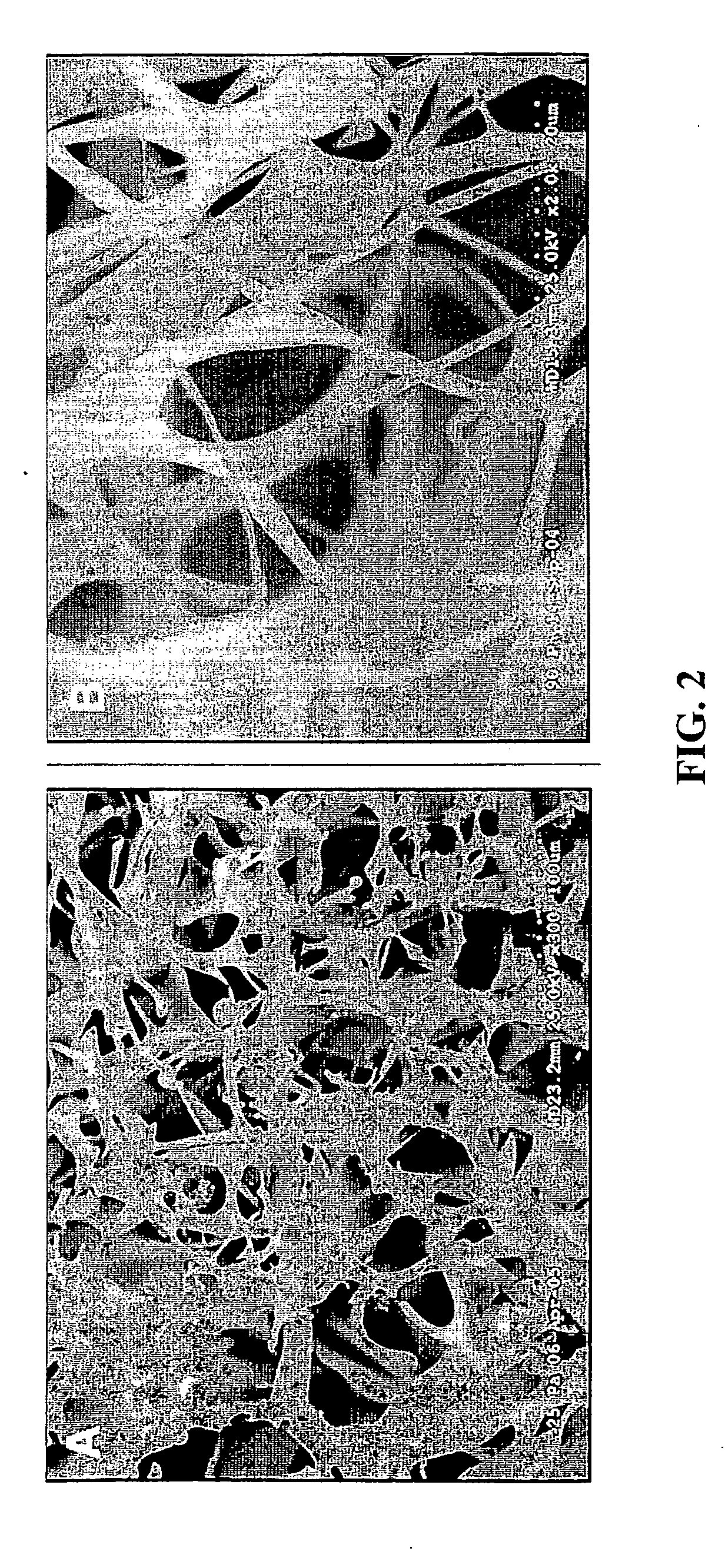
[0067] Keratins were extracted from human hair. 25 g of clean dry hair was oxidized with a 2 w / v % solution of peracetic solution at a liquid:solid ratio of 40:1. The oxidation was carried out at 37° C. with constant gentle stirring for 12 hours. After oxidation, the hair was recovered by sieve and rinsed with copious amounts of deionized (DI) water. The oxidized hair was extracted with 1 L of 100 mM tris base at 37° C. with gentle stirring for 12 hours. The hair was recovered by sieve and extracted again with 1 L of DI water at 37° C. with gentle stirring for 12 hours. Concurrently, the tris base solution was neutralized to pH 7.4 by dropwise addition of hydrochloric acid and refrigerated. The hair from the DI water extraction was separated by sieve and discarded. The liquid was recovered, neutralized to pH 7.4, and combined with the first extract. The extract solution was then centrifuged, filtered, and dialyzed against DI water with a dialysis cartridge having...
example 2
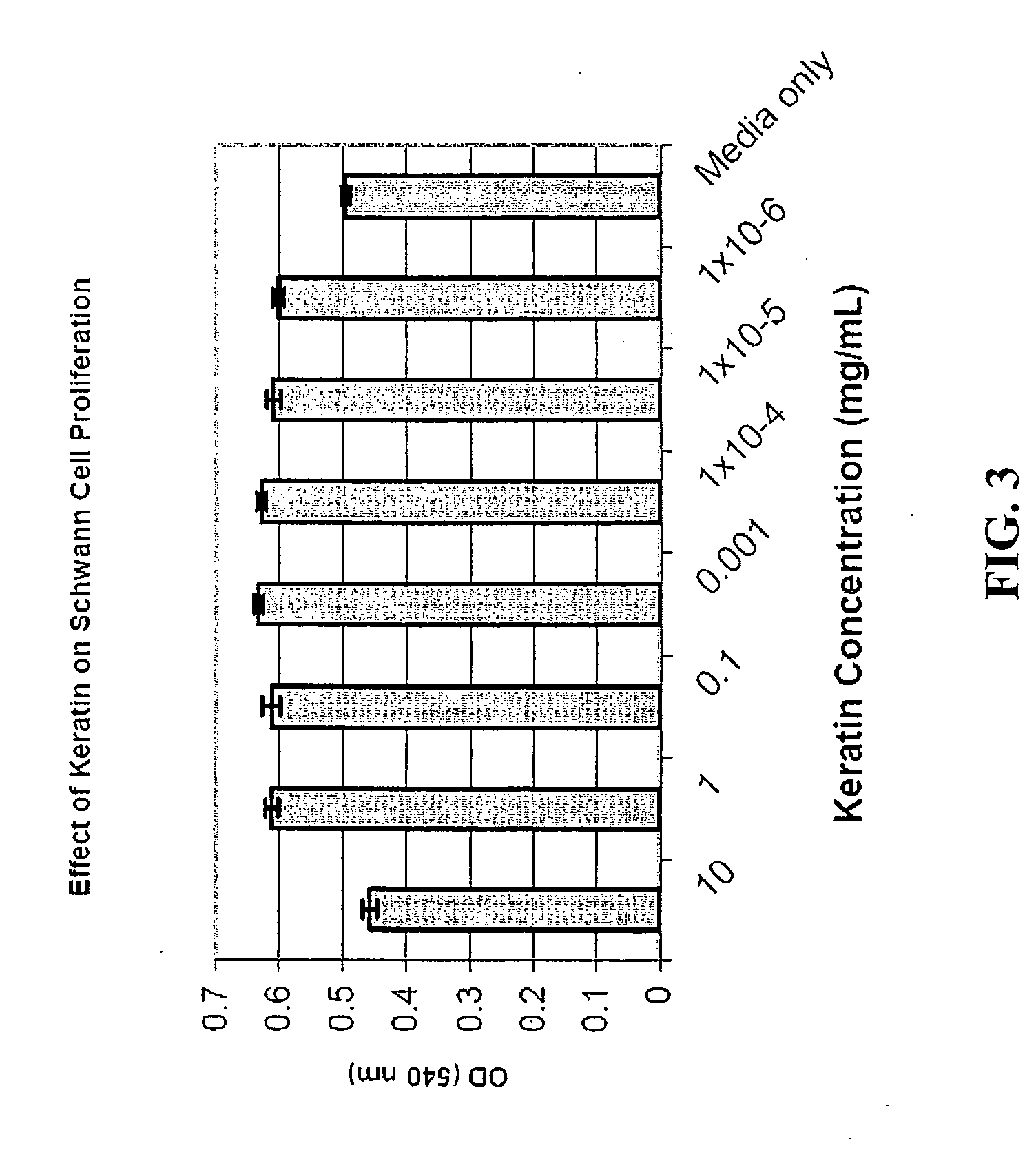
[0068] The ability of keratin to accelerate nerve regeneration, enhance functional recovery, and increase gap bridging capabilities was evaluated. This was accomplished by comparing the functional recovery (electrophysiology and muscle force generation) and structure (histologically) of a transected peripheral nerve repaired with an empty nerve conduit with a peripheral nerve repaired with a conduit filled with a keratin hydrogel matrix.
[0069] Eleven adult male Swiss Webster mice (20 g) were used in this study. Each animal was randomly placed into Group 1 (n=6) empty nerve conduit or Group 2 (n=5) keratin filled nerve conduit. The nerves from all animals were harvested at 6 weeks.
[0070] Before surgery, each animal was anesthetized with isoflurane (1-1.5 vol %), and the operative site was shaved and cleansed with Betadine® antiseptic. All surgical procedures were performed using aseptic technique. A 1.5 cm incision was made on the dorsum of the left thigh and the sciat...
example 3
Placement of Nerve Conduit & Tissue Engineering Matrix
[0071] Fixation of the nerve conduit began with the introduction of the suture needle into the conduit lumen 1.5 mm from the distal end. The suture was then threaded through the lumen until approximately 4 mm of the suture tail remained outside of the conduit wall. The suture needle was then placed into the end of the distal nerve segment and, while running parallel to the axon fibers, driven 1 mm into and out of the nerve, securing the epineurium and perineurium. Rotating the nerve fiber 120°, the suture needle was reintroduced into the distal nerve and while running parallel to the axon fibers, driven 1 mm back through the perineurium and out the distal nerve end. The needle was then reintroduced into the lumen of the nerve conduit, exiting the conduit wall 1.5 mm from the distal end, near the suture tail. The distal nerve end was then pulled 1.5 mm into the conduit by the suture ends, and the suture was tied with a surgeon's ...
PUM
| Property | Measurement | Unit |
|---|---|---|
| concentration | aaaaa | aaaaa |
| concentration | aaaaa | aaaaa |
| diameter | aaaaa | aaaaa |
Abstract
Description
Claims
Application Information