A method for enzymatically and efficiently preparing corneal acellular matrix tissue engineering scaffold

A technology of tissue engineering scaffold and acellular matrix, which is applied in tissue regeneration, medical science, prosthesis, etc., can solve the problems of destroying telopeptides, low toughness and tension, and difficulty in shaping, so as to achieve good biocompatibility and promote reconstruction , the effect of good biological function

Active Publication Date: 2020-05-19
SHANDONG UNIV
View PDF2 Cites 0 Cited by
  • Summary
  • Abstract
  • Description
  • Claims
  • Application Information

AI Technical Summary

Problems solved by technology

However, trypsin destroys the telopeptide of collagen, and cannot completely remove the genetic material released after degradation during decellularization or acellularization, and sometimes it is difficult to avoid possible immune rejection
In addition, the corneal decellularized matrix mainly composed of collagen has poor mechanical properties, low toughness and tension, rapid degradation and metabolism in the body, and is difficult to shape under water-containing conditions, which is not conducive to the reconstruction of human organs

Method used


Image

  • A method for enzymatically and efficiently preparing corneal acellular matrix tissue engineering scaffold
  • A method for enzymatically and efficiently preparing corneal acellular matrix tissue engineering scaffold
  • A method for enzymatically and efficiently preparing corneal acellular matrix tissue engineering scaffold

Examples


Embodiment 1

[0046] Embodiment 1: Extraction and purification of Myroilysin

[0047] 1. the preparation method of elastase Myroilysin specifically comprises the steps:

[0048] Preparation of crude enzyme solution of Myroilysin

[0049] According to the method established in the laboratory, the deep-sea psychrotrophic bacteria Myroides profundi D25 was inoculated in the liquid seed medium, and cultured with shaking at 15°C for 2 days to activate the strain.

[0050] The bacteria species activated in the above steps were inoculated in the liquid fermentation medium at an inoculum size of 1% (v / v), and cultured with shaking at 15° C. and 180 rpm for 72 hours to obtain a fermentation broth.

[0051] According to the method already established in the laboratory, the fermented broth prepared above was subjected to ammonium sulfate precipitation, resuspension, dialysis, and DEAE anion exchange to prepare Myroilysin crude enzyme liquid.

[0052] 2. Preparation of Myroilysin Concentrated Enzyme ...

Embodiment 2

[0054] Example 2: Preparation method of corneal decellularized scaffold

[0055] The preparation method of porcine corneal decellularized scaffold specifically comprises the following steps:

[0056] Take fresh pig eyeballs, and fix the pig eyeballs on the chopping board, with the pig cornea facing straight up. Use a negative pressure trephine to make a circular incision with a depth of about 200 μm in the center of the pig cornea, peel off the corneal lamellar layer with a tunnel knife and a blade, and place it in normal saline;

[0057] The concentrated Myroilysin solution was diluted to 200 μg / ml with 50 mM Tris-HCl (pH 9.0) with a glycerol content of 20% of the total volume. Place the corneal lamina in the Myroilysin solution, and treat it at 37°C for 16 hours with a rotation speed of 80rpm to prepare porcine corneal decellularized scaffolds;

[0058] Porcine corneal decellularized scaffolds were rinsed with distilled water, and then washed with physiological saline for ...

Embodiment 3

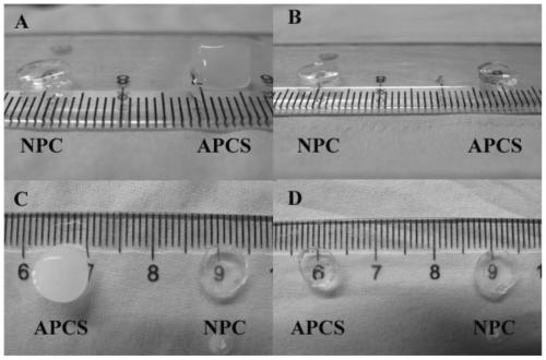
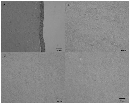
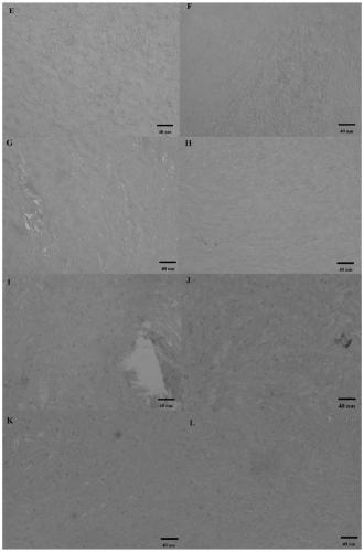
[0062] Example 3: Physicochemical and Physiological Properties Detection of Acellular Corneal Scaffold

[0063] 1. Transparency detection of porcine corneal decellularized scaffolds

[0064] Place porcine corneal decellularized scaffolds and natural porcine corneas on a 96-well plate, and detect the transparency changes of porcine corneal decellularized scaffolds in the wavelength range of 300-800nm ​​at an interval of 10nm;

[0065] 2. Stability testing of porcine corneal decellularized scaffolds

[0066] The buffer for type Ⅰ collagenase contains 5 mM Ca 2+ Dissolve 500 μg / ml type Ⅰ collagenase in the type Ⅰ collagenase solution in PBS buffer solution to obtain type Ⅰ collagenase solution.

[0067] The porcine corneal decellularized scaffolds and natural porcine cornea were soaked in type Ⅰ collagenase solution and type Ⅰ collagenase buffer respectively, and the residual mass of porcine corneal decellularized scaffolds and natural porcine cornea was measured with a microba...


PUM

PropertyMeasurementUnit
concentrationaaaaaaaaaa

Abstract

A method for effectively preparing a cornea acellular matrix tissue engineering scaffold by enzymatic method is as follows: (1) a porcine corneal matrix plate layer is put into diluted Myroilysin for decellularization, and flushed respectively with normal saline and distilled water to obtain a porcine corneal acellular scaffold board layer; (2) the porcine corneal acellular scaffold is put into a drill floor, a riboflavin solution is added dropwise, and the porcine corneal acellular scaffold is irradiated by an UV lamp, and then flushed respectively with the normal saline and the distilled water to obtain a riboflavin-crosslinked porcine corneal acellular scaffold; and (3) the riboflavin-crosslinked porcine corneal acellular scaffold obtained by the step (2) is dehydrated, sterilized and stored at 4 DEG C for standby use. The prepared acellular scaffold still retains enough mechanical strength, certain pores are formed, and after dehydration, the acellular scaffold has high transparency and is favorable for cell growth and migration.

Description

technical field [0001] The invention relates to a method for efficiently preparing corneal acellular matrix tissue engineering scaffold by enzymatic method, which belongs to the field of biotechnology. Background technique [0002] The blinding rate of corneal diseases occupies the second place in ophthalmic diseases, and there are millions of corneal blind patients in my country every year. The early treatment of corneal disease is mainly symptomatic treatment according to the etiology. In the late stage, due to the scars and ulcers in the cornea, surgery is required to replace the diseased cornea, that is, keratoplasty. Keratoplasty is the most important method for the treatment of corneal blindness. There are millions of corneal blind patients in our country every year, and most of them can be lifted out of blindness through keratoplasty. The vast majority of donor corneas for keratoplasty are human corneas, so the number is extremely limited. In the end, there are only...

Claims


Application Information

Patent Timeline
Patent Type & Authority Patents(China)
IPC IPC(8): A61L27/36A61L27/50
CPCA61L27/3633A61L27/3641A61L27/3687A61L27/3691A61L27/50A61L2430/16A61L2430/40
Inventor 张玉忠李悦陈秀兰石梅赵芳孙鹤敏宋晓妍
Owner SHANDONG UNIV
Why Eureka
Try Eureka