
Method for preparing cartilage tissue engineering scaffold from human umbilical cord Wharton jelly and cartilage tissue engineering scaffold

A cartilage tissue, Wharton's glue technology, applied in tissue regeneration, prosthesis, medical science, etc., can solve the problems of stem cell differentiation ability is inferior to type II collagen, unfavorable nutrients and waste transport, small pore size and porosity, etc. Excellent scaffold properties, loose pore arrangement, and easy absorption

Pending Publication Date: 2021-10-01
XIANGYA HOSPITAL CENT SOUTH UNIV
View PDF12 Cites 0 Cited by
  • Summary
  • Abstract
  • Description
  • Claims
  • Application Information

AI Technical Summary

Problems solved by technology

At present, the decellularized matrix materials in the field of cartilage tissue engineering have different functions due to different sources, but there are some problems. For example, the decellularized scaffolds of articular cartilage have biomimetic cartilage extracellular matrix composition and proteins, which are considered to be ideal for tissue engineering cartilage. However, it was found that the pore size and porosity of the decellularized scaffolds of articular cartilage are often too small, which makes it difficult to plant cells and is not conducive to the transport of nutrients and waste; the dermal decellularized matrix material is mainly type I collagen, which induces stem cells The ability to differentiate into chondrocytes is inferior to that of type II collagen
In addition, such materials are often obtained by decellularization and deimmunization of donated cadaveric tissue, and the source of materials is very limited

Method used


Image

  • Method for preparing cartilage tissue engineering scaffold from human umbilical cord Wharton jelly and cartilage tissue engineering scaffold
  • Method for preparing cartilage tissue engineering scaffold from human umbilical cord Wharton jelly and cartilage tissue engineering scaffold
  • Method for preparing cartilage tissue engineering scaffold from human umbilical cord Wharton jelly and cartilage tissue engineering scaffold

Examples


Embodiment 1

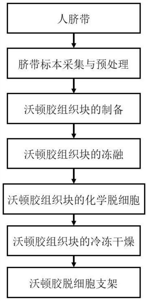
[0041] In this embodiment, a method of preparing cartilage tissue engineering brackets in human umbilical cord Warten is as follows (such as figure 1 Disted):

[0042] (1) Umbilical belt specimens collection and premium:

[0043] Inject 300ml antibiotic (penicillin: 10,000 units / 100ml, streptomycin: 50,000 units / 100ml) in the 500ml plastic grooming, will be taken in the obstetric surgery The umbilical belt specimens of the uterinestormation immediately placed in the spanking bottle; in the umbilical cord extent 30min, washed repeatedly with PBS solution until the umbilical cord surface and the internal naked eyes were unable to see blood clots and foreign objects; pull the clean umbilical cord straightened , Put into ordinary refrigerator cold freezer - 20 ° C for 1 h with sterile wrap

[0044] (2) Preparation of Wharton gum tissue block

[0045] The longitudinal long axis of the umbilical cord after pre-opening is cut into a cylindrical tissue tissue tall higher than about 10...


PUM


Abstract

The invention provides a method for preparing a cartilage tissue engineering scaffold from human umbilical cord Wharton jelly and the cartilage tissue engineering scaffold. The method for preparing the cartilage tissue engineering scaffold from the human umbilical cord Wharton jelly comprises the following steps: (1) collecting and pretreating of an umbilical cord specimen; (2) preparation of Wharton jelly tissue blocks; (3) freeze thawing of Wharton jelly tissue blocks; (4) chemical decellularization of Wharton jelly tissue blocks; and (5) freeze-drying of the Wharton jelly tissue blocks. The scaffold prepared by the invention can retain extracellular matrix components of Wharton jelly, especially related proteins for promoting cartilage differentiation and original structure characteristics, to the maximum extent, and at the same time effectively removes cell components. The Wharton jelly decellularized scaffold has the structural characteristics of being loose and porous, good in biocompatibility, conforming to the biodegradation law and highly similar to the cartilage extracellular matrix structure and component, and a new material system is provided for construction of cartilage tissue engineering scaffolds.

Description

Technical field [0001] The present invention relates to cartilage tissue engineering techniques, and more particularly to a method of preparing cartilage tissue engineering brackets with human umbilical cord Wardon glue, and cartilage tissue engineering brackets. Background technique [0002] Joint cartilage is easily damaged due to trauma or degenerative variations. Because the articular cartilage does not contain angioplasty and nerve, the cells mainly dependent the tissue penetration to obtain nutrients, and the end of cartilage cells differentiate, the metabolic ability is low, so the self-repair capacity after cartilage damage is very poor. The difficulty of regeneration after joint cartilage is a major challenge, and it is also a hot spot in the current research. The method used to repair joint cartilage is: joint cavity damage cartilage cleaning surgery, micro-fracture repair, autologous or allogeneic cartilage plantation and autologous bone soft bone graft, artificial joi...

Claims


Application Information

Patent Timeline
IPC IPC(8): A61L27/36A61L27/22A61L27/24A61L27/56A61L27/58
CPCA61L27/3604A61L27/3687A61L27/56A61L27/24A61L27/225A61L27/3654A61L27/58A61L2300/414A61L2430/06
Inventor 钟达王成功苏士龙高发维杨婷
Owner XIANGYA HOSPITAL CENT SOUTH UNIV
Learn More