
Trojan horse immunotherapy

a technology of immunotherapy and trojan horses, applied in the field of trojan horses immunotherapy, can solve the problems of lack of effective treatment options, physical and mental impairment, and substantial loss of brain cells, and achieve the effects of reducing the number of patients

Inactive Publication Date: 2008-02-14
THE BOARD OF TRUSTEES OF THE LELAND STANFORD JUNIOR UNIV
View PDF3 Cites 2 Cited by
  • Summary
  • Abstract
  • Description
  • Claims
  • Application Information

AI Technical Summary

Benefits of technology

[0002] Understanding of neuronal damage and loss due to neurological disease and injury has increased significantly during the past several decades, including an increase in our understanding of the molecular pathways that control neuron death. In tissue culture and animal models of neurological insults, delivery and enhanced expression of certain neuroprotective genes or of the encoded protein products themselves, can significantly reduce the extent of neuron death in these conditions.

Problems solved by technology

Neurological diseases can be devastating pathological conditions, with resultant damage to brain tissue that significantly compromises the CNS and leads to both physical and mental impairments.
Both forms of neurological disease cause a substantial loss of brain cells, including neurons.
These debilitating conditions lack effective treatment options.
The clinical applicability of therapeutic strategies based on these research findings is hindered, however, by the difficulty of delivering target genes or proteins to the CNS.
Specifically, gene or protein delivery to a site of injury in the CNS is hindered because access to brain tissue from the circulation is restricted by the ‘blood brain barrier’ (BBB).
This is highly invasive, requiring drilling a hole in the skull and controlled penetration of the brain with a syringe.
In addition, it is difficult, if not impossible, to deliver the gene or protein therapeutic agent to all of the affected sites.
This approach is sub-optimal because only a small percentage of the gene or protein therapeutic agent arrives at the injury site; the vast majority ends up elsewhere in the body and may lead to unwanted side effects.
Moreover, techniques that open up the BBB are often damaging in and of themselves, and cause non-specific opening, resulting in delivery of the therapeutic agent throughout the brain, often with untoward side effects.

Method used


Image

  • Trojan horse immunotherapy
  • Trojan horse immunotherapy

Examples


example 1

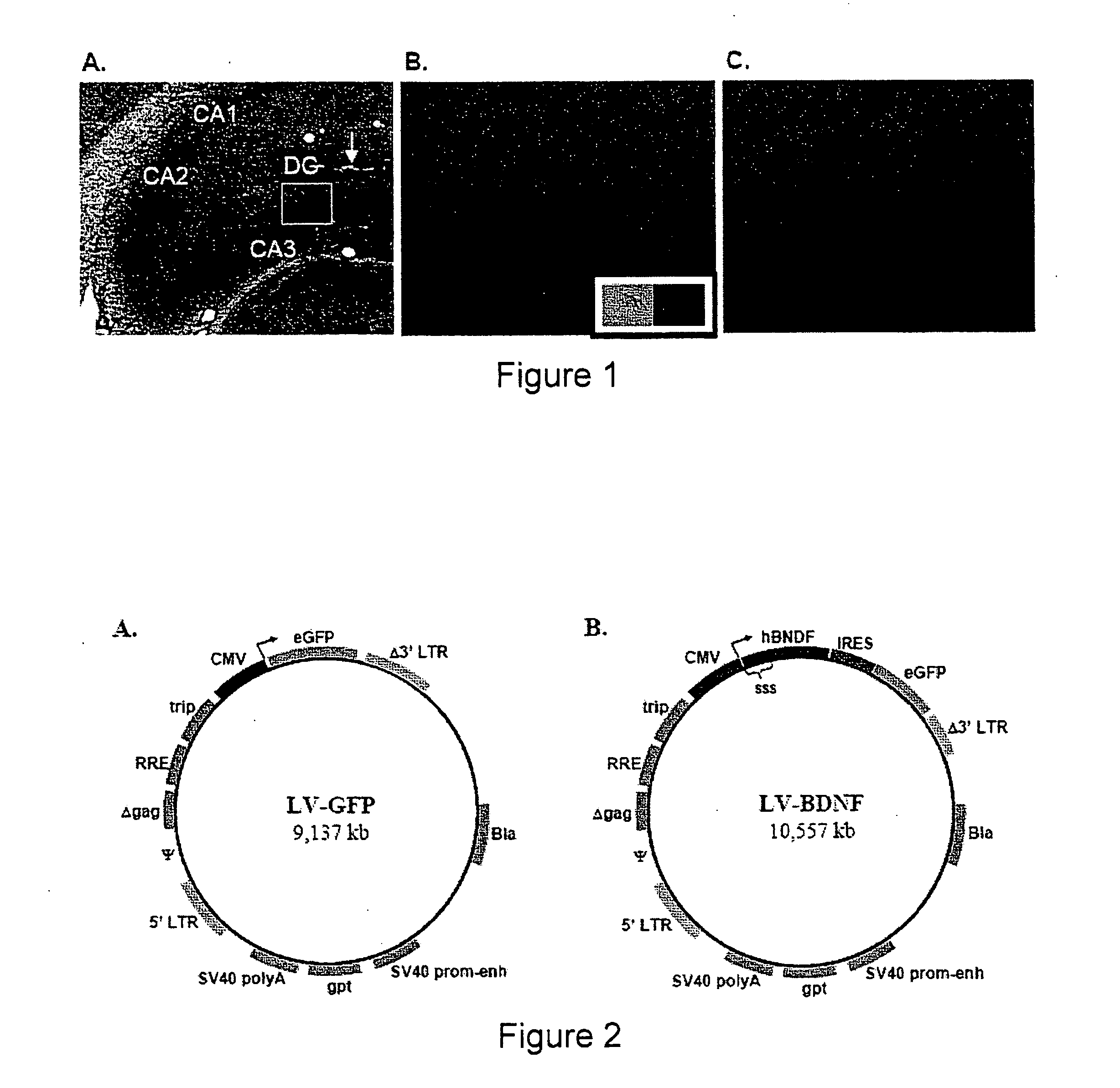
DCs Generated In Vitro Migrate to Site of CNS Lesion

[0072] DCs for this experiment were derived from bone marrow precursor cells, as described previously (Grauer et al. (2002) Histochem. Cell Biol. 117(4): 351-62). Briefly, cell isolates from the femur of inbred, male Lewis rats were cultured in nutrient-rich media containing interleukin-4 (IL-4), granulocyte / macrophage-colony stimulating factor (GM-CSF), and Flt-3 ligand. Under these conditions, DCs form semi-adherent cell aggregates, which are further enriched by removal of non-adherent lymphocytes.

[0073] After seven days, resultant DC content was measured by flow cytometry. Cultured DCs were scored for expression of MHC class II (OX6), integrin-α-X (CD11c), and α-E2 integrin (OX62) using monoclonal antibodies, the expression of maturation-indicative co-stimulatory surface molecules CD80 and CD86. For each antibody, matched isotype controls were run in parallel to account for non-specific binding. In addition to analysis of cell...

example 2

Lentiviral Vectors and DC Infection / Transduction

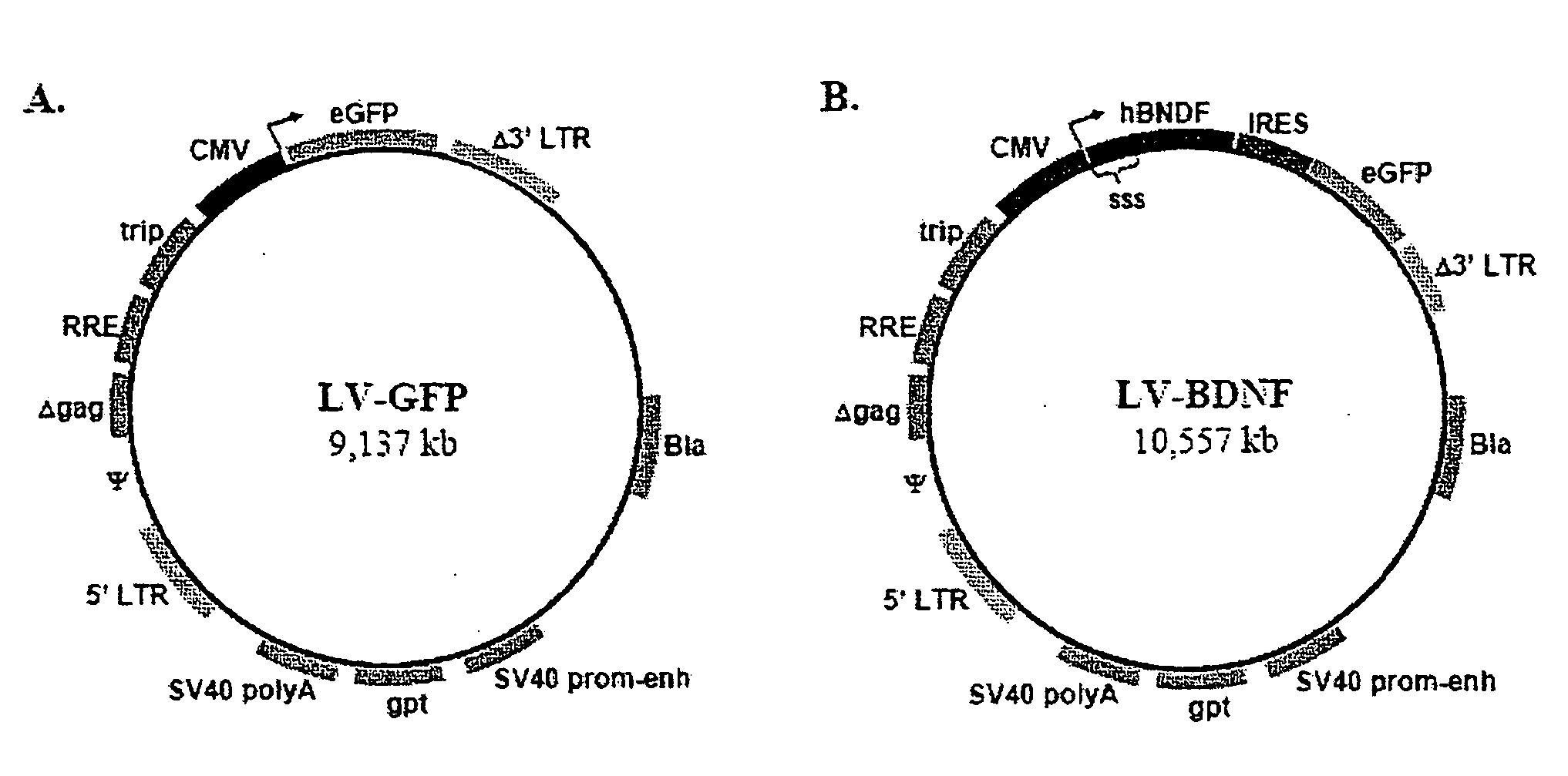
[0075] Lentiviral vectors (LV) that express the reporter gene, eGFP (LV-GFP) are employed to determine optimal transduction conditions of DC cell cultures (FIG. 2A). LV-GFP vector is prepared as previously described (Kafri et al, (1999) J. Virol. 73(1): 576-84). Production of LV particles occurs in 293T kidney cells, allowing in trans expression of LV packaging proteins, the VSV-G envelope protein, and the actual transfer vector. By this approach, resultant LV particles only carry the transfer vector, which contains the chosen transgene and a minimal set of LV genes required for reverse transcription and nuclear integration (FIG. 2A). Omission of LV packaging proteins and the VSV-G envelope protein limits the activity of LV particles to one round of DC infection and transgene integration. In addition, the transfer vector is made ‘self-inactivating’ by a deletion in the U3 region of the 3′ long terminal repeat (LTR) to minimize the ris...

example 3

Assay for In Vitro Secretion of BDNF by Transduced DCs

[0079] BDNF normally exists as an extracellular protein, and its cDNA contains an endogenous signal secretory sequence. Consequently, LV-mediated integration of unmodified BDNF allows proper secretion from transduced DCs. LV vectors are designed to contain myc-tagged human BDNF followed by an IRES and eGFP (LV-BDNF, FIG. 2B).

[0080] DC cultures are transduced with LV-BDNF or LV-GFP according to the methods described above. Flow cytometry is used to assay the percentage of GFP-positive DCs, thus providing a measure of transduction efficiency. At 0, 12 and 24, and 48 hours post-transduction, the supernatant from DC cultures is collected and used for sandwich ELISA. Briefly, each sample of DC supernatant is added to wells coated with a monoclonal antibody for BDNF. After 1 hour of incubation, a second, biotinylated antibody for BDNF is added to each well and assayed for relative absorbance at the appropriate wavelength. Purified BD...


PUM

PropertyMeasurementUnit
physicalaaaaaaaaaa
areaaaaaaaaaaa
fluorescent emissionaaaaaaaaaa

Abstract

The present invention provides methods for the delivery of therapeutic proteins to an area of inflammation and neuronal damage within the central nervous system (CNS). In this invention, immunocytes that can cross the blood brain barrier and access sites of CNS injury / inflammation, are genetically engineered to express and secrete a therapeutic protein of interest and are transplanted into a subject having a CNS injury. These genetically modified “carrier immunocytes” home to the site of a CNS lesion and secret the therapeutic agent, thereby ameliorating disease symptoms and pathology.

Description

[0001] Neurological diseases can be devastating pathological conditions, with resultant damage to brain tissue that significantly compromises the CNS and leads to both physical and mental impairments. Neurological diseases can arise from acute conditions, such as stroke or seizure, or from long-term neurodegeneration, as is the case in Alzheimer's and Parkinson's disease. Both forms of neurological disease cause a substantial loss of brain cells, including neurons. These debilitating conditions lack effective treatment options. [0002] Understanding of neuronal damage and loss due to neurological disease and injury has increased significantly during the past several decades, including an increase in our understanding of the molecular pathways that control neuron death. In tissue culture and animal models of neurological insults, delivery and enhanced expression of certain neuroprotective genes or of the encoded protein products themselves, can significantly reduce the extent of neuro...

Claims


Application Information

Patent Timeline
Patent Type & Authority Applications(United States)
IPC IPC(8): A61K48/00A61P25/00A61P37/00
CPCA61K38/185A61K2039/5156A61K2039/5154A61K39/0011A61P25/00A61P37/00A61K35/15A61K39/4622A61K39/4615A61K39/464838A61K39/46443
Inventor SAPOLSKY, ROBERT M.MANLEY, NATHAN C.BROOKE, SHEILADINKEL, KLAUS M.MUNHOZ, CAROLINA D.RIEPEL, ANGELA L.NICHOLAS, ANDREA C.
Owner THE BOARD OF TRUSTEES OF THE LELAND STANFORD JUNIOR UNIV
Learn More