Eureka AIR delivers breakthrough ideas for toughest innovation challenges, trusted by R&D personnel around the world.

Implantable article, method of forming same and method for reducing thrombogenicity

a technology of implantable devices and polymer materials, applied in the field of implantable devices, can solve the problems of reducing the thrombogenicity of polymer materials used in the production of implantable devices, unable to overcome problems, and unable to achieve the effect of reducing the thrombogenicity of implantable devices

Inactive Publication Date: 2008-04-24
NANYANG TECH UNIV
View PDF99 Cites 16 Cited by
  • Summary
  • Abstract
  • Description
  • Claims
  • Application Information

AI Technical Summary

Benefits of technology

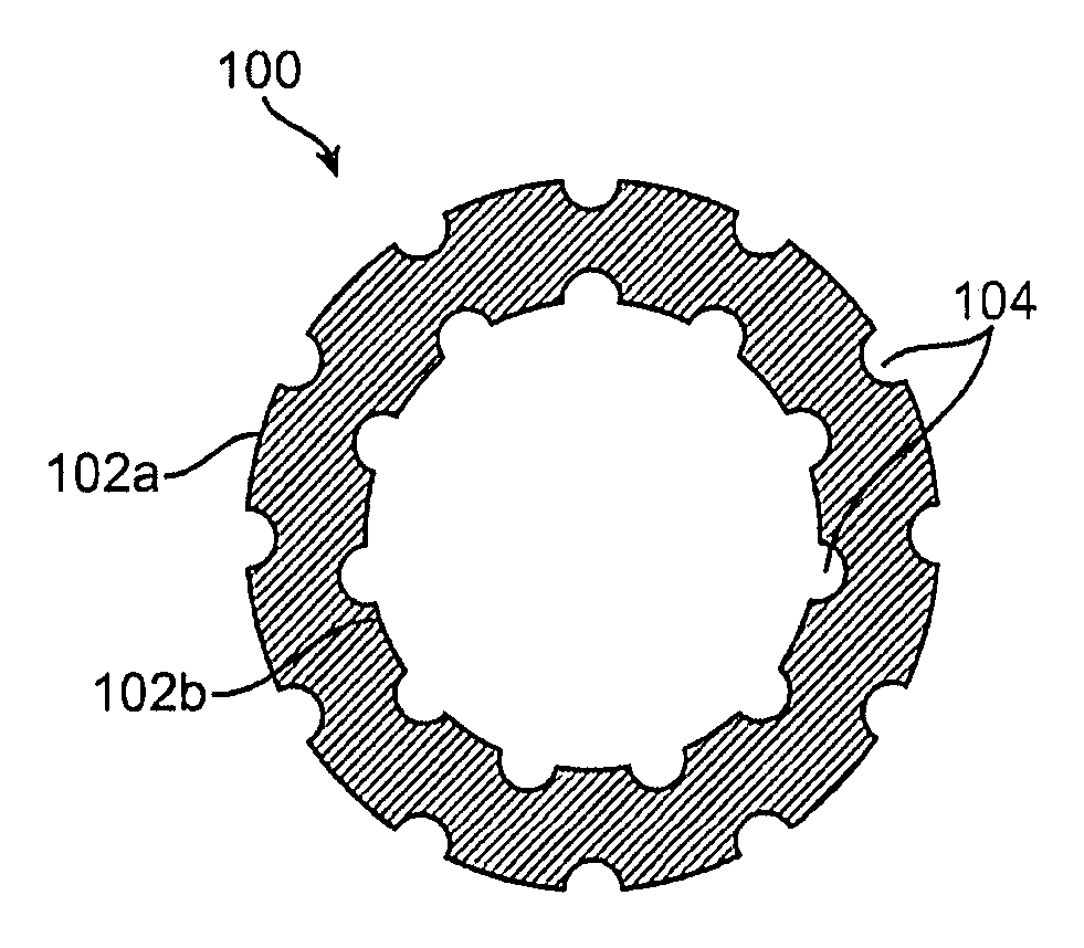
[0012] According to a further aspect of the invention, there is provided a method of reducing thrombogenicity of an implantable device having at least one surface for contacting bodily fluid or tissue, or promoting or enhancing endothelialization of an implantable device having at least one surface for contacting bodily fluid or tissue, comprising: providing on said at least one surface a plurality of nano-craters that enhance or promote endothelialization of said at least one surface.
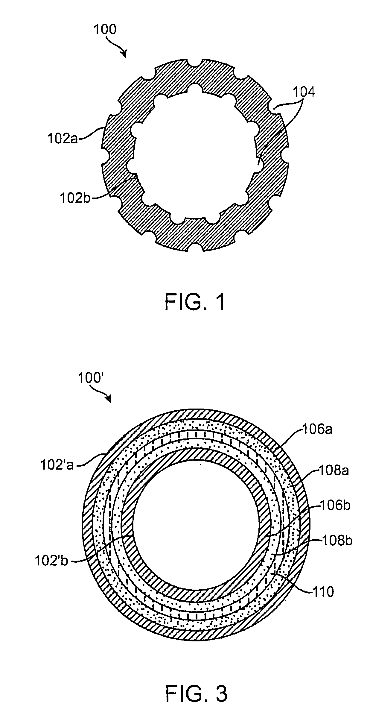
[0014] According to still another aspect of the invention, there is provided a method of reducing thrombogenicity of an implantable device having at least one surface for contacting bodily fluid or tissue, comprising: providing at least one first degradable layer which provides said at least one surface and which is disposed about a central core, and at least one second degradable layer between said first degradable layer and the central core, wherein said first degradable layer has a first degradation rate and said second degradable layer has a second degradation rate such that said at least one first degradable layer degrades more rapidly than said at least one second degradable layer on contact with bodily fluid or tissue.

Problems solved by technology

Progress in this area has, however, been hampered somewhat by the thrombogenicity of many polymer materials.
Efforts to overcome the problems associated with thrombogenicity of polymer materials used in the production of implantable devices have not met with a great deal of success to date.
This is because many of the chemical and physical modifications of the device surfaces have limited shelf-life, both ex vivo and in vivo.
Moreover, the methods involved in the production of implantable devices using these approaches are both elaborate and intricate.

Method used


Image

  • Implantable article, method of forming same and method for reducing thrombogenicity
  • Implantable article, method of forming same and method for reducing thrombogenicity
  • Implantable article, method of forming same and method for reducing thrombogenicity

Examples


example 1

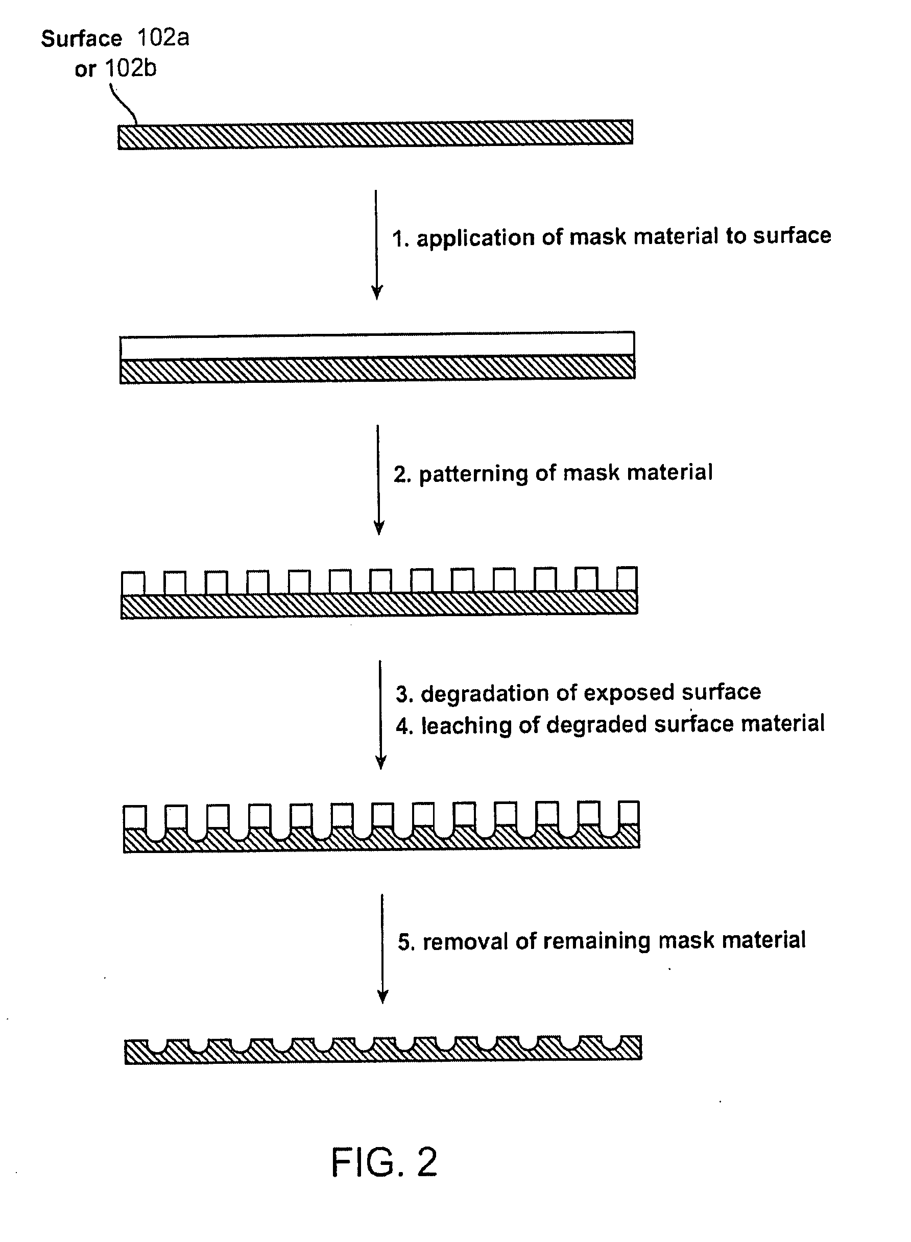
[0097] For the following examples, each polymer was first dissolved in chloroform. Nano-sized salt particles were ground and sieved, and then dispersed in the polymer solution with constant stirring until the particles were visually uniformly dispersed. The polymer concentration was chosen such that it had sufficiently high viscosity to maintain a stable dispersion. The dispersion was then cast as a film of required thickness using a coater. The film was dried in an oven at 37° C., and then left at room temperature for several days in a dry environment. The dried films were immersed in water for 14 days, with constant exchange of the water. The salt nano-particles were thus leached out, and the resulting film was dried again at 37° C. and at room temperature.

[0098] Control films were prepared as pure polymer films without any surface modification.

[0099] PLLA and PLGL films having nano-craters in the surface were obtained by leaching out incorporated nano-particles of NaCl, as indi...

example 2

[0102] In another example of a method for modifying a surface of a polymer for implantation within a patient body, porogen leaching of surfaces may be utilized to yield a surface which enhances endothelial cell growth over a defined range of surface features. In this particular example, surface pores were created by filling polymers such as Poly caprolactone (PCL), Poly L-lactide (PLLA), Poly (lactide-co-glycolide), etc. (although any of the other suitable polymers described herein may be utilized) with leaching agents of sugar and gelatin.

[0103] The sugar and gelatin particles ranged in size from 20 to 90 microns in diameter (although particles as small as 5 microns may also be utilized) where the average particle sizes typically ranged from 20, 45, and 90 microns. The leaching agents were added in concentrations ranging from 1 to 10% by weight in the polymer. More particularly, the leaching agents were added in concentrations ranging from 1%, 5%, and 10% by weight in the polymer....

example 3

[0109] As mentioned above, chemicals such as sodium hydroxide may be used to dissolve PLA in regions unprotected by an alkali-resistant mask material where the dissolved material may be rinsed away in water to form nano-craters. In another example, the polymer surface may be first irradiated prior to etching with the sodium hydroxide to enhance the etching process.

[0110] In this example, samples of PLGA, PCL, and PLLA (other suitable polymers described above may alternatively be utilized) were first irradiated with an electron beam and then etched using the sodium hydroxide, as described above, for a period of 16 hours to create surface features. The average surface roughness of the samples was measured using an atomic force microscope (AFM) and the etched samples were then exposed to endothelial cells. Growth was quantified over a period of 15 days and the irradiated and etched samples were compared to control samples after 4 days, 8 days, and 15 days. Table 2 shows a comparison o...


PUM

PropertyMeasurementUnit
diameteraaaaaaaaaa
distanceaaaaaaaaaa
distanceaaaaaaaaaa

Abstract

Endothelialization of a bodily fluid or tissue-contacting, particularly blood-contacting, surface may be accomplished to render that surface substantially non-thrombogenic. Thrombosis may also be mitigated or eliminated by providing an eroding layer on the surface that results in the removal of any thrombus formation as the layer erodes. An implantable device may utilize at least one surface having a plurality of nano-craters thereon that enhance or promote endothelialization. Additionally, an implantable device may have at least one first degradable layer for contacting bodily fluid or tissue and disposed about a central core, and at least one second degradable layer between the first degradable layer and the central core. The first degradable layer has a first degradation rate and the second degradable layer has a second degradation rate which degrades more slowly than the first degradable layer on contact with bodily fluid or tissue.

Description

CROSS-REFERENCE TO RELATED APPLICATIONS [0001] This application claims the benefit of priority to U.S. Provisional Patent Application Ser. No. 60 / 808,558 filed May 26, 2006, which is incorporated herein by reference in its entirety.FIELD OF THE INVENTION [0002] The present invention relates generally to implantable devices, such as implantable medical devices, and methods for the manufacture thereof. The invention also relates to methods for enhancing and promoting endothelialization and for minimizing thrombus formation on the surface of the implantable device. BACKGROUND OF THE INVENTION [0003] In recent years there has been growing interest in the use of artificial materials, particularly materials formed from polymers, for use in implantable devices that come into contact with bodily tissues or fluids particularly blood. Some examples of such devices are artificial heart valves, stents and vascular prosthesis. Progress in this area has, however, been hampered somewhat by the thr...

Claims


Application Information

Patent Timeline
Patent Type & Authority Applications(United States)
IPC IPC(8): A61F2/02A61F2/06
CPCA61F2/0077A61L2420/08A61F2210/0004A61L27/18A61L27/50A61L27/56A61L33/0011A61L2400/18A61F2/06A61L2420/06A61L27/58A61L29/08A61L27/34A61L31/10A61L31/08C08L67/04
Inventor VENKATRAMAN, SUBRAMANIANBOEY, YIN CHIANG
Owner NANYANG TECH UNIV
Why Eureka
Eureka Blog
Learn More