Implantable medical articles having pro-healing coatings

a technology of medical articles and coatings, applied in the direction of prosthesis, antithrombosis treatment, blood vessels, etc., can solve the problems of excessive proliferation of ecs, device failure in vivo, inflammation, fibrosis, etc., and achieve the effect of improving the function of the articl

Inactive Publication Date: 2008-10-02
WILLIAMS STUART K +3
View PDF2 Cites 51 Cited by
  • Summary
  • Abstract
  • Description
  • Claims
  • Application Information

AI Technical Summary

Benefits of technology

[0014]The present invention generally relates to implantable medical articles having coatings that improve the function of the article in vivo. The invention also relates to methods for using these coated medical articles in a subject. Generally, the coated medical articles promote one or more physiological events associated with a pro-healing response. The medical articles of the present invention include a coating having at least one adhesion factor (e.g., matrix proteins, active portions thereof, or binding members thereof) formed on the surface of the device in a manner that provides a particularly desirable endothelial cell response, which can occur on the blood contacting surface of the device.
[0016]In vivo studies associated with the invention show that coatings that include a adhesion factor immobilized using photogroup chemistry provide particularly desirable levels of endothelialization following a period of implantation. In other words, the coatings promote attachment of endothelial cells, but do so in a manner that also results in limiting the proliferation of other cell types on the surface. This can be important, particularly for medical devices, such as stents, that are implanted for a substantial period of time for the treatment of a medical condition.
[0017]The coatings of the present invention can be formed on coronary stents to provide a pro-healing response. This pro-healing response is characterized by a modulated formation of an endothelial cell layer that also can limit the proliferation of smooth muscle cells. This in turn can reduce the incidence of restenosis an improve stent function and lifetime.
[0018]In some aspects of the invention, the photogroup and adhesion factor are used in conjunction with polymeric material that forms a coated layer and a bioactive agent that is elutable or releasable from the coated layer. The adhesion factor, which is immobilized by the photogroup, improves an otherwise sub-optimal or abnormal endothelial cell response, which is observed on devices when the bioactive-releasing layer is used as the coating alone. This aspect of the invention is advantageous as it can improve therapy for devices with drug-eluting coatings, which typically require a prolonged systemic anticoagulation therapy.
[0021]The coatings can be formed on the surface of devices that would otherwise promote an undesirably high level of endothelialization (such as a bare metal surface of a stent). The coating including the photogroup and adhesion factor can also be used to modulate the endothelial response on the surfaces of these types of implantable devices. In some aspects, the coatings of the invention are used to modulate the endothelialization on surfaces that, for example, after a period of implantation, promote hyperproliferation of smooth muscle cells. Therefore, in some aspects, the coatings can provide a positive, lower level of endothelialization beneficial for the function of devices that are implanted in the body for a prolonged period of time.

Problems solved by technology

Despite being inert and nontoxic, implanted biomaterials associated with the device, such as various plastics and metals, often trigger responses such as inflammation, fibrosis, infection, and thrombosis.
If excessive, some of these reactions may cause the device to fail in vivo.
While moderate proliferation of ECs is desirable, excessive proliferation of ECs may also be associated with hyperproliferation of SMCs.
Hyperproliferation of SMCs can lead to hyperplasia and restenosis.
In other words, the coatings promote attachment of endothelial cells, but do so in a manner that also results in limiting the proliferation of other cell types on the surface.
The coatings can be formed on the surface of devices that would otherwise promote an undesirably high level of endothelialization (such as a bare metal surface of a stent).

Method used


Image

  • Implantable medical articles having pro-healing coatings
  • Implantable medical articles having pro-healing coatings
  • Implantable medical articles having pro-healing coatings

Examples


example 1

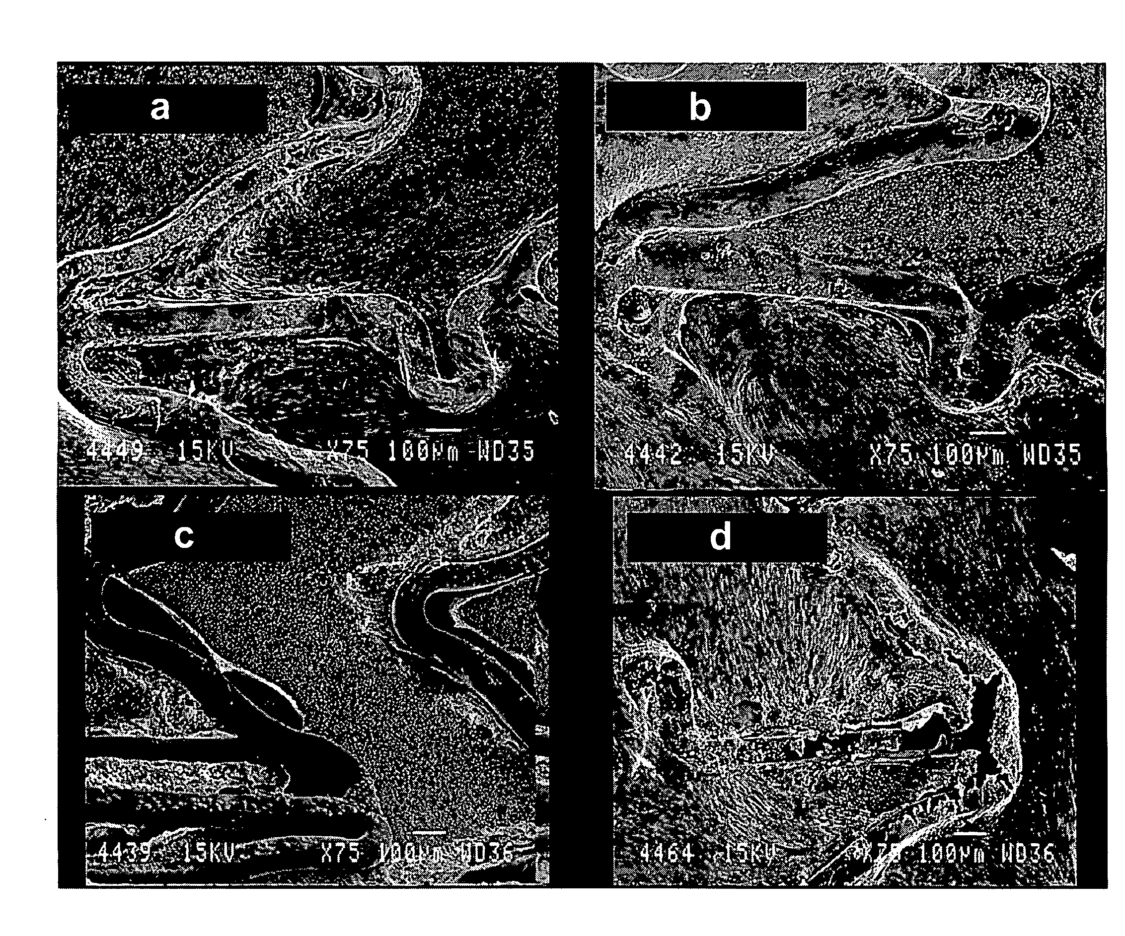
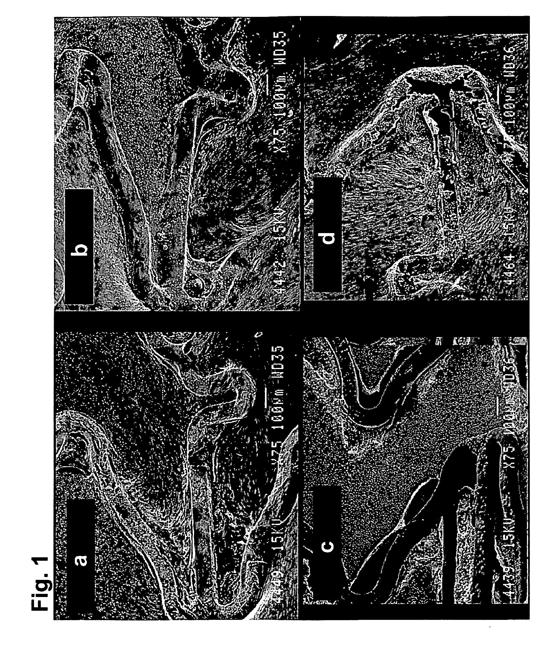
[0129]HBPR / protein-modified (HBPR COLI / LM5, etc) coronary stents (3×8 mm) were evaluated for healing responses in the iliac arteries of New Zealand white rabbits. The coatings were formed on either 3×8 mm cobalt chromium bare metal stents or 3×8 mm cobalt chromium metal stents having a silane / Parylene™ base coat and a drug-eluting pBMA / pEVA / paclitaxel coat. The coatings are summarized in Table 1 below. Prior to protein or photo-protein coating, but following any silane / Parylene™ or pBMA / pEVA / paclitaxel coating the stents were EtO sterilized. Aseptic techniques were then used to apply the protein or photo-protein coatings.

[0130]For some stent samples, coatings were formed using photogroup-derivatized matrix proteins (photo-collagen and photo-laminin). Photogroup-derivatized matrix proteins were prepared as described in U.S. Pat. No. 5,744,515 (Clapper). A stent was placed into a 10×75 mm glass test tube (1 stent per test tube) and 1 mL of photo-collagen-I at a concentration of 200 ug...

example 2

[0145]Collagen 1 coatings, including those formed from photo-collagen, were prepared in non-fibrillar and fibrillar forms.

[0146]A photo-collagen-I solution for formation of a fibrillar coating was prepared. An aqueous solution of 12 mM HCl was cooled on ice and photo-collagen-I was added to provide a concentration of 3 mg / mL, and the solution kept on ice. The photo-collagen solution was diluted 1:3 with cold 12 mM HCl resulting in a concentration of 1 mg / ml.

[0147]The photo-collagen-I solution in an amount of 1.5 mL was centrifuged for 5 min at 14,000 RPM. Supernatant in an amount of 600 uL was transferred to a new tubes. A collagen-I (non-photo, lyophilized material) solution was prepared in chilled 12 mM HCl at 1 mg / mL and subjected to the same centrifugation and supernatant removal.

[0148]To 600 uL of the collagen-I and photo-collagen-I solutions were added 600 uL 0.1 M carbonate / bicarbonate buffer (CBC), pH 9.0, resulting in a pH>9.0. A collagen-1 solution was also prepared at pH ...

example 3

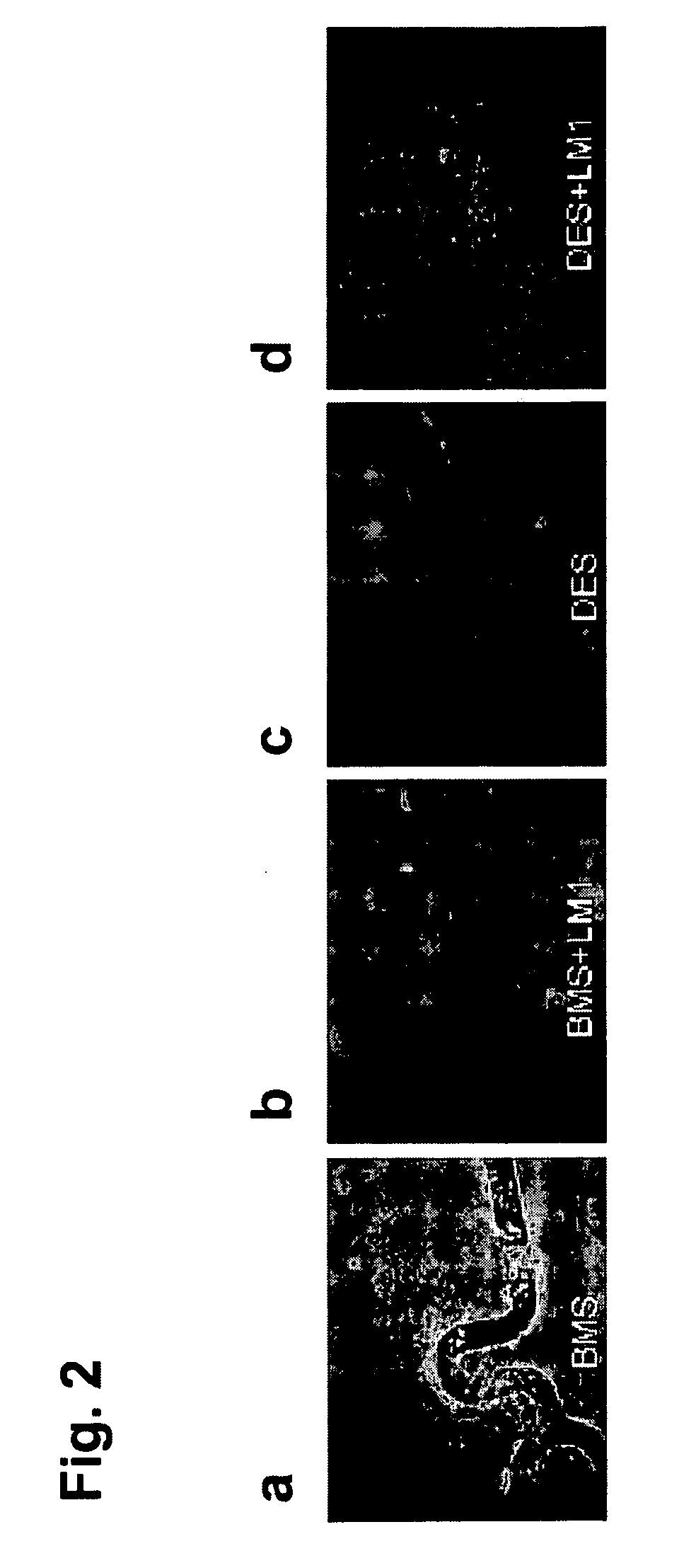
[0151]The thrombotic effect of the bare metal surfaces, photo-collagen coated surfaces and regular collagen surfaces was examined in a porcine ex-vivo AV shunt model, similar to the procedure as described in Hanson S. R., et al., (1980) “In vivo evaluation of artificial surfaces using a nonhuman primate model of arterial thrombosis,”J Lab Clin Med 95, 289-304.

[0152]For the photo-collagen coating, a Parylene-coated stent was soaked in photo-collagen-I (200 ug / ml, 12 mM HCl) for 1 hr at 4 C while shaking followed by an in-solution illumination for 3 min. The stent was then rinsed in water and dried.

[0153]Results of the ex-vivo study are shown in FIGS. 7a-7c, showing excessive thrombosis on the collagen-coated stents (7c), and moderate, acceptable level of thrombosis on the photo-collagen coated stents (7b).


PUM

PropertyMeasurementUnit
timeaaaaaaaaaa
timeaaaaaaaaaa
concentrationaaaaaaaaaa

Abstract

Coatings including adhesion factors for the surfaces of implantable medical articles are disclosed. The coatings are used to improve the function of the device by promoting a pro-healing response following implantation. The coatings can modulate endothelialization of the article surface to reduce the risk of adverse tissue responses that may reduce the functionality of the device.

Description

CROSS-REFERENCE TO RELATED APPLICATIONS[0001]The present non-provisional Application claims the benefit of commonly owned provisional Application having Ser. No. 60 / 819,091, filed on Jul. 7, 2006, and entitled IMPLANTABLE MEDICAL ARTICLES HAVING PRO-HEALING COATINGS; and commonly owned provisional Application having Ser. No. 60 / 848,588, filed on Sep. 29, 2006, and entitled IMPLANTABLE MEDICAL ARTICLES HAVING PRO-HEALING COATINGS; which Applications are incorporated herein by reference in their entirety.FIELD OF THE INVENTION[0002]The invention relates to coatings for implantable medical article and methods for promoting a pro-healing response.BACKGROUND OF THE INVENTION[0003]Until more recently, the primary focus of advances in implantable medical article technology has been to alter a structural characteristic of the article to improve its function within the body. However, it has become appreciated that function of the implanted device at the site of implantation can be greatly en...

Claims


Application Information

Patent Timeline
Patent Type & Authority Applications(United States)
IPC IPC(8): A61F2/06
CPCA61F2/91A61L27/34A61L29/085A61L31/10A61L33/0029A61L33/122A61L33/128C08L89/00C08L89/06A61L2420/08
Inventor WILLIAMS, STUART K.BABCOCK, DAVID E.CHINN, JOSEPH A.CLAPPER, DAVID L.
Owner WILLIAMS STUART K
Why Eureka
Try Eureka