Anti-angiogenic cellular agent for cancer therapy

a cancer therapy and cellular agent technology, applied in the field of antiangiogenic cellular agent for cancer therapy, can solve the problems of limited in vivo efficacy, large toxicities of il-2 administration, and massive endothelial destruction, and achieve the effect of minimal risk of vls associated with eat cell administration

Inactive Publication Date: 2008-10-23
HOPE ERNEST G
View PDF0 Cites 9 Cited by
  • Summary
  • Abstract
  • Description
  • Claims
  • Application Information

AI Technical Summary

Benefits of technology

[0014]Many types of EAT cells directly attack tumor cells in addition to attacking tumor vasculature. Thus, such EAT cells can be particularly effective for the treatment of cancer. Because EAT cells attack tumor vasculature they can be therapeutically effective for treatment of vascularized cancer even if they do not substantially kill the tumor cells themselves either in vitro or in vivo. Thus, cells that do not kill cells of a particular cancer cell line in vitro can nonetheless be effective therapeutically for treating that cancer because they can attack tumor vasculature.
[0023]EAT cells selectively damage tumor vasculature, reducing the flow of blood and nutrients to the tumor, arresting or reducing the growth potential of the tumor.

Problems solved by technology

However, their in vivo efficacy is limited by their limited proliferative potential and the requirement for the co-application of IL-2 (Rosenberg et al.
Systemic administration of IL-2 is associated with considerable toxicities, including massive endothelial destruction (Siegel et al.
However, TILs must be isolated from a surgical specimen of the patient and their generation tends to be cumbersome and yields low cell numbers.

Method used


Image

  • Anti-angiogenic cellular agent for cancer therapy
  • Anti-angiogenic cellular agent for cancer therapy
  • Anti-angiogenic cellular agent for cancer therapy

Examples


example 1

Generation and Maintenance of Cells and Cell Lines

(A) CIK Cells

[0081]Human CIK cells were prepared from peripheral blood lymphocytes (PBL) obtained from whole venous blood and buffy coats of healthy community donors (Stanford Blood Center, Stanford, Calif.). PBLs were isolated using Ficoll-Hypaque (Pharmacia Fine Chemicals, Uppsala, Sweden) density gradient centrifugation. PBLs isolated according to this method were resuspended at a density of 0.5-2×10⁶ cells / ml in RPMI 1640 (GIBCO-BRL / Life Technologies, Grand Island, N.Y.) containing 50 μm β-mercapto-ethanol (ME), 100 IU penicillin-G / ml, 100 IU streptomycin / ml, and 10% fetal calf serum (FCS) (Sigma Chemical Co., St. Louis, Mo.). Subsequently, an enriched population of large granular lymphocytes (LGL) and T-cells was obtained by the exclusion of plastic and nylon wool adherent cells. The enriched population was cultured in a humidified incubator with 5% carbon dioxide at 37° C.

[0082]The enriched cell population was stimulated at day...

example 2

51Cr-Release Cytotoxicity Assays

[0087]Target cells used in cytotoxicity assays were metabolically labeled with 51Cr (DuPont-New England Nuclear, Boston, Mass.) by incubating 1×10⁶ cells with 300 mCi 51Cr at 37° C. for 1-1.5 hours. Following the incubation, the cells were washed three times with phosphate buffered saline (PBS) containing 0.1% bovine serum albumin. The labeled target cells were then distributed in triplicate in flat-bottomed 96 well microtiter plates at a concentration of 2×10⁴ cells / well. Effector (CIK) cells were added at the indicated ratios. Monoclonal antibodies were incubated with target cells for 15-30 minutes at room temperature prior to the addition of effector cells. The final volume of the assay mixture in each well was 0.2 ml. After 4 hours at 37° C., the cells were collected by centrifugation and an aliquot of the supernatant was counted in a gamma counter (Micromedic Systems, Horsham, Pa.). The percentage of specific 51Cr release was calculated according...

example 3

Ex Vivo Expansion and Cytotoxicity Profile of CIK Cells

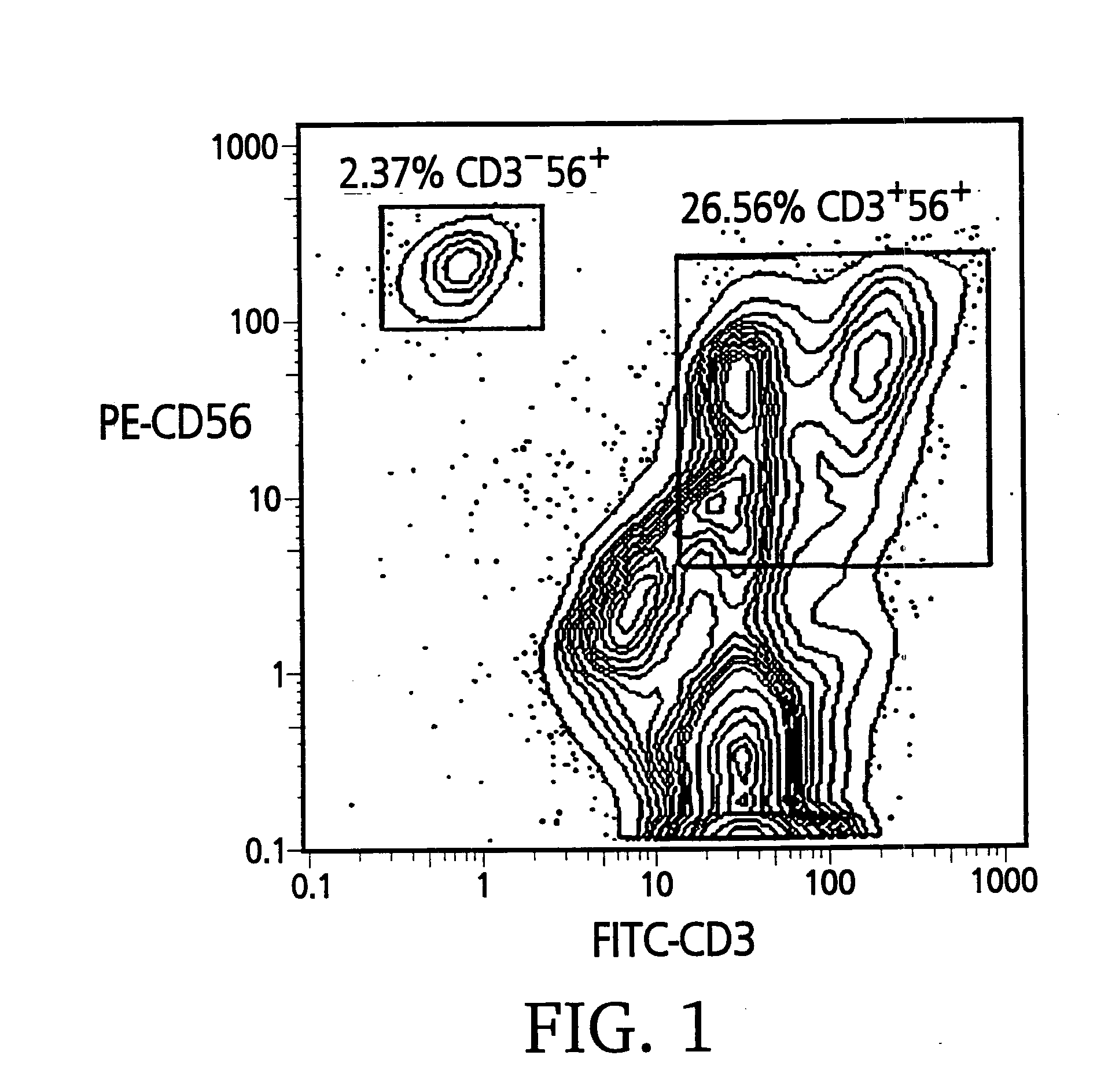
[0089]Three to five percent of unmanipulated peripheral blood mononuclear cells (PBMC) present a CD3+56+ phenotype. Preferential expansion of CD3+56+ cells is observable as early as day 10 of culture (using the CIK cell culture conditions of Example 1), when of 12-15% of cells stain positive for the CD3+56+ phenotype. Day 14 to day 28 cultures typically yielded 30-50% of CD3+56+ cells.

[0090]Cell surface expression of CD3 and CD56 was analyzed for mature (day 21) CIK cells that were expanded ex vivo in bioreactors. Approximately 106 CIK cells were stained with anti-CD56-PE and anti-CD3-FITC monoclonal antibodies according to the manufacturer's recommendations and analyzed on a FACS-Star® (Beckton-Dickinson, San Jose, Calif.). The contour graph of a typical batch of CIK cells (FIG. 1) shows that 26.5% of the CIK cells are CD3+56+ and 2.3% are CD3−56+.

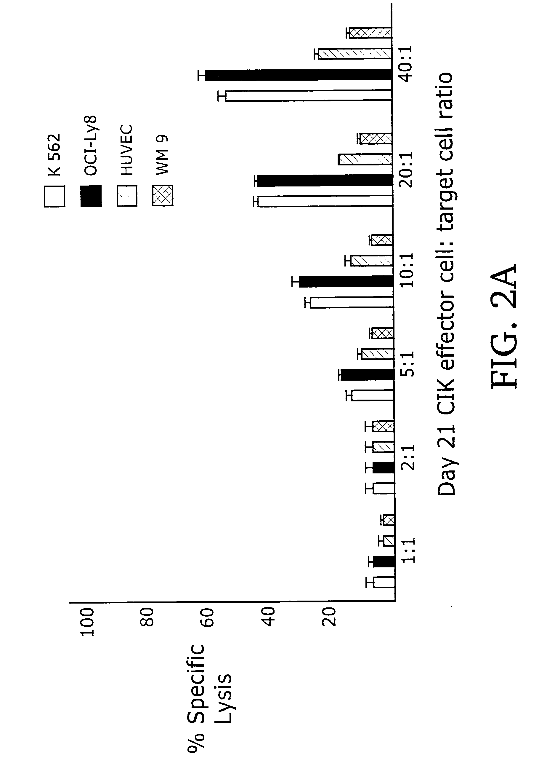
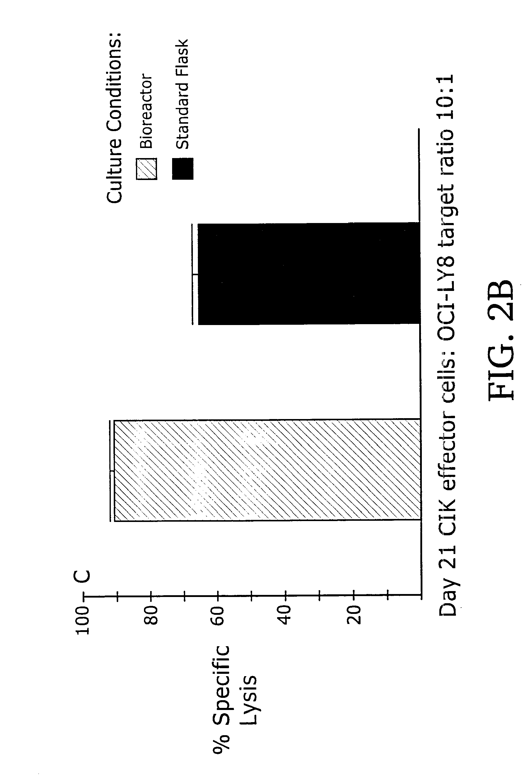
[0091]When used as effectors in 51Cr-release cytotoxicity assays, day 21 CIK cell...


PUM

PropertyMeasurementUnit
temperatureaaaaaaaaaa

Abstract

The invention provides cytokine induced killer (CIK) cell populations and methods of using CIK cells to treat cellular proliferative disorders. CIK cells generated in vitro include both bulk cultures and clones. Individual CIK cell clones display distinct but overlapping lytic specificities for tumor cells and endothelial cells in vitro. When injected in vivo, bulk CIK cell cultures selectively attack tumor tissue. CIK cells can be used to treat a variety of cellular proliferative disorders, including early and late stage cancers as well as hematopoietic cell and solid tissue tumors.

Description

CROSS REFERENCE TO RELATED APPLICATIONS[0001]This application claims priority from U.S. Provisional Application No. 60 / 167,513, filed Nov. 24, 1999. The content of this application is incorporated herein by reference in its entirety.FIELD OF THE INVENTION[0002]The invention relates to methods of using immune system cells in the treatment of cellular proliferative disease and cancer.BACKGROUND OF THE INVENTION[0003]Adoptively transferred cellular immunity constitutes a major factor controlling relapse in some cancer patients undergoing allogeneic hematopoietic cell transplantation. Immune-mediated benefits of bone marrow transplantation (BMT) were first described as the “graft-versus-leukemia” (GvL) effect (Sullivan et al. (1989) Blood 73:1720; Weiden et al. (1979) N. Engl. J. Med 300:1068). Both natural killer (NK) cells and T cell subsets contribute to GvL (Antin (1993) Blood 82:2273; Hauch et al. (1990) Blood 75:2250). Depletion of T cells from the donor hematopoietic cell populat...

Claims


Application Information

Patent Timeline
Patent Type & Authority Applications(United States)
IPC IPC(8): A61K35/12C12N5/00C12N5/10A61K35/14A61K39/00A61K48/00A61P35/00C12N5/02C12N5/07C12N5/0783C12N5/09
CPCA61K39/0011A61K48/00A61K2039/515A61P35/00A61P35/04A61K35/17
Inventor HOPE, ERNEST G.
Owner HOPE ERNEST G
Why Eureka
Try Eureka