Method and its kit for quantitatively detecting specific analyte with single capturing agent

a technology of specific analyte and kit, applied in the field of biotechnology, can solve the problems of limited broader application of sandwich elisa, usually takes a great effort and a long time to develop such antibody pairs against the same analyte, and achieves low background noise, high specificity, and reduced background noise

Inactive Publication Date: 2009-01-22
XUZHOU LINGXIN BIOSICENCIES
View PDF12 Cites 23 Cited by
  • Summary
  • Abstract
  • Description
  • Claims
  • Application Information

AI Technical Summary

Benefits of technology

[0029](3) High specificity and low background noise. There are two steps in the SALRA method to safeguard assay specificity and reduce the background noise. First, the Capture Device captures the specific analyte in the sample. When the analyte is being labeled, most of nonspecific materials are already removed. Therefore, instead of total proteins and all other materials in the sample, only the analytes captured by the capturing reagents can be labeled. Second, in the process of recapturing the labeled analyte on the Detection Device, nonspecific materials are further removed by washing steps so that they have no place to stay on the Detetion Device.
[0030](4) High sensitivity. The step of labeling the analytes in the SALRA method is also an important step of signal amplification. For example, when NHS-biotin is used to label the captured antigen, because of the presence of at least several or even tens of lysine residues in most of proteins, each protein molecule can be labeled with several or even tens of biotin moieties, resulting in increased signal intensity and increased detection sensitivity. In addition, the foregoing described 2 steps that ensure assay specificity also provide rooms to increase assay sensitivity: because of the low background noise, milder washing conditions can be used to increase the binding between antibodies and antigens. This is particularly important for antibody-antigen pairs that have relatively low affinities. Moreover, because the Capture Device and the Detection Device are separated, the analyte captured on the Capture Device can be properly concentrated before adding to the Detection Device to increase the assay sensitivity. For example, wells of larger surface areas of a microtiter plate can be used to make the Capture Device to increase the surface area for analyte capturing, meanwhile the surface area of the Detection Device can be reduced to make it possible to increase the concentration of the analyte on the Detection Device.
[0031](5) The SALRA method is particularly useful for multiplexed assays of many proteins at the same time. In multiplexed assays, many analytes can be measured by coating the Capture Device with a mixture of multiple capturing reagents and individually spotting the capturing reagents on the Detection Device. The SALRA method makes it possible to perform multiplexed assays with small quantities of biological samples. It is particularly suitable for multiplexed assays and proteomic analysis of small samples (such as biopsy samples).

Problems solved by technology
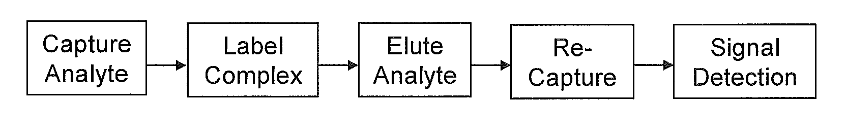
While Sandwich ELISAs are usually specific and sensitive (usually the sensitivity is at or below 0.5-2 ng / ml), this method has three major limitations.
These requirements have, to a large extent, limited the broader application of the Sandwich ELISA.
This is because it usually takes a great effort and a long time to develop such antibody pairs against the same analyte.
For proteins (or protein domains) of low molecular weights or with just a few antigenic epitopes, it is even harder to establishing such coordinately paired antibodies.
This is not only a huge and tedious task, but also the labeling of antibodies may have different efficiency in different lots for each antibody and among different antibodies, causing variations in the detection.
In addition, the chemical modification of the detection antibodies by the labeling process may also affect the antibody-antigen binding.
Third, in performing multiplexed assays, the Sandwich ELISA requires mixing all the labeled detection antibodies together, thus greatly diluting each individual antibody, decreasing the assay signals and increasing nonspecific binding and the background noise.
These limitations prevent broader use of the Sandwich ELISA, particularly making it difficult to use Sandwich ELISA as a major technical platform in the proteomic (for example, using protein microarrays) studies.
First, there are frequently thousands types of molecules of different natures and sizes in a biological sample, and many of them may directly or indirectly interfere with the pre-labeling of the specific analyte in question.
Proteins present at low concentrations may not be labeled efficiently.
Second, the process of pre-labeling may modify the specific antigenic determinant region on protein molecules, lowering or even completely abolishing its ability to bind to the specific capture antibody on the solid phase.
It has been shown that assays based on the analyte pre-labeling method usually have low sensitivity and high background noise.
Although these methods can significantly increase the assay sensitivity, the experimental procedures are rather complicated and tedious, and the cost is high.
More importantly, the limitations of the Sandwich ELISAs mentioned earlier are still present with the immuno-PCR method.

Method used


Image

  • Method and its kit for quantitatively detecting specific analyte with single capturing agent
  • Method and its kit for quantitatively detecting specific analyte with single capturing agent
  • Method and its kit for quantitatively detecting specific analyte with single capturing agent

Examples


specific example 1

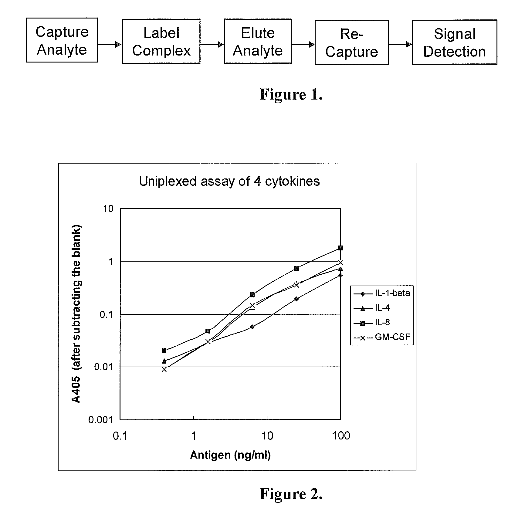
Uniplexed Assay (Detecting One Protein)

[0037]In this Specific Example, the Capture Device and the Detection Device were both made of 96-well microtiter plates. The Capture Device was coated with a single antibody for detecting one antigen. The analytes were 4 human cytokines: IL-1-beta, IL-4, IL-8 and GM-CSF. Their corresponding antibodies were all monoclonal antibodies from Biolegend, USA).

[0038](1) Capturing the Antigens

[0039]a. Coating with the capture antibodies: Two 96-well microtiter plates (flat-bottom, with high binding capacity to proteins) were used, one (the Capture Device) for capturing the antigens in samples, and the other (the Detection Device) for detecting the labeled antigens. To each well, 100 μl of a capture monoclonal antibody at 0.5 μg / ml (diluted in phosphate-buffered saline, PBS) was added. On each plate, 8 wells were coated with each of the monoclonal antibodies against IL-1-beta, IL-4, IL-8 and GM-CSF. The plates were incubated at 4° C. overnight.

[0040]b. B...

specific example 2

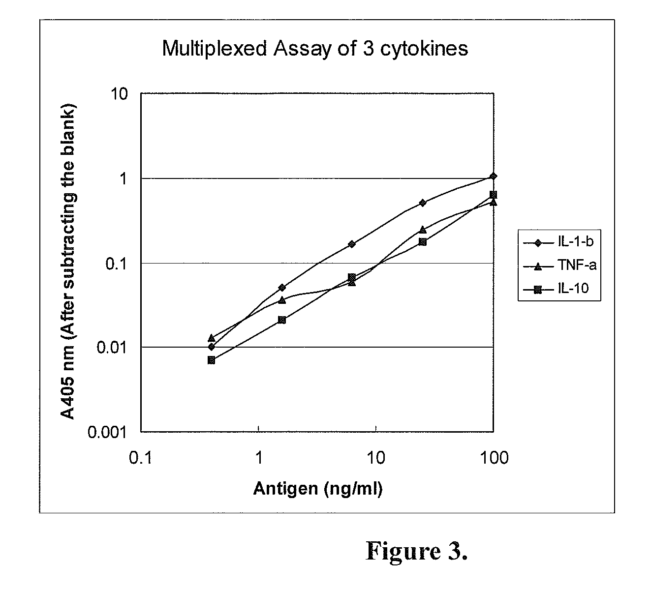
Multiplexed Assay (Detecting Three Proteins Simultaneously)

[0051]In this specific example, the Capture Device and the Detection Device were both made of a 96-well microtiter plates. The Capture Device was coated with a mixture of three antibodies for detection of three antigens simultaneously. The analytes to be tested were 3 human cytokines: IL-1-beta, TNF-alpha and IL-10. Their corresponding antibodies were all monoclonal antibodies.

[0052](1) Capturing the Antigens

[0053]a. Coating with the capture antibodies: Two 96-well microtiter plates (flat-bottom, with high binding capacity to proteins) were used, one for capturing the antigen (the Capture Device) and another for detecting the signal (the Detection Device). The wells in the Capture Device were coated with 100 μl of a mixture of the 3 monoclonal antibodies against human IL-1-beta, TNF-alpha and IL-10, each at 0.5 μg / ml (in PBS). Eight wells were coated. The wells of the Detection Device were separately coated with just one of ...


PUM

PropertyMeasurementUnit
concentrationsaaaaaaaaaa
subcellular structureaaaaaaaaaa
fluorescentaaaaaaaaaa

Abstract

The invention provides a method and its kit for quantitatively detecting a specific analyte with a single capturing agent. The quantitative detection of a specific analyte with a single capturing agent comprises: firstly combining the tested analyte with a solid phase capturing agent, then labeling analyte which has been trapped by the capturing agent with a report molecule; secondly eluting the labeled analyte from the complex, recombining the tested analyte with a new solid phase capturing agent, and ascertaining the content of analyte by detecting the report molecule's label signal. The kit of the invention comprises a capturing device, a detecting device, a report molecule for labeling and an analysis substance eluate. The advantages of the invention are the need of one single capturing agent, the capability of detecting for many analytes which can't be tested at present, wide application, high sensibility and low noise. The invention can be applied to diagnosis, medical expertise, new medicine development, application of protein micro array and chip, and fundamental research.

Description

TECHNICAL FIELD[0001]This invention relates to the field of biotechnology; particularlly, it relates to a new method of using single capturing reagents to quantitatively detect specific analytes, and to reagent kits based on the method.BACKGROUND TECHNOLOGIES[0002]Measuring a specific protein factor(s) in a biological (including human) sample is of great importance in applications and basic researches in medical, biological, agricultural and environment protection fields. For example, detection of specific protein components of pathogenic microorganisms is currently an important tool for the diagnosis of infectious diseases. Similarly, measurement of early specific protein biomarkers in cancers and cardiovascular diseases is extremely important for early diagnosis, early treatment and monitoring treatment efficacy of these diseases. Depending on the purpose of the applications, the analytes to be tested may be a single protein or a few protein factors, or may be a group of proteins ...

Claims


Application Information

Patent Timeline
Patent Type & Authority Applications(United States)
IPC IPC(8): G01N33/543C12Q1/68G01N33/577
CPCG01N33/54306
Inventor SUN, DONGXU
Owner XUZHOU LINGXIN BIOSICENCIES
Why Eureka
Try Eureka