Chemokine receptor antagonists as therapeutic agents

a technology of chemokine receptor and antagonist, which is applied in the field of chemokine receptor antagonists as therapeutic agents, can solve the problems of remain elusive, and the molecular mechanism used by immature dcs or other putative tolerogenic apcs to suppress t cell responses is unclear, so as to reduce immune tolerance

Inactive Publication Date: 2009-02-12
MUNN DAVID H +2
View PDF0 Cites 1 Cited by
  • Summary
  • Abstract
  • Description
  • Claims
  • Application Information

AI Technical Summary

Benefits of technology

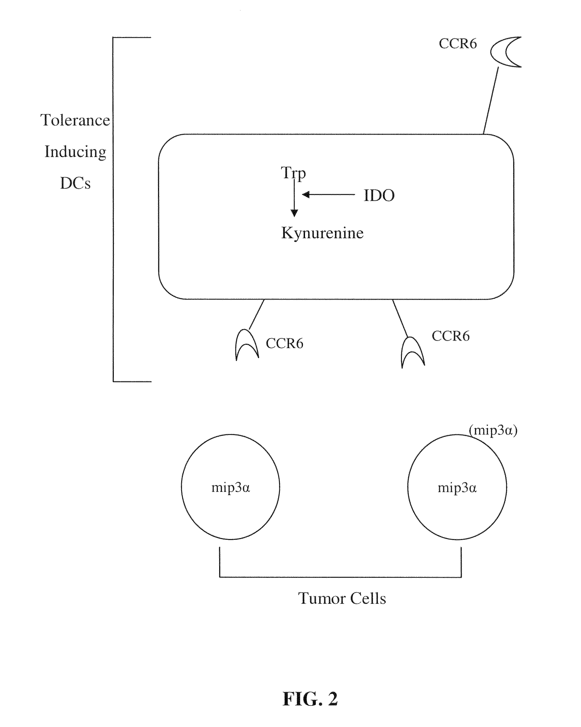
[0010]Embodiments of the present invention recognize that tolerance-inducing (suppressive) antigen-presenting cells (APCs) may express certain cytokine receptors on their surface whereas non-tolerogenic APCs may express a different complement of cytokine receptors on their surface. For example, in one embodiment, the cytokine receptor that is preferentially expressed on tolerogenic APCs is CCR6. The present invention also recognizes that the agents that prevent binding of cytokine receptors expressed on the tolerogenic APCs to a ligand present at the site of APC recruitment may be used to reduce immune tolerance in a subject.
[0012]In another embodiment, the present invention comprises a method to reduce tumorgenicity in a subject comprising administering a composition to the subject to reduce recruitment of tolerance-inducing antigen-presenting cells (APCs) or their precursors to a tumor and / or a tumor draining lymph node in the subject.
[0015]There are many advantages associated with the present invention. For example, in an embodiment, the present invention provides methods and compositions to reduce immune tolerance in a subject at sites where tolerance is detrimental.
[0016]Also, the present invention provides methods and compositions for enhancing the immune response by preventing migration of tolerogenic APCs to a site requiring an immune response.
[0017]The present invention may be used to enhance the immune response to tumors, infectious agents, and / or other pathologies that may trigger inflammation or an immune response. In an embodiment, recruitment of tolerogenic APCs to a tumor and / or a tumor draining lymph node may reduced. For example, the present invention provides methods and compositions to prevent migration of tolerogenic APCs that express the CCR6 chemokine receptor to tumors that express a ligand for CCR6. Additionally, and / or alternatively, recruitment of tolerogenic APCs to a site of viral infection may be reduced.

Problems solved by technology

However, in humans and mammals other than mice, the identity of tolerogenic APCs, and the mechanisms they use to induce tolerance, remain elusive.
However, the molecular mechanism used by immature DCs or other putative tolerogenic APCs to suppress T cell responses is unclear.

Method used


Image

  • Chemokine receptor antagonists as therapeutic agents
  • Chemokine receptor antagonists as therapeutic agents
  • Chemokine receptor antagonists as therapeutic agents

Examples


example 1

Cell Culture

[0163]Human monocytes and lymphocytes were isolated by leukocytapheresis and counterflow elutriation (D. H. Munn et al., J. Exp. Med. 189, 1363-1372 (1999)). Monocytes (typically >95% purity) were cultured in 100 mm tissue culture petri dishes in RPMI-1640 medium with 10% newborn calf serum (Hyclone) and including penicillin / streptomycin and glutamine. Cultures received either MCSF (200 U / ml, Genetics Institute) on day 0, or GMCSF (50 ng / ml, R&D Systems)+IL4 (50 ng / ml, R&D Systems) on days 0, 2 and 4. For experiments where CCR6 expression was of interest, cultures received a single dose of GMCSF+IL4 (100 ng / ml each) on day 0, with no further supplementation. Loosely adherent dendritic cells (GMCSF+IL4) were harvested by gentle aspiration; adherent macrophages (MCSF) and non-dendritic APCs (GMCSF+IL4) were harvested with EDTA. Other cultures were conducted in serum-free medium (X-vivo 15; BioWhitaker, Walkersville, Md.) plus cytokines.

example 2

Production of Antibodies

[0164]All antibodies were obtained commercially except for polyclonal antiserum against human IDO which was manufactured as a work for hire by ZCB Inc., Hopkinton, Mass. All commercial antibodies and reagents were from BD Biosciences-Pharmingen (San Jose, Calif.) unless specified otherwise. For detection of cell surface antigens, DCs were triple-stained with anti-CD123-biotin (clone 7G3; it was found that clone 9F5 gave suboptimal results with dendritic cells) followed by streptavidin-perCP, plus anti-CD11c-allophycocyanin (clone S—HCL-3) or anti-CCR6-fluorescein (clone 53103.111, R&D systems, Minneapolis, Minn.). CCR6 results were also confirmed using a second anti-CCR6 antibody (clone 11A9; Pharmingen). For detection of IDO, cells were fixed and permeablized (Cytofix / Cytoperm), and then stained with rabbit anti-IDO antibody prepared against the peptide followed by polyerythrin-labeled anti-rabbit secondary antibody (Jackson Immunoresearch, West Grove Pa.) c...

example 3

Co-Expression of IDO with Cell Surface Markers CCR6, CD123, and CC11c in APCs

[0166]Expression of IDO in immature monocyte-derived (myeloid) dendritic cells (Dhodapkar, M. V., et al., J. Exp. Med. 193: 233-238 (2001)) and in immunosuppressive monocyte-derived macrophages (Munn, D. H., et al., J. Exp. Med. 189: 1363-1372 (1999)) was analyzed. FIG. 3 shows the expression of IDO and CCR6 by myeloid antigen-presenting cells which express the cell surface antigen CD123 (CD123+). Human monocytes were cultured as described above (Example 1) for 7 days with GMCSF+IL4 to produce myeloid dendritic cells (FIGS. 3A and 3C), or for 7 days in MCSF to produce macrophages (FIG. 3B) (Munn, D. H., et al., J. Exp. Med. 189: 1363-1372 (1999)). Prior to analysis, cells were treated with interferon-γ (INFγ) for 18 hrs to induce maximal expression of IDO. Harvested cells were triple-stained for CD123, CD11c and IDO. For FIG. 3D, cells were cultured as in Example 1 except in a commercial, FDA-approved serum...


PUM

PropertyMeasurementUnit
concentrationaaaaaaaaaa
diameteraaaaaaaaaa
coloraaaaaaaaaa

Abstract

The present invention provides methods and compositions to reduce immune tolerance at specific sites. In one aspect, the present invention comprises methods and compositions to reduce tumorigenicity. In an embodiment, the present invention reduces recruitment of tolerance-inducing antigen presenting cells (APCs) or their precursors to a tumor and / or tumor draining lymph node by decreasing binding of at least one tumor-associated ligand to a chemokine receptor present on the tolerance-inducing APCs or APC precursors. In an embodiment, the chemokine receptor is CCR6 and the tumor-associated ligand is mip-3α. In another aspect, the present invention comprises methods and compositions to reduce immune tolerance to a virus. In an embodiment, the virus is HIV. The present invention further provides for the development of CCR6 antibodies and antagonists as therapeutic agents to prevent or reduce immune tolerance.

Description

RELATED APPLICATIONS[0001]This application claims priority to Provisional Application 60 / 409,804, filed Sep. 11, 2002, and is a divisional application of U.S. patent application Ser. No. 10 / 660,131, filed Sep. 11, 2003. The entire disclosures of Provisional Application 60 / 409,804 and U.S. patent application Ser. No. 10 / 660,131 are incorporated in their entireties herein.FEDERAL FUNDING[0002]The studies described herein were supported at least in part by Federal grants from the National Institutes of Health (NIH R01HL60137; NIH R01 HL57930; NIH R01 AI44219; NIH R21AI49849; NIH R21AI44759; NIH CA 103220; and NIH K08 HL03395), the National Institutes of Health and National Cancer Institute (NIH / NCI / RAID), and the Mason Trust Foundation. Thus, the Federal government may have rights in this invention.NOTICE OF COPYRIGHT PROTECTION[0003]A section of the disclosure of this patent document and its figures contain material subject to copyright protection. The copyright owner has no objection...

Claims


Application Information

Patent Timeline
Patent Type & Authority Applications(United States)
IPC IPC(8): A61K39/395A61KA61K38/00A61K39/00C07K14/00C07K16/28C07K16/40G01N33/50G01N33/53G01N33/68
CPCC07K16/2866C07K16/40G01N2333/52G01N33/5047G01N33/6863C07K2316/96C07K2317/76
Inventor MUNN, DAVID H.MELLOR, ANDREW L.PEIPER, STEPHEN C.
Owner MUNN DAVID H
Why Eureka
Try Eureka