Co-detection of single polypeptide and polynucleotide molecules

a technology applied in the field of co-detection of single polypeptides and polynucleotides, can solve the problems of limited vitro methods for co-detection of polypeptides and polynucleotides, significant time-consuming and laborious,

Inactive Publication Date: 2009-04-02
SINGULEX
View PDF99 Cites 7 Cited by
  • Summary
  • Abstract
  • Description
  • Claims
  • Application Information

AI Technical Summary

Benefits of technology

[0047]FIG. 3. Discrimination of a protein and a nucleic acid based on fluorescence intensity. A) Fluorescence intensity is plotted as a function of the time offset for detection of fluorescence at the two detectors. Each spot represents measurements taken on a single molecule. A value of 500 photons/msec was u

Problems solved by technology

Known in vitro methods for co-detection of polypeptide and polynucleotide molecules are limited in that they require large amounts of sample, complex and expensive equipment, or a significant amount of time to perform.
These measurements obscure valuable information about properties of individual molecules.
One of the major disadvantages of these techniques is the need for large (pg to ng) quantities and high concentrations of target molecules (Haugland, 2002).
Also, the run and analysis takes a long time (from a few hours to overnight).
Further, quantitation requires densitometry measurements that are generally not precise, making it difficult to obtain accurate information.
Although these techniques are fast to perform and a large number of events can be analyzed, detectable fluorescence intensity from single molecules cannot be achieved because it requires multiple bound target molecules and only single cells or particles are detected.
Another disadvantage of this technology is that it does not allow for measurement of isolated molecules but requires they be attached to cells or beads.
Precise quan

Method used


Image

  • Co-detection of single polypeptide and polynucleotide molecules
  • Co-detection of single polypeptide and polynucleotide molecules
  • Co-detection of single polypeptide and polynucleotide molecules

Examples


example 1

Discrimination of a Protein and Nucleic Acid Directed Labeled with the Same Fluorescent Dye—Electrophoresis with Linear Polyacrlyamide

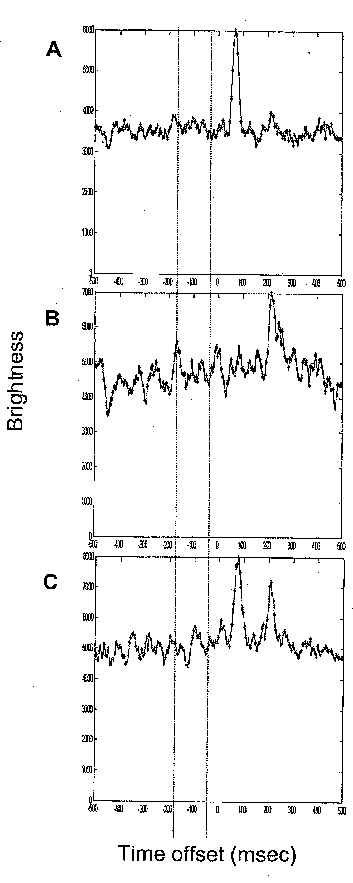
[0138]Samples of Alexa Fluor® 647-labeled IgG and 1.1 kb PCR product were prepared in 18 mM tris, 18 mM glycine, pH 8.6 with 0.2% linear polyacrylamide (LPA, 5,000,000-6,000,000 mw), 0.01% sodium dodecyl sulfate and 1 μg / ml each bovine serum albumin, Ficoll®, and polyvinylpyrrolidone. Samples were pumped into the SMD capillary, the pump was stopped, and an electric field was applied (300 V / cm). Cross-correlation of the molecules was determined as a function of time offset. One minute data sets were collected and analyzed.

[0139]Examples of the histogram plots of the molecule cross correlations are shown in FIG. 1. A. 26 fM Alexa Fluor 647 labeled IgG (cross-correlation peak at 75 msec). B. 10 fM Alexa Fluor labeled PCR product (peak at 220 msec). C. 13 fM Alexa Fluor 647 labeled IgG and 5 fM Alexa Fluor labeled PCR product (peaks at 75 and 215 msec).

example 2

Discrimination of a Protein and Nucleic Acid Directed Labeled with the Same Fluorescent Dye—Electrophoresis

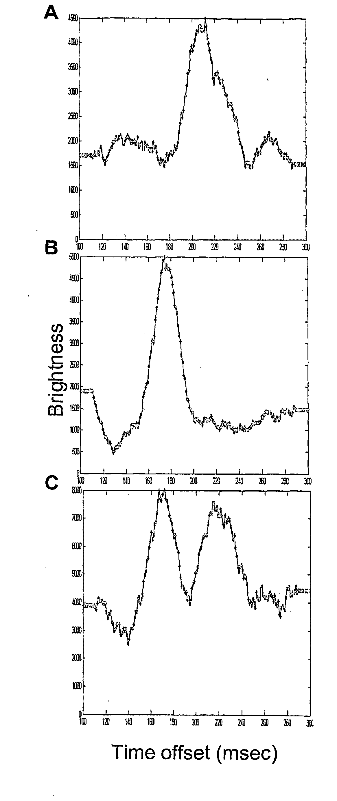
[0140]SMD electrophoretic separation of a protein and a nucleic acid. Samples of Alexa Fluor® 647 labeled IgG and 1.1 kb PCR product were prepared in 18 mM tris, 18 mM glycine, pH 8.6 with 0.01% sodium dodecyl sulfate and 1 μg / ml each bovine serum albumin, Ficoll®, and polyvinylpyrrolidone. Samples were pumped into the SMD capillary, the pump was stopped, and an electric field was applied (150 V / cm). Cross-correlation of the molecules between channels 1 and 2 was determined as a function of time offset. One minute data sets were collected and analyzed. a: 52 fM Alexa Fluor 647 labeled IgG (cross-correlation peak at 210 msec). b: 20 fM Alexa Fluor labeled PCR product (peak at 175 msec). c: 26 fM Alexa Fluor 647 labeled IgG and 10 fM Alexa Fluor labeled PCR product (peaks at 170 and 215 msec).

example 3

Differences in the Characteristic Intensity of Fluorescence Emission were Used to Distinguish a Protein Complex and a Nucleic Acid within a Mixture

[0141]This example also demonstrates that the concentration of sample components can be determined by comparing the counts detected to a standard curve. The protein, PBXL-3, emits at a generally high intensity, and the nucleic acid, linearized pUC19, emits at a generally lower intensity. PBXL-3 is an intrinsically fluorescent protein complex. The pUC19 DNA was labeled with Alexa Fluor® 647 following the protocol of a ULYSIS® nucleic acid labeling kit (Molecular Probes, Inc. Eugene, Oreg.). PBLX-3-strepavidin was purchased from Martek Biosciences Corp. (Columbia, Md.). Phosphate Buffered Solution (PBS) (10 mM NaPO4, 150 mM NaCl, pH 7.2) was supplemented with 0.01% Casein Acid Hydrolysate and used to make dilution series (2.5, 5, 7.5, 10 and 20 fM) of protein alone, nucleic acid alone or mixtures of both. Samples were moved through the anal...


PUM

PropertyMeasurementUnit
Pressureaaaaaaaaaa
Volumeaaaaaaaaaa
Intrinsic viscosityaaaaaaaaaa

Abstract

The invention relates to a method for co-detecting and distinguishing individual polypeptide and polynucleotide molecules in a single sample analysis. The polypeptide and polynucleotide targets which emit electromagnetic radiation are labeled with probes that emit electromagnetic radiation at the same emission wavelength, are suspended in fluid and moved through an interrogation volume allowing co-detection of low concentrations of individual molecules and discrimination based on characteristics other than emission wavelength.

Description

CROSS-REFERENCE TO RELATED APPLICATIONS[0001]This application claims priority from U.S. Provisional Application Ser. No. 60 / 492,137 filed on Jul. 31, 2003, which is incorporated herein by reference in its entirety.STATEMENT REGARDING FEDERALLY SPONSORED RESEARCH OR DEVELOPMENT[0002]Not Applicable.INCORPORATION-BY-REFERENCE OF MATERIAL SUBMITTED ON A COMPACT DISC[0003]Not Applicable.BACKGROUND OF THE INVENTION[0004]1. Field of the Invention[0005]The invention relates to co-detecting single polypeptide and polynucleotide molecules in a single sample analysis.[0006]2. Description of Related Art[0007]The ability to co-detect proteins and nucleic acids is important for determining reactions and interactions during complex processes such as those that occur in biological systems. Most organisms contain both of these biomolecules, and the biological processes often involve measurable changes in both proteins and nucleic acids that help to elucidate their function. Known in vitro methods fo...

Claims


Application Information

Patent Timeline
IPC IPC(8): G01N33/483G06F19/00C07H21/02C07H21/04C12NC12Q1/68G01N33/00G01N33/53G01N35/00
CPCG01N33/5308C12Q1/6825
Inventor FUKUSHIMA, NOELLE H.PUSKAS, ROBERT S.
Owner SINGULEX
Why Eureka
Try Eureka